A person’s feet and heels perform an important shock-absorbing function; thanks to this structure of the feet, a person can move on two legs. But if pain appears in the foot area, this may be a sign of a serious pathology that requires treatment.
There are many causes of pain in the feet; if heel pain occurs in a child, then this may be a sign of apophysitis of the calcaneus. This pathology causes a lot of inconvenience to the child; he cannot walk or exercise normally, so at the first signs of the disease, the child must be shown to a specialist.
Definition
Calcaneal apophysitis is an inflammatory disease that occurs before the age of 14 years. In a child, all bones have cartilaginous layers called growth plates, including in the heel area. In adults, cartilage is replaced by bone tissue, the bone becomes solid.
During the active growth of a child, the bones lengthen, and the muscles that cannot keep up with it become tense, resulting in tension in the Achilles tendon. If the child is also subject to increased physical activity, inflammation and pain occur in the heel area.
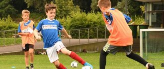
Most often, the disease occurs in child athletes, and its exacerbation occurs in the autumn, when the child returns after the summer holidays and begins to actively train. In general, calcaneal apophysitis does not cause serious complications, but it can cause very severe pain, so the pathology requires attention.
Osteochondrosis of the foot and lower limb
The faster a child develops, the more vulnerable growth points become. This is especially noticeable in boys aged 4, 7-8 and 12 years. That is, at the age when the next growth spurt occurs and children begin to actively engage in sports. And parents worry about their sports success.
What to pay attention to, how not to miss the onset of the disease or its complications was discussed with Olga Chizhevskaya, executive director of the League for the Promotion of Podiatry.
Signs of susceptibility to osteochondrosis
Bone develops from hyaline cartilage, in which the processes of ossification and calcification gradually occur. And by the time of birth, the only areas where hyaline cartilage remains are the articular surface and the growth plate area. Moreover, in the latter we observe the presence of cartilage even during the period of skeletal maturation. That is, this area remains vulnerable even in adulthood.
There are three main factors that influence the development of osteochondrosis: the intensity of growth, the weight of the child and his activity.
The child's weight is associated with the vertical load on the foot and lower limb. When the optimal body weight is exceeded, the foot experiences additional stress, which can lead to both the development of flat feet and more serious problems, including osteochondrosis.
“Growth spurts often coincide with the start of a particular sport. If swimming or cycling unload the foot, then running or football with increased vertical loads, on the contrary, contribute to the development of degenerative changes.
In this case, it is useful to alternate between different sports. This will help not only reduce the risk of developing osteochondrosis, but also harmoniously develop the musculoskeletal complex and even allow the child to achieve more significant sports results,” says Vladimir Nechaev, orthopedic traumatologist, chiropractor, sports doctor, osteopath.
Diagnosis of osteochondrosis
The first thing a doctor should pay attention to when diagnosing osteochondrosis of the lower extremities is the patient’s age. Due to the physiology of the development of the skeletal system, different parts and bones are most vulnerable at different periods of life.
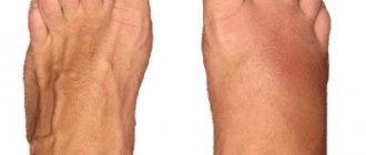
The second is clinical signs. Most often this is pain without signs of inflammation. If a local increase in temperature, redness of the skin and swelling is observed in the affected area, more serious inflammatory diseases may develop.
The third diagnostic criterion is the results of x-ray examination. But they can only be valid if there is a clinical picture.
Kohler's disease – osteochondrosis of the scaphoid bone
This is an uncommon variant of osteochondrosis, occurring in boys aged 2 years and older. At this age, the child begins to walk, the foot begins to experience weight bearing, and secondary foci of ossification appear in the scaphoid bone.
With this disease, the child simply refuses to put any weight on the affected leg and refuses to walk. Attempts to examine and palpate also cause severe pain and crying. In this situation, only careful superficial palpation is possible, during which pain can be detected specifically in the area of the scaphoid bone.
The main danger of this condition is that aseptic necrosis may develop. It is also always worth remembering the risk of complications, including infectious ones. Therefore, if the examination reveals not only pain, but also signs of inflammation, it is worth taking a particularly careful approach to diagnosis.
The simplest treatment method is to apply a plaster immobilizing bandage for 4-6 weeks. Also, depending on the individual foot condition, orthoses may be used to relieve weight on the affected limb. When selecting treatment, you should always pay attention to the severity of the pain syndrome and the rate of restoration of function of the lower limb when the weight load is reduced.
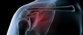
Sever's disease – apophysitis of the calcaneus
The most common type of osteochondrosis of the foot. The disease develops in children aged 7-14 years. It is at this age that a secondary ossification center appears in the calcaneus. These children will complain of heel pain after playing sports and occasional limping. If the heel is squeezed during examination, the child will feel pain.
Clinically, the doctor must confirm that it is the apophysis that is affected, and not the heel bone or ankle joint itself. If there are signs of inflammation or the child complains of pain not only after playing sports, but also during other periods, then it is worth taking a more careful approach to the diagnosis to exclude complications, including idiopathic juvenile arthritis.
During treatment, it can be recommended to stretch the calf muscle group and use shoes with high heel pads to relieve the necessary parts of the foot and growth plate. With the correct selection of shoes, the child can return to sports and continue training.
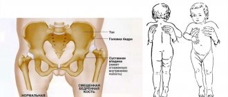
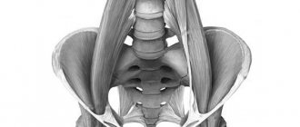
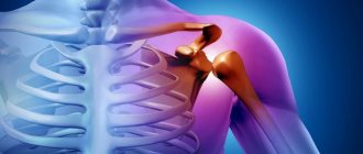
Perthe disease – osteochondrosis of the hip joint
This disease develops from age 4 and affects children under 10 years of age. Boys get sick 4 times more often than girls. As a rule, the joint is affected only on one side, although bilateral disease is possible.
Clinically manifested by lameness. Upon examination, the child has a reduced range of motion in the affected hip joint compared to the healthy one. The radiograph shows fragmentation of the articular surface.
Also, this type of osteochondrosis can manifest itself as pain in the knee. Therefore, if, with characteristic complaints during examination, there are no structural changes specifically in the knee area, it is worth paying attention to the hip joint.
Management of such a patient involves mainly rest and unloading of the affected limb. But it is difficult for a child at this age to give up activity. In this case, you can replace running with swimming to relieve the load on the hip joint.
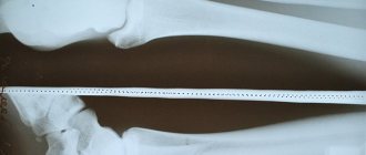
Osgood-Schlatter disease – osteochondrosis of the tibial tuberosity
Just as the heel is the most affected bone of the foot, the knee is the most common osteochondrosis of the lower limb.
This condition develops in children as young as 8 years old, with an average age range of 8-15 years. This disease is called a self-limiting condition, that is, it is believed that by the age of 15-18 it goes away on its own. But recent research suggests that not everything is so simple.
Approximately 40% of children still have signs of the disease 2 years after the onset of the disease. That is, the child is still limited in his physical activity. In 25% of children they persist even 8-10 years after manifestation.
Clinically, the child experiences pain when running, playing sports, and any physical activity that requires sudden extension movements of the knee. Upon examination, the area of the tibial tuberosity is sharply painful on palpation. The patella can stand quite high.
It is recommended to ensure rest during the first month. It is also worth changing the type of activity from running or other sports with a high load on the knee to swimming. Good results are achieved by training aimed at strengthening the quadriceps femoris muscle.
When making differential diagnoses, it should be remembered that similar signs can also result from more serious diseases. The most dangerous of them is osteogenic sarcoma. In this case, a characteristic symptom will be pain at night.
When diagnosing osteochondrosis of the lower extremities, it is worth paying attention not only to the clinical picture, but also to the age of manifestation, which is characteristic of certain diseases. It is also important to watch for developing complications such as idiopathic juvenile arthritis, infectious complications, trauma, osteosarcoma, or avascular necrosis.
As Olga Chizhevskaya, executive director of the League for the Promotion of Podiatry, emphasizes, when treating osteochondrosis in children, it is necessary to provide rest to the affected joint by limiting physical activity or applying a splint. You can also relieve the joint with orthopedic shoes or orthoses, change the type of physical activity and alternate between different sports.
Causes
As a rule, the disease does not appear for any specific reason, but is a consequence of the impact of several negative factors on the child’s body:
- Increased physical activity, frequent training. In children under 14 years of age, the heel bone is not yet formed, so the loads should be selected according to age.
- Rapid growth of the child. This feature occurs in many children; in this case, a sharp growth of bones occurs, resulting in overstretching of the muscles and Achilles tendon.
- Congenital pathologies of bone tissue, serious skeletal diseases.
- Lack of calcium in the body.
- Large excess weight. In this case, the unformed bone constantly undergoes enormous loads.
- Incorrectly selected shoes. In this case, very tight shoes that rub and press can provoke the disease. If you have the disease, you should not wear ballet flats, sneakers or other flat-soled shoes, as the pain will worsen.
- Incorrect gait, when the child does not stand on the entire foot, but mainly on the heel, as a result of which the load on it increases.
- According to statistics, boys aged 7 to 14 years are more susceptible to the disease.
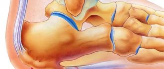
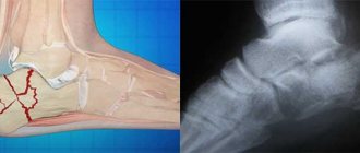
Anatomy of apophysitis
It can be noted that after the birth of a child, his bones are some kind of cartilage, some of which develop into hard bones. As calcaneal osteogenesis develops, one large area in the central part of the heel cartilage can be noted to become ossified. This area of the heel is the main site for the development of bone tissue, which will gradually replace cartilage. The second zone of bone formation can be called the apophysis - the back part of the heel bone.
In the space between the two areas there is a cartilaginous layer, which disappears when a person reaches 16 years of age, after which these two bone areas merge into one.
Diagnostics
Pathology on X-ray
Despite the pronounced symptoms, only a doctor can make a correct diagnosis. It is worth understanding that heel pain can also occur due to injury, for example, if a child lands poorly when jumping on his heel.
To identify the cause of the pain, the patient is sent for an x-ray; it is carried out in several projections to examine the heel bone in detail from all sides. This study helps to identify any abnormalities in the affected area and prescribe the correct treatment.
What are the dangers of foot deformities?
Foot deformities cannot be ignored. Without treatment and correction, over time they will lead to disorders in the ankle, knee, hip joints, incorrect position of the pelvis, and overload of individual muscles. They can cause premature wear of overlying joints in adulthood, the development of osteochondrosis, arthrosis, curvature of the spine, and poor posture.
Foot deformities affect the general condition of the child, cause pain, fatigue, make walking difficult, and contribute to improper development of the musculoskeletal system. They lead to other foot problems: ingrown toenails, crooked toes, calluses, corns.
Treatment
Apophysitis of the calcaneus is treated only with conservative methods. First of all, the patient is advised to remain calm and active training is prohibited during the recovery period. Massage and special physical therapy are also prescribed, this will help relieve pain and speed up recovery.
It is worth understanding that physical therapy is prescribed individually, taking into account the child’s condition, and a specialist monitors the exercises. Loads should be minimal, and you can start training only after prescribing exercise therapy; until then there should be no amateur activity. An equally important part in the treatment of calcaneal apophysitis is shoes; they must be properly selected. Special orthopedic insoles can be useful here, they will relieve the heel and relieve pain when moving. It is forbidden to wear shoes with flat soles, since in this case the pressure on the heel is maximum.
If the child is experiencing severe pain, then drug treatment is indicated. As a rule, non-steroidal anti-inflammatory drugs, for example Nurofen (Ibuprofen), are prescribed to relieve pain and inflammation. The child is also prescribed vitamin complexes to quickly restore the affected area.
For heel apophysitis, the intake of ascorbic acid, calcium, and vitamin D is indicated. The child is also recommended to have a proper and balanced diet, but preference should be given to dairy products, fish, vegetables and fruits, and the consumption of sweets, salty and junk foods should be greatly limited. During the treatment period, physical therapy is also prescribed; it will help get rid of inflammation faster and relieve pain. Calcaneal apophysitis is often treated with balneotherapy, that is, therapeutic salt baths. In this case, baths according to folk recipes, with herbal decoctions, also help well.
Folk
Folk remedies are often used for calcaneal apophysitis; they help to quickly relieve pain and inflammation and alleviate the condition. But it is impossible to cure the disease with folk recipes; therapy must be comprehensive and under the supervision of a specialist.
Before using the product, you need to make sure that the child does not have an allergic reaction to it, and you should not apply external ointments and decoctions to damaged skin. The following remedies will help relieve pain from calcaneal apophysitis.
Contrasting baths have a wonderful effect. To do this, hot and cold water is poured into 2 basins, but it should not be icy or too hot. Then the child puts his feet in hot water for 5 minutes, transfers them to cold water for 10 seconds, and again to hot water for 1 minute, then again to cold water for 10 seconds, and again to hot water for a minute. In general, the duration of the procedure is no more than 15 minutes. A warm salt bath helps with heel pain; to prepare it you need to use natural sea salt. The duration of the procedure is 10-15 minutes.
Another effective remedy is a warm compress of potatoes and lugol. It is necessary to boil the potatoes and mash them into a thick puree, then add lugol to it and mix quickly. Place the warm puree in a bowl and place your heels in it for 5-7 minutes, and after the procedure, put warm woolen socks on your feet.
Horseradish root is very effective for heel pain. It is crushed in a blender or on a grater, and the resulting pulp is applied to the heel for 2 hours, the product must be secured on top with cling film and socks must be put on. A compress with radish also helps with heel pain. To prepare them, take black radish, wash it well and grate it, apply the pulp to the heel and wrap it with cling film, put warm socks on top and leave for 3-4 hours.
You can remove the inflammatory process with a compress of salt, honey, and iodine. For one bottle of iodine take 1 tbsp. honey and 1 tsp. salt. The product is mixed well and applied to a napkin, then applied to the sore spot for several hours, secure the product with cling film on top and put on socks.
If, while using a folk remedy, a child complains of a strong burning sensation, the skin should be rinsed with running water and the recipe should not be used again. If rashes and itching appear after using the product, you should definitely consult a doctor for advice and abandon traditional medicine recipes.
Treatment: effective drugs and auxiliary methods
The goal of therapy is to transfer the acute inflammatory process into remission. For this, rheumatologists prescribe complex treatment. It includes:
- absolute rest for the feet;
- medications;
- herbal medicine;
- physiotherapeutic procedures;
- massage.
It is useful for children with this pathology to eat vegetable dishes.
Quartz irradiation and foot baths have a good effect. Bandaging and dressings eliminate overload of the foot. Arch supports ensure correct positioning of the leg and faster healing. Nutritionists actively correct patients’ diets, recommending juices and smoothies, vegetable dishes enriched with vitamins and minerals. The table presents pharmacological medications for the treatment of calcaneal apophysitis and their therapeutic effect:
| Drug groups | Therapeutic effect | Medication |
| NSAIDs | Reduce inflammation and relieve pain | "Nurofen" |
| "Nise" | ||
| "Sulindak" | ||
| Analgesics | Eliminate pain attacks | "Paracetamol" |
| "Panadol" | ||
| "Calpol" | ||
| Calcium preparations | Necessary for bone formation | "Kalcemin" |
| "Calcium gluconate" | ||
| "Complivit Calcium D3" | ||
| Vitamin D | Regulates phosphorus-calcium metabolism, ensures the strength of bone and cartilage tissue | "Vigantol" |
| "Ergocalciferol" | ||
| "Alpha D3-Teva" |
Prevention
It is quite possible to prevent the occurrence of the disease; to do this, you must follow the following recommendations:
- Choose the right shoes for your child, not too tight, preferably made from natural materials and not with flat soles, but with small heels.
- If a child plays sports, training should not be too intense; exercise should be done taking into account the child’s age.
- Sports children should definitely massage their feet in the evening after training to normalize blood circulation in the feet and prevent tissue destruction.
- Swimming is a good prevention of bone diseases, so it is recommended to send children to the pool.
- A child's diet should be healthy and balanced; overeating and weight gain should not be allowed. If a child is obese, you need to contact a nutritionist as soon as possible and begin treatment according to his recommendations. Putting your child on a diet on a whim is not recommended; it can be dangerous.
In general, disease prevention is a healthy lifestyle and moderation in everything, that is, in food, in sports.
Parents need to understand that there must be a golden mean in everything, and the child will achieve success even with less intense training, and there will be no harm to health. Share:
Is it possible to prevent osteochondropathy?
There are some recommendations that you can follow to prevent the development of this disease:
- The right shoes: not too narrow, not too wide, with a small heel.
- Optimal physical activity, which should not be excessive.
- Daily massage with rubbing, pressing movements.
- Classes in the pool.
In any case, pain in the foot does not occur on its own, and to find out its nature, it is recommended to consult a doctor.
How to warn?
To prevent foot deformities in children, doctors advise parents to follow simple recommendations:
- You shouldn’t put any weight on your baby’s legs before 7–8 months, don’t try to force him to stand on his legs, teach him to walk early. When the child is ready, he will do it himself;
- consult a pediatrician about preventing rickets, walk more in the fresh air on sunny days to ensure a sufficient supply of vitamin D;
- choose the right shoes for your child with a high, rigid heel that helps keep the heel on the axis of the shin, a bendable sole, and an instep support;
- make sure that the child is active, moves a lot, and does not gain excess weight;
- provide adequate nutrition, rich in vitamins and minerals necessary for the proper development of the musculoskeletal system;
- do leg exercises with your child;
- undergo regular examinations by an orthopedist (at 1, 3, 6 months, at 1 year, at 3 years, from 4 years - once a year), especially if there is a hereditary predisposition to orthopedic diseases.
child orthopedics