In the International Classification of Diseases (ICD), there is no separate group “breast cyst”. This type of formation is classified as fibrocystic diseases (ICD 10 code N60.0 - mastopathy).
The cyst looks like a cavity containing liquid secretion. It is predominantly benign in nature. It can be asymptomatic for a long time. It is a common pathology occurring in women of all ages.
Cystic cavities only in isolated cases resolve on their own, so it is necessary to treat the breast cyst.
We will tell you:
- Causes of breast cysts
- Risks of the disease
- Treatment of breast cyst
- Diagnostics
- Drug treatment
- Surgical methods of treatment
- Treatment without side effects
- The effectiveness of using Neurodoctor
Treatment of gums with salt
Causes of gum inflammation
Why salt helps
How to properly rinse your mouth with saline solution
Salt gum massage
Other methods for treating gums at home
Bleeding gums, increased sensitivity and bad breath indicate that your gums are not healthy. Why do they become inflamed? Is it necessary to see a doctor or can they be strengthened at home? How to use salt to relieve inflammation? Read more in the article.
Why is it important to treat breast cysts?
Small cyst formations are not dangerous. They do not manifest themselves and are felt only upon palpation, but they are capable of growing. The patient experiences chest discomfort and sometimes pain. Large tumors cause deformation (change in shape) of the mammary gland, which becomes visible externally.
If a cyst exists for a long time, it becomes inflamed, which leads to the development of infections and suppuration of the cystic contents. These processes can cause purulent mastitis and breast abscess. They are manifested by severe pain even without palpation of the chest, fever and swelling. The skin in the affected area turns red, body temperature rises, weakness appears, appetite disappears, and nausea and vomiting are possible.
Despite the benign nature of the disease, the possibility of malignant degeneration cannot be completely excluded, especially for intraductal cysts.
How to treat a breast cyst?
Diagnostic methods
By palpation, the doctor can identify medium and large cysts. Small formations are detected only by ultrasound or mammography.
In addition to size, these studies make it possible to establish the structure of the neoplasm and the presence of small chambers in the bladder. Using mammography, differential diagnosis with fibroadenoma is carried out.
How to treat a breast cyst? There are two directions:
- Conservative therapy.
- Surgical removal.
Drug treatment of breast cysts
Is it possible to cure a breast cyst by taking medications? Yes, the goal of drug treatment is to normalize metabolic processes in breast secretion and suppress the production of the lactotropic hormone prolactin.
An important condition for the effectiveness of non-surgical treatment of breast cysts at home is following a diet that excludes salt, caffeine, chocolate, fruits and vegetables high in fiber.
Most often doctors prescribe the following medications:
- hormonal: progestogens (Duphaston, Utrozhestan), estrogens (Femoston), inhibitors of pituitary gonadotropic function (Buserelin, Goserelin), inhibitors of prolactin secretion (Bromocriptine, Cabergoline);
- symptomatic: sedatives (Gelarium), choleretic and hepatoprotectors (Exhol, Antraliv, Essentiale), antiprostaglandins (Nimesulide, Naproxen), diuretics (Furosemide, Spironolactone), immunomodulators (Tiloron, Cycloferon);
- non-hormonal: vitamin complexes (Ascorutin, ascorbic acid, vitamin E), herbal medicine (Indinol, Mastiol Edas, Klamin, Mastodinon, Mastopol).
The addition of an inflammatory process is the basis for prescribing drugs from the NSAID group (Dicloberl, Diclofenac).
The behavior of the cyst is monitored using mammography and ultrasound.
Surgical treatment of breast cysts
Surgical treatment of a breast cyst is required if conservative therapy is ineffective, the formation is large or has spread to neighboring tissues.
There are three methods of surgical treatment:
1) The puncture is carried out through a puncture using a biopsy syringe and fluid is pumped out from the neoplasm. Subsequently, the walls of the cyst stick together and resolve. The procedure is performed under ultrasound guidance for approximately 1 hour. Local anesthesia is sufficient for pain relief.
2) The essence of the laser method is to expose the cyst to light: the doctor inserts an LED through a puncture in the chest - the laser beam has a destructive effect on the cyst capsule without affecting healthy tissue. The operation is low-traumatic and painless, requires local anesthesia, after it there are almost no scars, and there are no complications.
3) Abdominal surgery is performed if the cyst has growths inside the capsule or is large in size, as well as if a malignant disease is suspected. Under general anesthesia, the woman with the cyst and surrounding tissues (at least 1 cm around the formation) is excised through an incision, and the incision site is sutured. The tumor is sent for histology to exclude oncology.
Rehabilitation after cyst removal
In the first two days after surgery, analgesics are prescribed to relieve pain, as well as antibiotics to reduce inflammation and prevent the spread of infection. After 2–3 days, the drainage is removed, and the sutures are removed on days 7–10.
During the rehabilitation period, the following rules should be observed:
- refuse physical activity;
- adhere to proper nutrition, increase the amount of protein foods;
- follow the doctor’s recommendations and take prescribed medications;
- wear underwear made from natural fabrics that do not rub or irritate the skin of the breast (a sports bra is excellent);
- see a mammologist.
Is it possible to cure a breast cyst without the risk of side effects?
Possible side effects of hormonal therapy for pathology:
- from the genital organs and mammary gland: irregular menstrual cycle, oligomenorrhea, amenorrhea, menorrhagia, metrorrhagia, increased sensitivity, swelling and/or tenderness of the mammary glands, decreased libido;
- from the digestive system and liver: nausea, gastric dysfunction, bloating, vomiting, liver dysfunction;
- neoplasms: increase in size of progestogen-dependent formations;
- other: weight gain, edema, headache, migraine, dizziness, drowsiness, depression, hemolytic anemia, hypersensitivity reactions to the drug, urticaria.
In addition, drug therapy has a number of contraindications, including periods of pregnancy and lactation, suspected breast cancer or a history of oncology, vaginal bleeding of unknown etiology, untreated endometrial hyperplasia, etc.
Any, even minimally invasive, surgical intervention is stressful for the female body. If a course of medications did not help get rid of a breast cyst or there are contraindications to it, you should pay attention to the “Neurodoctor” - a device awarded the Nobel Prize in Physiology or Medicine.
How it works?
Neurodoctor affects the control centers of the brain.
The brain begins to produce dozens of neurohormones and form “corrective” nerve impulses.
Neurohormones are many times more effective than the most powerful drugs in quickly curing the disease. Nerve impulses eliminate pathological processes at the cellular level and correct the functioning of other organs and systems of the body.
LEARN MORE ABOUT THE METHOD
How to get rid of a breast cyst? The effectiveness of Neurodoctor in treatment
Neurodoctor is an innovative device based on a light treatment method with a thousand-year history. It has long been established that each organ has a specific color. The operating principle of the device is the resonant interaction of colors that match the frequency parameters of human organs.
The device supplies light to the retina of the eye, from where it is immediately transmitted through the pineal gland to the pineal gland and other structures of the body, where it affects them. Vibrations weaken or increase, as a result of which the body’s work weakens or increases accordingly, which has preventive and therapeutic effects.
For breast cysts, Neurodoctor helps suppress the production of prolactin and normalizes metabolic processes in breast secretion. Under the influence of the device, an antioxidant and immunostimulating effect develops, promoting rapid recovery. As a result, cyst formation is gradually depleted, the cyst capsule sticks together and gradually resolves.
According to patient reviews, after just a few sessions of using the device, the tumor is significantly reduced in size. You can check Neurodoctor with a unique guarantee - 100% refund in case of ineffective treatment.
Causes of gum inflammation
Gum inflammation can occur for various reasons. Heredity, poor environment, bad habits, hormonal imbalance, etc. But most often, the inflammatory process in the gums occurs due to poor oral hygiene. The more plaque and soft deposits on the teeth, the higher the risk of inflammation. To prevent diseases of teeth and gums, professional hygiene is recommended, which must be done every six months (more often if necessary). The procedure removes deposits that the toothbrush did not reach, freshens breath and makes teeth noticeably whiter.
By the way, scientists conducted an interesting experiment. People who maintained good hygiene and had no dental problems stopped brushing their teeth within three weeks. The result showed that this time is quite enough for primary inflammation to appear on the gums.
Chapter 7. COMPLEX TREATMENT OF PATIENTS WITH OPEN TROPHIC ULCERS OF THE LOWER LIMB
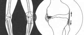
Venous trophic ulcers (VTU) of the lower extremities became the object of attention of doctors even during the existence of ancient civilizations. In the Ebers papyrus, dating from 1550 BC. e., there is a mention of varicose veins and trophic ulcers on the legs. Hippocrates believed that ulcers of the lower extremities are associated with enlarged veins, the treatment of which was proposed to be carried out by puncturing the enlarged veins and bandaging the lower extremities. The term “varicose ulcer” itself was first proposed in 1676 by R. Wiseman, a surgeon at the court of the English king Charles II. The formation of trophic ulcers occurs, as a rule, in areas subject to the greatest “hydrodynamic bombardment” by pendulum-like movements of excess blood volumes caused by the failure of the valve apparatus of the perforating and superficial veins. Therefore, the most common location for lower extremity ulcers is the lower third of the leg above the medial malleolus. There are frequent cases of circular tissue damage to the lower leg.
Violation of blood outflow through deep or superficial veins leads to its deposition. As a result, the pressure in the venous part of the capillary increases, reducing the arteriovenular gradient necessary for normal tissue perfusion. The consequence of these processes is first periodic and then constant tissue hypoxia, leading to destabilization of the intercellular relationships of the endothelium of the venous capillaries, triggering the activation of genes encoding the synthesis of various adhesion molecules. Red blood cells, which have a more stable and ergonomic shape, push white blood cells to the periphery of the vessel. Endotheliocytes respond to these changes and trigger a mechanism known as “leukocyte rolling.” With continued long-term exposure to the provoking factor, strong adhesion of leukocytes to endothelial cells occurs. The pathological cascade continues with the infiltration of leukocytes and their metabolic products, which maintains aseptic inflammation and ultimately leads to macroscopic changes in the venous bed.
The appearance of a trophic ulcer is preceded by a number of symptoms indicating decompensation of blood outflow from the lower limb. The progression of venous outflow disorders against the background of varicose veins or post-thrombotic disease leads to increased swelling of the lower leg, pain in the lower extremities, night cramps, and skin itching. Extravasation of formed elements and blood plasma proteins into soft tissues is clinically manifested by the formation of pigment spots and compaction of subcutaneous tissue. Subsequently, the zones of hyperpigmentation and liposclerosis merge, the skin thickens, becomes tense, immobile, and painful. The breakdown products of blood cells that accumulate in soft tissues, having antigenic properties, cause an inflammatory reaction, manifested by skin hyperemia and eczematous dermatitis. This is also facilitated by the direct damaging effect on tissue of reactive oxygen species and cationic proteins of neutrophil granulocytes. Destruction of the lymphatic plexuses leads to lymphedema. The skin of the lower leg takes on an “orange peel” appearance. Subsequently, in the area of the greatest pathological changes, a focus of exfoliation of the epidermis appears, which outwardly looks like a whitish spot, reminiscent of paraffin deposits. This pre-ulcerative condition is called white skin atrophy. Against this background, the slightest injury is enough for the formation of an ulcerative defect.
Diagnostic tasks that directly determine treatment tactics include assessing the nature and localization of disturbances in the venous outflow from the affected limb and assessing local manifestations, such as the location and size of the ulcer, characteristics of the tissues forming the bottom of the ulcer, the nature and type of wound discharge, and a general assessment of the somatic status patient.
Among the many existing methods for wound surface planimetry, the most convenient and easy to use is the method based on counting the number of marking squares of a given area. The rate of epithelization of trophic ulcers in dynamics was determined using the formula of L.N. Popova: S – Sn/t, where S is the initial area of the ulcer, Sn is the area of the ulcer during subsequent measurements, t is the number of days between measurements.
Identification and quantitative determination of microflora in the tissues of trophic ulcers, as well as dynamic morphological assessment of the course of the wound process, make it possible to analyze the effectiveness of the treatment and timely correction of therapeutic measures.
The timeliness and correctness of diagnosis of the disease, in which the formation of trophic ulcers of the lower extremities occurs, largely depends on a detailed and comprehensive analysis of anamnestic information, the results of examination and instrumental examination of patients.
The main goal of treatment tactics for patients with CVI is to eliminate dynamic phlebohypertension and interrupt the cascade of pathological changes leading to the formation of trophic ulcers. The ultimate goal of treatment is to heal trophic ulcers, prevent their recurrence and restore ability to work with improving the patient’s quality of life.
Currently, there is no universal set of therapeutic measures that effectively affects the healing of ulcers. In practical surgery, a certain stereotyped approach to the treatment of venous trophic ulcers dominates, the main postulate of which is the need for healing of ulcers and only then performing radical surgery (including phlebectomy). The low effectiveness of existing local treatments for venous trophic ulcers requires long-term therapy, which leads to the postponement of surgical intervention indefinitely. To date, a new position has been formed on the issue of the order of treatment measures in relation to trophic ulcers and pathological venovenous refluxes. To reduce the time of treatment and rehabilitation of patients with CVI of the lower extremities with open trophic ulcers of the legs, the rational organization of diagnostic and treatment processes is increasingly important.
Among all currently known methods of conservative treatment of trophic ulcers, the main position is occupied by compression therapy, carried out using elastic bandages or compression hosiery, which is widely represented on the domestic market. The use of compression therapy leads to a reduction in edema, a reduction in vein capacity, acceleration of venous blood flow, improvement in the function of the venous pump and microcirculation, and an increase in the drainage function of lymphatic collectors. The highest possible working pressure is created when using multi-layer (3-4 bandages) compression bandages or a special set of anti-ulcer jersey, which is a set of three compression socks of two types. One pair of socks with a pressure at the ankle level of 20 mm Hg. Art. serves to provide round-the-clock compression. This moderate resting pressure is well tolerated by patients for 24 hours. A distinctive feature of this pair of knee socks is elemental silver integrated into the structure of the yarn. Even in low concentrations, silver neutralizes a wide range of microbes for an extended period of time. That is why knee socks have antibacterial and antifungal effects even against antibiotic-resistant microorganisms. This prevents the formation of unpleasant odors, counteracts the tendency of patients to become socially isolated and increases their sense of self-confidence. With a pair of knee socks, the patient will always have the opportunity to put on a clean knee socks on the affected limb, and wash the dirty ones. Another additional golf course included in the set, also with a pressure of 20 mm Hg. Art., is worn over one of the knee socks described above and is used during the day during periods of prolonged activity of the patient. In this case, the level of resting pressure in the ankle area rises to 40 mmHg. Art. - this pressure is optimal for the treatment of trophic ulcers. At the same time, the increasing surface hardness of the materials ensures high operating pressure.
One of the types of compression bandages, now rarely used in the treatment of VTU, is the zinc-gelatin bandage, better known as the “Unna boot,” proposed more than 100 years ago by dermatologist P. G. Unna. The therapeutic effect of the zinc-gelatin dressing lies in the high degree of pressure created when the patient is in orthostasis. Glycerin and gelatin have a soothing and analgesic effect on the skin and ulcers, and zinc oxide has a bactericidal effect on certain types of microflora that colonize ulcer tissue. Treatment of trophic ulcers with zinc-gelatin dressings, in addition to being effective, is also economically beneficial. The patient remains able to work, the frequency of dressings is reduced to once every 2-3 weeks. After healing, for more stable epithelization of the ulcer, it is advisable to apply one or two more bandages for a period of 3 weeks.
Zinc-gelatin dressing (Fig. 7.1, 7.2) as an independent method of treatment is used in patients with contraindications to surgical treatment and ulcers that are refractory to other methods of local treatment.
Rice. 7.1.
Stage of formation of the “Unna boot”: filling the cavity of a trophic ulcer of the leg with a layer of cotton wool
Rice. 7.2.
View of the lower limb after the formation of the “Unna boot”
The conditions for applying a zinc-gelatin bandage are identical to the regulated rules for the use of compression bandages, but at the same time they have a number of differences:
- Before applying a bandage in a hospital setting, stay in bed with the leg end elevated for 24 hours. You cannot apply a bandage if there is soft tissue swelling.
- The correct application of the bandage is from the base of the fingers to the knee joint.
- To ensure uniform compression of all parts of the foot and lower leg, it is advisable to pre-fill the peri-ankle grooves with cotton swabs soaked in paste. The cavity of the trophic ulcer is sealed with a tampon with heated paste. The filling should rise 5 mm above the skin level.
- The bandage is made of different thicknesses depending on the level: for the foot - 2 layers; in the zone of trophic ulcer (lower third of the leg) - 4-5 layers; upper third of the shin - 2-3 layers.
- The bandage should not limit the flexion of the limb at the knee joint.
During the hot season, the amount of zinc and gelatin in the composition is slightly increased so that the bandage is drier and firmer and does not soften around the ankles and on the foot. In winter, the dressing turns out to be too dry and hard, so the zinc oxide content in it should be reduced.
The tissues of a trophic ulcer are always colonized by microflora, the mere presence of which cannot serve as a sufficient basis for making a decision on antibacterial therapy. Indications for antibiotic therapy are:
- An acute infectious and inflammatory process in the tissues of the periulcerous area.
- Generalization of the inflammatory process.
Long-term non-healing venous trophic ulcers, in addition to microbial associations, are usually contaminated with fungi, most often of the genus Candida, so their verification and the prescription of an appropriate antimycotic are necessary.
Any method of treating infected wounds should be aimed at maximizing the reduction of the alteration and exudation phases, the early formation and growth of full-fledged granulation tissue and stimulation of the regeneration phase.
A fundamental feature of trophic ulcers from most wounds is the lack of a clear staged nature of the wound process and a pronounced inhibition of tissue reparative processes. Within one ulcerative defect, signs corresponding to all three phases of the wound process can be simultaneously detected (Fig. 7.3).
Rice. 7.3.
Infected circular trophic ulcer of the leg, on the surface of which there are layers of fibrin, necrosis of the skin and fiber, granulation tissue
The most radical method of eliminating devitalized tissue and sanitizing the source of infection is surgical treatment. One of its varieties is the use of a “laser scalpel” to treat purulent wounds (Fig. 7.4).
Rice. 7.4.
Treatment of trophic ulcer tissue with a CO2 laser
The advantages of laser surgery for purulent wounds are the rapid and immediate removal of affected tissue with minor blood loss.
The VER SAJET hydrosurgical system has similar advantages, allowing the surgeon to simultaneously hold, excise and remove damaged and contaminated tissue from traumatic and chronic wounds, burns and other soft tissue injuries in one stage.
However, in some cases it is impossible to achieve complete removal of purulent-necrotic tissues and wound microflora using only surgical treatment, for example in the area of neurovascular bundles and other vital formations, due to the danger of their wounds.
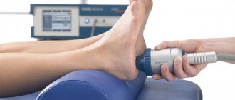
V. Falanga (2002) formulated a strategy for treating the base of a chronic wound “Wound Bed Preparation” with the aim of converting it into an acute one. In this case, both the necrotic component and the altered cells of the edges and base of the wound are removed. For this purpose, a variety of proteolytic enzymes are also used, which have a number of advantages, including the rapid and high-quality removal of necrotic tissue, suppression of wound infection, and to some extent stimulation of regenerative processes in the wound. However, it should be noted that many of them have a short duration of action at acidic pH values of the wound environment. Recently, in the practice of purulent-septic surgery, the method of ultrasonic wound cavitation has firmly occupied its niche (Fig. 7.5, 7.6).
Rice. 7.5.
Ultrasonic cavitation device Soring (Sonoca)
Rice. 7.6.
The effect of ultrasound on wound tissue
It is used as an adjunct to surgical debridement. The sanitizing effect of ultrasound is carried out due to the destruction of the cellular elements of wound discharge and the release of lysosomal enzymes, chemotactic factors, bactericidal proteins, and is enhanced when solutions of antibiotics and antiseptics are used as an acoustic medium. Due to its aggressive effects, the ultrasound cavitation method requires preliminary anesthesia.
Among the methods of chemical exposure, nitrogen monoxide has successfully proven itself in the local treatment of trophic ulcers (Fig. 7.7), which has a direct local bactericidal effect, stimulating phagocytosis, improving trophism.
After a period of long oblivion, various ozone therapy techniques have found their place in clinical practice (Fig. 7.8).
Rice. 7.7.
Impact on a trophic ulcer with an air-plasma NO flow produced by the apparatus “Scalpel-coagulator stimulator SKSVP/NO-01 “Plazon”
Rice. 7.8.
Aeration of a trophic ulcer with an ozone-oxygen mixture in a plastic insulator. The ozone-oxygen mixture is generated by the UOTA-60-01 apparatus
The therapeutic effect of ozone is based on a universal anti-inflammatory effect, which consists in the activation of antioxidant protection, stimulation of energy and plastic metabolism, a fairly powerful bactericidal effect against microorganisms, including those that are multi-resistant to most antibiotics and antiseptics, immunomodeling and antihypoxic effects. It is necessary to note the ability of ozone to stimulate the processes of oxygenation and tissue regeneration in the area of trophic disorders. The advantage of the methods of exposure to nitric oxide and local ozone therapy is the possibility of influencing large wound surfaces without the risk of traumatizing important anatomical structures.
After completion of the first phase of the wound process in a trophic ulcer, the primary task is to stimulate reparative processes and protect the ulcer tissue from re-infection. Traditionally used dressings, when exposed for a long time on the wound surface as a result of impregnation with wound discharge, tend to easily harden when dry. The developing granulation tissue grows through the fibers of the dressing material and is inevitably injured when changing the dressing, increasing the pain syndrome and slowing down the reparative processes of the ulcer tissue. Nowadays, so-called atraumatic dressings are used, often in combination with various medicinal impregnations.
The choice of one or another dressing requires mandatory consideration of the phase of the wound process and the degree of exudation. Dressings designed to implement a therapeutic effect during the inflammation phase, due to the structure of their material, inactivate wound exudate and eliminate microorganisms, toxins and tissue detritus, while stimulating the process of necrolysis.
In turn, wound coverings used in the treatment of sanitized and healing ulcers in phases 2 and 3 of the wound process are required to maintain the necessary parameters of the wound microclimate, protection from mechanical damage and secondary contamination, and stimulation of reparative processes.
Currently, the practice of treating wounds and ulcers, including varicose veins, is dominated by the treatment proposed and justified in the 60s of the twentieth century. G. Winter and J. Maibach concept of healing in a moist environment. Currently, within the framework of the concept of moist healing, a variety of wound coverings are used, providing:
- autolytic cleaning of ulcers and absorption of wound exudate;
- a moist environment for cell migration, proliferation, differentiation and formation of new vessels;
- thermal protection and temperature stability of the wound surface;
- prevention of secondary wound infection and contamination of environmental objects;
- adequate gas exchange between the wound and the atmosphere;
- painless and atraumatic removal of the bandage;
- hypoallergenic.
These coatings are easy to use and are responsible for a specific phase of the wound process.
Alginate dressings, namely Algipor, Teralgim, Algimaf, etc., promote the transformation of calcium alginate fibers into a gel-like amorphous mass that binds wound detritus and pathological exudate. In this case, a wound environment with a high level of humidity is formed, and microorganisms and toxins are reliably bound in the gel structure. A prerequisite for the use of calcium alginate-based dressings is the presence of fluid in the wound.
Sponge dressings
(Zeruvit, Cosmopor, Gelevin, Comprigel, Mepore, etc.) create a balanced environment on the wound surface, stimulate the growth of granulation tissue and isolate the wound from secondary infection.
Hydrocolloid dressings
(Hydrocoll, Tender Wet, Comfeel ulcus, Coloplast, etc.) are effective in phase I of the wound process and especially during its transition to phase II, for the treatment of moderately and slightly exuding wounds, as well as wounds with areas of “dry” necrotic formations. Due to the properties of the hydrogel, a plasticizing effect on wound tissue is ensured, softening of necrotic formations during the diffusion of the gel under them and facilitating the removal of non-viable tissue.
The purpose of these hydroactive dressings is to stimulate the growth of vascular granulation tissue in the wound and prepare it for plastic closure.
Hydrogel dressings
(Hydrosorb, Intrasait, Gelepran) are used when a vascular wound bed has been formed for the purpose of rehydration and rejection of dense necrotic scab or stimulation of epithelization of flat granulating (including donor) wounds.
Atraumatic mesh dressings
(Autrauman, Branolin, Voskopran, Parapran, etc.) do not stick to the wound and do not interfere with the outflow of excess wound fluid. In the presence of abundant exudate, it is advisable to use mesh dressings simultaneously with secondary sorption dressings.
Despite the variety of drugs used to treat patients with VTU, the number of drugs that effectively affect the formation of granulation tissue and epithelization remains small, so at present the patient’s own skin is actively used to close ulcers.
In the light of publications on the vascular endothelium as a paracrine organ that, even under hypoxic conditions, retains the ability to secrete various growth factors that not only provide angiogenesis, but also indirectly stimulate the proliferation of fibroblasts and keratinocytes, leading to the healing of trophic ulcers, those removed by phlebectomy can be used with some success. autovein as a temporary wound covering.
In our clinical practice, we used a temporary autovenous graft in the complex treatment of patients with an open trophic ulcer of the leg in order to close the ulcerative surface, which, in our opinion, is an alternative to the method of closing autoskin ulcers and can reduce the degree of surgical trauma and reduce the risk of re-infection of trophic ulcers.
Why does salt help?
Salt is considered one of the most effective methods for treating periodontal disease. It stimulates the restoration of periodontal tissue and relieves inflammation. The effectiveness of salt is explained by the fact that it has the ability to draw out moisture. Once in the oral cavity, salt “takes” liquid from the inflamed area, thus depriving bacteria and microorganisms of their natural habitat. When treating, it is better to use sea salt, as it is rich in iodine.
Salt rinses
Saline solution is considered one of the most effective methods of treating teeth and gums.
It is often used as a first aid remedy before visiting the dentist. Table 1. Types of salt rinse solutions
| View | Preparation |
| Salt + soda | Suitable for those for whom regular saline solution is not suitable. Mix one teaspoon of baking soda and salt in a glass of water. Can be used after tooth extraction, but carefully so as not to injure the socket. |
| Salt + soda + iodine | 1 tsp salt and soda, 2 - 3 drops of iodine per glass of warm water. |
| Salt + vodka | Add 2 - 3 tbsp to a glass of water. vodka and 1 tsp. salt. Rinse with caution so as not to burn the mucous membrane. |
| Salt + herbal decoctions | Decoction recipes:
|
Recommendations for topical treatment of diabetic foot ulcers
Despite progress in modern medicine, the problem of treating non-healing wounds still remains extremely difficult. Various mechanisms are involved in the healing process. Understanding these mechanisms to restore tissue integrity and maintaining an optimal wound environment allows for more effective wound treatment. The most important aspects that impede wound healing include: impaired blood supply, impaired venous outflow, excess pressure on the affected area, infections, and injuries.
Rice. 1 a). Appearance of a diabetic ulcer of the right foot in the area of greatest pressure (Wagner stage 3);
b). Gangrene of the big toe of the left foot in a patient with severe type 2 diabetes mellitus.
There are many topical preparations. At the same time, significant attention these days is paid to means of local treatment of wound defects, such as modern interactive dressings for the treatment of wounds in a humid environment.
Successful treatment of chronic diabetic ulcers requires the following conditions:
· Compensation of carbohydrate metabolism;
· Unloading the affected limb;
· Fighting possible infection;
· Timely selection of the correct tactics for ischemia correction;
· Treatment of concomitant diseases;
· Use of modern means of local wound treatment.
This set of treatment methods is primarily aimed at restoring the trophism of damaged tissues, preventing critical ischemia and further spread of infection.
An important component of treatment is wound dressings with properties that provide the most favorable microclimate in the wound, necessary to ensure optimal functioning of cellular healing mechanisms.
Tasks of the dressing:
· Protection from mechanical influences (pressure, impact, friction), from contamination and chemical irritation;
· Protection against secondary infection;
· Protection against drying out and loss of physiological fluids (electrolytes);
· Maintaining adequate temperature.
In addition to protecting the wound, the dressing can also actively influence the healing process by cleansing the wound, creating a microclimate that promotes healing and maintaining the wound at rest.
Among modern dressings for the treatment of necrotic and/or infected wounds and trophic ulcers in patients with DFS, we give preference to ready-to-use interactive dressings that long-term maintain optimal hydrobalance in the wound. Such dressings are also called hydroactive. Currently, it is possible to use various types of such modern hydroactive dressings.
Among them, we consider the sequential use of hydroactive dressings based on superabsorbents - TenderWet® active (plus), as well as protected hydrophilic sponge dressings with hydroactive properties - HydroTac® (comfort) and ready-to-use hydrogel dressings Hydrosorb® (comfort) to be very promising. Given appropriate indications, these dressings can be used both as local monotherapy and sequentially.
The TenderWet® active (plus) dressing is a multi-layer wound dressing that has the appearance of a pad containing cellulose and, as a sorption element, polyacrylate superabsorbent powder. The sorption element of the dressing is factory activated by soaking it in Ringer's solution, which is then continuously released into the wound for 24-72 hours. Thus, dressings based on a superabsorbent provide continuous “washing” of the wound and, thereby, contribute to the softening and self-separation of areas of necrosis. At the same time, the wound exudate is absorbed by the sorbent. This exchange is due to the closer affinity of the polyacrylate with the proteins of the wound discharge than with an isotonic saline solution. In this regard, the wound exudate absorbed by the sorbent displaces Ringer's solution from the absorber, which, in turn, is continuously supplied to the wound bed, stimulating autolytic cleansing of the wound, its microbial decontamination, as well as the growth and development of full-fledged granulation tissue.
Rice. 2 (a-c) Local treatment of an extensive infected ulcer of the right leg in a patient with type 2 diabetes mellitus. Positive dynamics of the wound process when using interactive dressings TenderWet® active (plus).
Currently, both “classical” superabsorbent-based dressings, activated with Ringer’s solution under production conditions, and their more modern analogues are used. Thus, a ready-to-use dressing based on a super absorbent with a duration of action of up to 72 hours is called TenderWet® plus. All variants of superabsorbent-based dressings can be used both for packing deep and for treating superficial, especially chronic wounds.
Rice. 3 (a-c) Local treatment of an infected arteriovenous trophic ulcer of the left leg in a patient with severe type 2 diabetes mellitus. Dynamics of the wound process when using TenderWet® plus interactive dressings.
When treating exuding chronic wounds after cleansing the wound surface of necrotic detritus, interactive sponge hydrophilic dressings with high sorption capacity are effectively used. They are made of special polyurethane foam, which has a unique open pore structure. The size of these pores gradually decreases in the direction from the layer in contact with the wound to the surface of the dressing. The composition of the hydrophilic sponge additionally includes a hydrophilic matrix. It enhances the vertical absorption of wound fluid and reduces its desorption. Such dressings allow their use for the treatment of chronic wounds with severe exudation. This speeds up the cleansing of the wound surface from infected exudate and fibrin.
Rice. 4 (a-c) Treatment of an extensive exuding stage 4 pressure ulcer located in the sacral region in a patient with severe type 2 diabetes mellitus using PermaFoam® and PermaFoam® cavity hydrophilic sponge dressings. Positive dynamics of the wound process are noted.
There are anatomical varieties of hydrophilic sponge dressings for the treatment of sacral, heel and elbow pressure sores with an additional self-fixing membrane and a special version of this sponge dressing created for packing wound cavities. The indication for changing the dressing is its complete saturation with exudate. This is manifested by marginal leakage of fluid or deformation of the outer membrane layer of the dressing. Reducing wound exudation creates a risk of excessive drying of the wound bed, damage to granulations and the development of pain due to natural wound adhesion. Therefore, to improve the hydrobalance in the wound and prevent tissue trauma during dressing, a fundamentally new dressing with improved properties was developed. Such hydrophilic sponge dressings, protected by a hydroactive gel layer, are innovative and are intended for the treatment of wounds in the granulation and epithelization phase with moderate and even slight exudation. Thanks to these unique properties, the dressing can remain on the wound until epithelialization and complete closure of the wound defect. Thus, the main advantage of protected sponge dressings is that they are non-traumatic and can be used almost regardless of the level of exudation immediately after cleansing the wound and until it is completely closed.
Rice. 5 a). External view of the working surface of the hydrophilic sponge dressing HydroTac®, protected by a hydroactive gel layer.
b). Continuation of local treatment of an infected arteriovenous trophic ulcer of the left leg in a patient with severe type 2 diabetes mellitus using a HydroTac® dressing. Positive dynamics of the wound process. Healing of a wound defect.
Hydrosorb® hydrogel dressings are a gel made of polyurethane polymers fixed to a semi-permeable membrane that can retain its original shape upon contact with liquid and have a pronounced moisturizing and moderate absorbent ability. The gel contains up to 60% water, so this dressing provides a moist wound environment for several days and at the same time absorbs excess amounts of wound discharge, which is bound by the gel structure. This ensures an optimal level of humidity for wound healing and, as a result, accelerates the processes of granulation and epithelization of the wound. Moreover, the surface of gel dressings is impermeable to microorganisms and water. Therefore, they reliably protect the wound from secondary infection. Hydrosorb® hydrogel dressings do not stick to the wound bed even with prolonged use and are easily removed without traumatizing the regenerative layer of wound tissue. At the same time, their structure, unlike hydrocolloids, is not destroyed under the influence of absorbed wound discharge. Therefore, no residue remains on the wound and its condition can be easily assessed without first washing the wound surface. A significant advantage of modern ready-to-use Hydrosorb® hydrogel dressings is their transparency, which remains even after prolonged stay on the wound. This allows you to examine the wound without changing the dressing and thus provide not only the peace necessary for wound healing, but also high efficiency due to less frequent dressing changes.
Rice. 6 (a, b) Treatment of a long-term non-healing donor wound located on the posterior surface of the right thigh in a patient with severe type 2 diabetes mellitus using a hydroactive dressing Hydrosorb®comfort. Positive dynamics of the wound process are noted.
Of course, bandages equipped with a special adhesive contour adapted for fixation to the skin are more convenient to use and more comfortable for the patient. This role is usually performed by the outer layer of the interactive dressing - a special membrane treated with hypoallergenic glue. This form of the bandage allows you to quickly and conveniently attach it to the skin of the peri-wound area only if the skin is relatively healthy, i.e. is not thinned, affected by dermatitis or sensitive to the components of the adhesive base. If the skin in the wound area is problematic, which often accompanies long-term chronic wounds, then it is necessary to use the main types of dressings that do not have an additional adhesive contour. How can such bandages be fixed? There are additional options such as adhesive, i.e. associated with gluing and non-adhesive fixation of standard dressings. For long-term and regular adhesive fixation of dressings when treating patients with chronic wounds, including those with profusely weeping wounds, the hypoallergenic Omnifix® elastic plaster can be effectively used.
Rice. 7 (a, b, c) Appearance and sequential use of the adhesive hypoallergenic fixing patch Omnifix® elastic to secure the bandage on the plantar part of the foot in a patient with severe diabetes mellitus.
Thanks to through pinholes on the surface, it has greater permeability to moisture vapor than the membranes of most interactive dressings. At the same time, it is thin, strong and elastic enough to not cause additional trauma to the skin. The structure of the Omnifix® elastic patch allows it to be effectively used on anatomically complex surfaces, such as the foot of a patient with diabetes. A complex surface of the foot in a patient with diabetes mellitus can be formed as a result of the development of its deformation, the so-called Charcot foot, or as a result of operations for purulent-necrotic processes that complicate the course of diabetic foot syndrome against the background of progressive diabetes mellitus. In these cases, adhesive fixation is not always convenient, and is often contraindicated for problem skin. The modern fixing cohesive bandage Peha-haft® with elastic properties allows you to successfully cope with this situation.
Rice. 8 (a,b,c). Appearance and sequential use of the Peha-haft® fixation cohesive bandage to secure the lower leg bandage in a patient with severe diabetes mellitus.
Rice. 8 g. Appearance of a bandage secured to the foot using a Peha-haft® bandage.
One and a half to two rounds of the bandage are enough to form a reliable circular bandage, which will help to successfully fix the dressing material for a long time even to the most anatomically complex surface, will not require tying and, moreover, can retain its functions in patients even if there is an increase during the day. swelling. Thus, the original blood circulation is preserved and the development of the tourniquet effect is prevented, which is usually associated with increasing edema and possible compression of the limb segment with an inextensible and knotted bandage.
Thus, based on practice, dressing material and means of its fixation, selected in accordance with the stage of the wound process, location, size and degree of microbial contamination of the wound, can be considered effective. All of them together must be able to maintain a moist environment, control wound discharge and prevent maceration of the edges, be well fixed and safe, as well as comfortable when used independently against the background of adequate and necessary unloading of the limb.
How to properly rinse your mouth with saline solution
To prepare the solution you need 1 – 2 teaspoons of salt and a glass of boiled water. However, there are a number of subtleties that need to be taken into account.
- Before rinsing, you should brush your teeth.
- The solution should be warm. Hot can burn the mucous membrane, and cold can increase the pain.
- It is recommended to rinse your mouth after every meal.
- After using the saline solution, you do not need to rinse your mouth with water. If there is a need, you should wait at least 5 minutes.
Despite its effectiveness, the salt solution is not an alternative to professional treatment. If you have any problems with your gums, we recommend that you consult a doctor.
Remember that the health of your teeth depends on the condition of your gums, so try not to delay your visit for too long. You can make an appointment with our doctors right now: 220-86-30
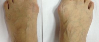
for tendon sprains - saline dressings
Saved by salt dressings
Material by Vadimir Sachenko, published in the Healthy Lifestyle Bulletin, No. 13 (435), July, 2011 (not available in electronic form)
“Once, while abroad, in Ukraine, I severely twisted my leg and apparently suffered a sprain. It was during the day, after that I walked for some time, limping, but when I came to my place, I decided to lie down to calm my leg. After a couple of hours of rest, I could no longer step on it. The leg is swollen and red. It seemed problematic to receive treatment in what was once my native and now foreign country, and in two days I had to leave.
I remembered the publications in the newsletter on the use of saline solutions. I prepared a salt solution and applied a gauze bandage with this solution throughout the evening and the following night. I changed it as it dried out.
The next morning I was able to move around the apartment, and in the evening I walked freely along the street. Since then, I have offered this method of treatment to my friends in similar situations. There was a case of a severe fracture of the foot, the person was bedridden,
lay in traction. The swelling on the leg and at the fracture site did not go away for a long time. And again, the use of saline solution allowed me to quickly get rid of the swelling.
This method is very effective, especially for sprains and the associated swelling.”
In connection with this message, a healthy lifestyle repeats the previously published recipe for a hypertonic solution of table salt, drawing the attention of readers to the fact that in medical practice a 10% solution of table (rock and no other) salt is usually used, that is, 100 g per 1 liter of water. Salt dressing effectively cures inflammatory processes and tissue swelling.
Not all water is suitable for preparing a hypertonic solution. Spring, artesian, and sea water are not suitable for this purpose, especially water containing iodide salts, which neutralize table salt in the solution. It is better to use distilled water (from a pharmacy) to prepare the solution, or, in extreme cases, purified rain or snow.
The salt dressing is prepared from hygroscopic material - you can use repeatedly washed, not new, not kitchen or starched “waffle” towels in 3-4 layers and thin medical gauze in 8-10 layers, also absorbent cotton wool.
If large joints are swollen, they are bandaged with large gauze bandages with 10% saline solution at night every day for two weeks. Not only the joints themselves are bandaged, but also the limbs above and below the joint by 10-15 cm.
Popularity: 20%
Tags: hypertonic solution, stretching, saline solutions
Share…
Other methods for treating gums at home
- Oral baths with decoctions and tinctures, which include Kalanchoe, chamomile, St. John's wort, calendula and other anti-inflammatory herbs and extracts.
- Special diet - just increase the number of foods that contain a large amount of calcium (cheese cheese, cottage cheese, beans, almonds, etc.) and vitamin C (orange, kiwi, strawberries, black currants, etc.).
- To give up smoking. Cigarettes worsen the condition of teeth and gums and provoke the development of inflammation.
Home methods for treating gums bring good results, but they can never replace full-fledged dental treatment.