directions
Shoulder pain is not always associated with injuries caused by falls or accidents. The shoulder joint experiences a huge daily load, which, due to its anatomical structure, leads to injuries such as dislocations, subluxations, sprains, etc. The shoulder joint itself is a rather complex structure of the human body, consisting of fibers not strengthened by muscles, which allows the arms to be mobile and perform the functions of movement, rotation, tilt, extension, and flexion. At the same time, such a design leads to frequent injuries and diseases of the joint.
Currently, work is underway on the website to change the price list; for current information, please call: 640-55-25 or leave a request, and an operator will contact you.
In what cases is ultrasound of the hip joints prescribed?
Diagnostics can be prescribed by a traumatologist, surgeon or orthopedist. Typically, ultrasound examination of the hip joints is indicated in the following cases:
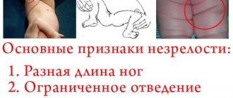
- Limited mobility, difficulty getting out of bed after sleep;
- The appearance of stiffness when moving;
- Painful sensations;
- Suspicion of a dislocation or dislocations in the past;
- Change in skin color in the hip joint area;
- The appearance of a distinct crunch in the joint during physical activity;
- Frequent muscle spasms in the buttocks and thighs;
- Different lengths of the lower limbs.
Advantages and disadvantages of ultrasound of the hip joints
Today, there are many different diagnostic procedures, such as x-rays, computed tomography or magnetic resonance imaging. Of course, these studies provide more detailed information, but such methods have a higher cost. Ultrasound examination, in turn, is cheaper, completely safe and allows you to identify pathological processes even in the early stages. In addition, the diagnostic result can be obtained within 10 minutes after the end of the examination.
Let's look at a few more benefits:
- The procedure is non-invasive;
- It is painless;
- It is also possible to study the condition of the blood vessels in the area under study.
How is the procedure carried out?
The patient removes clothing below the waist (except underwear) and lies down on the couch. Diagnosis is made using a sensor, with which the doctor reads information about the condition and density of bone, cartilage and soft structures. To obtain a complete clinical picture, the doctor uses several access options.
Anterior approach
The ultrasound doctor asks the person to lie on his back, and a small cushion is placed under the femoral surface. This position helps to better examine the structural elements of the hip joint. By observing the image on the screen, the specialist receives information about the condition of the ilium, femoral head, ligaments, muscles and lymph nodes of the groin.
Rear access
For such an examination, the specialist asks the patient to lie on his side and bend his knees slightly closer to his chest. During the study, it is possible to identify the functioning and characteristics of the sciatic nerve, obtain information about the surface of hyaline cartilage, and examine the superficial and internal structure of muscle fibers in the gluteal region.
Medial access
The person lies on his back, bends the lower limb and moves it to the side. This position allows for a detailed examination of the pelvic flexor muscles and articular ligaments if a pathological process is suspected.
Lateral access
The person lies on his side, and the specialist asks him to rotate his hip inward. In this position, the protruding elements of the tibia and the trochanteric bursa are clearly visualized.
On average, this manipulation lasts no more than 30-40 minutes. The patient is given the results of the examination; in some cases, additional images are attached so that the doctor can independently assess the clinical picture.
The procedure for performing an X-ray examination of the shoulder joint
When performing an X-ray of the shoulder joint, the patient must undress to the waist. Depending on the type of projection (direct or lateral), the patient lies on a special table, or the picture is taken in a standing position. The patient is covered with a lead apron, which protects the person from the negative effects of X-rays. The duration of the procedure is several minutes. After this, the image is processed, a radiologist describes the image and issues a conclusion, the patient is sent to the attending physician or to recommended specialists.
Decoding the results
An orthopedist, surgeon, or orthopedic surgeon can interpret the results. The research protocol usually contains the following information:
- the presence of pathological processes, a detailed description of the deviation.
- congenital or acquired changes inside the joint;
- condition of ligaments and nerve bundles;
- increase in the thickness of the joint capsule;
- functioning of adjacent muscles;
- features of cartilaginous structures;
- the amount of synovial fluid inside the joint.
- the presence of neoplasms, if any, their shape, structure, size and individual characteristics;
- information about the ilium, labrum and femoral head;
- metastasis of nearby tissues;
- presence of hematomas.
Please note that the result of an ultrasound examination is not a basis for making a diagnosis. If any inconsistencies or pronounced pathological processes are detected, it is additionally necessary to perform a computed tomography scan and pass various tests for laboratory research. In some cases, a biopsy may be necessary (for example, if the doctor suspects a malignancy). The final diagnosis and necessary treatment are prescribed only after additional studies.
What does an x-ray of the shoulder joint show?
On an X-ray of the shoulder joint, the radiologist will be able to see various damage caused by injuries or diseases, confirming or refuting the diagnosis. For example, X-ray signs of diseases such as arthrosis, arthritis, etc. clearly visible on an x-ray of the shoulder. The doctor will be able to examine in detail and evaluate the condition of the humerus, scapula, the distance between the bones that form the joint, the contours and condition of the joint space, etc. Based on the X-ray findings, the attending physician makes a diagnosis and develops a treatment regimen.
Normal hip joints according to ultrasound
If the patient does not have various pathological processes, the protocol indicates that the hip joints have a clear and smooth surface, without deformations and bone growths. The joint capsules should have folds and branches; their structure is normally hypoechoic.
The following information is also usually indicated:
Right joint:
The bony part of the acetabulum is normal. No major changes. Rectangular bony protrusion. The overlap of the cartilaginous part of the roof of the femoral head is sufficient. The limbus is projected laterally from the femoral head, is presented, and has a normal angle of inclination. The head of the femur in the acetabulum is located correctly. The ossification nucleus is located. Angle a is less than 60 degrees. Angle b is greater than 55 degrees.
Left joint:
the roof of the acetabulum is moderately flattened, without structural changes. The femoral head is centered correctly. The ossification nucleus is located. Angle a is more than 60 degrees. Angle b is more than 55 degrees. The angular parameters of the joint are not changed. When performing functional tests, the joint is stable.
Please note that these indicators may vary depending on age - deviations from them do not always indicate the presence of diseases or some problems.
Joint space: definition of the concept, norm of indicators in different joints
1. X-ray examination of diseases and injuries of the musculoskeletal system
First, let’s look at the articles of the “Resolutions”, for which the largest number of discrepancies and expert errors arose in practical work. This is a military medical examination of diseases and injuries of the musculoskeletal system.
| Disease schedule article | Name of diseases, degree of dysfunction | Category of suitability for military service | |||
Igraph | IIgraph | IIIgraph | IVgraph | ||
| 64. | Arthropathy of infectious and inflammatory origin, systemic lesions of connective tissue: | ||||
| a) with significant dysfunction, persistent and pronounced changes; | D | D | D | NG | |
| b) with moderate dysfunction and frequent exacerbations; | D | D | IN | NG | |
| c) with minor dysfunction and rare exacerbations | IN | IN | B | NG | |
The article covers rheumatoid arthritis, ankylosing spondylitis, Reiter's disease, periarthritis nodosa, Wegener's granulomatosis, psoriatic arthropathy and other arthritis associated with infection, and other systemic connective tissue diseases.
Point “a” includes:
- systemic connective tissue diseases, regardless of the severity of changes in organs and systems, the frequency of exacerbations and the degree of functional impairment;
- rheumatoid arthritis and ankylosing spondylitis (ankylosing spondylitis) with significant dysfunction or their systemic forms with persistent loss of ability to perform military service duties.
Point “b” includes:
- slowly progressive forms of inflammatory diseases with moderately pronounced exudative-proliferative changes and functional failure of the joints in the absence of systemic manifestations;
- initial forms of rheumatoid arthritis and ankylosing spondylitis in the presence of clinical and laboratory signs of process activity.
Point “c” includes chronic diseases of the joints and spine with rare (once a year or less) exacerbations.
Under point “c”, military personnel undergoing conscription service with a protracted (4 months or more) course of acute inflammatory arthropathy with persistent exudative-proliferative changes in the joints, laboratory signs of process activity and unsuccessful treatment are examined.
In case of chronic infectious and inflammatory arthritis, the category of fitness for military service is determined by point “a”, “b” or “c” depending on the damage to other organs and systems, the state of joint function. The function of the joints is determined in accordance with Table 3 “Table for assessing the range of motion in the joints.”
Chronic forms of reactive arthritis in the absence of exacerbation of the disease for more than 5 years and without dysfunction of the joints are not grounds for the application of this article and do not interfere with military service or admission to military educational institutions and colleges.
After acute inflammatory diseases of the joints, examination is carried out according to Article 86 of the schedule of diseases.
There are no particular difficulties in issuing an expert radiological opinion on this article. The radiologist only needs to establish the fact of the disease. The technique, scope of x-ray examination, as well as x-ray symptoms of rheumatoid arthritis, ankylosing spondylitis, psoriatic arthropathy and other arthritis associated with infection are described in the relevant guidelines for x-ray diagnostics. It should be noted that Reiter's disease, periarthritis nodosa, and Wegener's granulomatosis do not have pathognomonic radiological symptoms.
| Disease schedule article | Name of diseases, degree of dysfunction | Category of suitability for military service | |||
Igraph | IIgraph | IIIgraph | IVgraph | ||
| 65. | Surgical diseases and lesions of large joints, cartilage, osteopathy, chondropathy: | ||||
| a) with significant impairment of functions; | D | D | D | NG | |
| b) with moderate dysfunction; | IN | IN | IN | NG | |
| c) with minor dysfunction; | IN | IN | B | NG | |
| d) in the presence of objective data without dysfunction | B-3 | B | A Airborne Forces, PS, MP, SS - IND | NG | |
The conclusion about the category of fitness for military service for diseases of bones and joints is made, as a rule, after inpatient examination and treatment. In this case, it is necessary to take into account the tendency of the disease to relapse or progress, the persistence of recovery and the characteristics of military service. In case of unsatisfactory results of treatment or refusal of it, a conclusion is made under point “a”, “b” or “c” depending on the function of the limb or joint.
Point “a” includes:
- ankylosis of a large joint in a vicious position, fibrous ankylosis, artificial joint;
- pathological mobility (non-supporting joint) or persistent contracture of the joint with significant limitation of movements;
- severe deforming (presence of coarse bone growths of the articular ends of at least 2 mm) arthrosis of large joints with frequent (2 or more times a year) relapses of exacerbations of pain, destruction of articular cartilage (the width of the joint space on an x-ray is less than 2 mm) and deformation of the axis of the limbs;
- bone defect more than 1 cm with limb instability;
- aseptic necrosis of the femoral head;
- osteomyelitis with the presence of sequestral cavities, sequestra, long-term non-healing or frequently (2 or more times a year) opening fistulas.
In case of ankylosis of large joints in a functionally advantageous position, with good functional compensation of the artificial joint, the category of fitness for military service of those examined under Column III of the disease schedule is determined by point “b”.
Point “b” includes:
- frequent (3 or more times a year) dislocations of large joints, resulting from minor physical exertion, with severe instability (looseness) or recurrent synovitis of the joint, accompanied by moderate atrophy of the muscles of the limbs;
- deforming arthrosis in one of the large joints (the width of the joint space on the radiograph is 2 - 4 mm) with pain;
- osteomyelitis (including primarily chronic) with annual exacerbations;
- hyperostoses that interfere with the movement of a limb or the wearing of military clothing, shoes or equipment;
- persistent contractures of one of the large joints with moderate limitation of range of motion.
Point “c” includes:
- rare (less than 3 times a year) dislocations of the shoulder joint, instability and synovitis of the joints due to moderate physical activity;
- osteomyelitis with rare (every 2-3 years) exacerbations in the absence of sequestral cavities and sequestra;
- persistent contractures of one of the large joints with slight limitation of range of motion.
What pathologies can be identified
Most often, during an ultrasound examination of the hip joints, the following diseases are detected:
- Arthrosis (it develops as a result of congenital dislocation of the hip, a disturbance in the circulatory system, or as a result of a fracture of the femoral neck);
- Arthritis (may occur due to infection of joint joints by pathogens);
- Synovitis, which is characterized by inflammation and fluid accumulation inside the joint;
- Necrosis of bone tissue (can occur due to frequent fractures, dislocations, bruises);
- Tears and spasms of muscle fibers;
- Swelling of the joint.
The hip joints are most susceptible to various pathological processes. Diseases can be either congenital or acquired. If you visit a medical facility as soon as the first symptoms appear, you can hope for a favorable outcome and complete recovery. Otherwise, your doctor may recommend surgery.
Our clinics in St. Petersburg
Structural subdivision of Polikarpov Alley Polikarpov 6k2 Primorsky district
- Pionerskaya
- Specific
- Commandant's
Structural subdivision of Zhukov Marshal Zhukov Ave. 28k2 Kirovsky district
- Avtovo
- Avenue of Veterans
- Leninsky Prospekt
Structural subdivision Devyatkino Okhtinskaya alley 18 Vsevolozhsk district
- Devyatkino
- Civil Prospect
- Academic
For detailed information and to make an appointment, you can call +7 (812) 640-55-25
Make an appointment
We draw your attention to the schedule of technological breaks in the CT and X-ray rooms.
The nature and extent of injury, the presence of disease and pathology can be determined by such an informative diagnostic study as radiography of the shoulder.
In St. Petersburg, you can have an X-ray of the shoulder joint done in almost any medical institution - from a clinic to a hospital and a multidisciplinary medical center. But if you want to save time without wasting it in queues, or you need urgent examination and emergency medical care due to an injury, if you want to have an X-ray of the shoulder joint done with good and safe equipment, then sign up and come to the medical office. The trauma department of the clinic is equipped with the latest digital X-ray device Clinomat (made in Italy), which allows us to quickly, efficiently and fully provide all types of X-ray examinations.