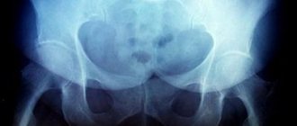
Transient, transient, or toxic synovitis is inflammation of the hip joint and usually causes pain when walking and lameness in children.
The disease is accompanied by pain usually in one hip joint, but can affect both. Sometimes the discomfort begins gradually, causing the child to limp, and then progresses until he can no longer walk at all. Other times it happens suddenly.
Causes of synovitis of the pelvic bones in a child
- Boys are 2-3 times more likely than girls.
- Affects 3% of children and adolescents
- Most often occurs between the ages of 5-7 years
- May occur before age 13
- Etiology not established
- Often a history of respiratory tract infections (40-50%)
- No bacterial or viral infection of the hip joints
- Legg-Calvé-Perthes disease can be caused by effusion or compression of the intra-articular vessels of the epiphysis.
Diagnosis of transient synovitis
Diagnosis of transient synovitis in children
Diagnosis of transient synovitis is difficult, so the doctor must first rule out other diseases. Conditions with similar symptoms include:
- septic arthritis;
- epiphysiolysis;
- Legg-Calvé-Perthes disease, which causes damage to the head of the femur;
- undiagnosed injury or musculoskeletal pain.
The doctor will check your symptoms, then do a physical examination. During it, he will determine the location of the pain.
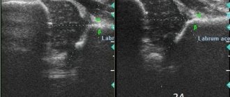
Additionally, an ultrasound examination of the hip joint is used to identify signs of inflammation and the presence of fluid in the joint cavity.
A blood test may show signs of a bacterial infection. X-rays help the doctor rule out epiphysiolysis and Legg-Calvé-Perthes disease.
Diagnosis of septic arthritis in adults
To rule out septic arthritis, your doctor may take a sample of fluid from the joint. A culture sample is also examined to check for bacterial flora that indicates septic arthritis.
Which method of diagnosing synovitis of the hip joint in children to choose: MRI, CT, X-ray, ultrasound
What will an ultrasound of the hip joint show for synovitis in a child?
- An anechoic effusion in the joint space is usually detected with widening of the medial parts of the joint space and balloon-like stretching of the capsule.
- In chronic cases, the capsule is often thickened, showing signs of synovitis.
Transient synovitis of the hip joints. Ultrasound of the hip joints. Anechoic joint effusion (1)
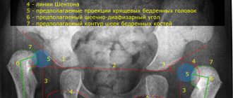
What will x-rays of the hip joints show when there is inflammation in a child?
- Used to rule out other bone pathology such as Legg-Calvé-Perthes disease
- The joint space is widened due to the presence of effusion (in 25% of cases)
- Effusion may also cause lateral displacement of the femoral head
- Regional osteoporosis is detected in 30% of cases.
In what cases does a child undergo an MRI of the hip joints for inflammation?
- Used to exclude early stage Legg-Calvé-Perthes disease or osteomyelitis
- T2-weighted fat-suppressed images demonstrate hyperechoic joint effusion
- Normal bone marrow signal
- T1-weighted images demonstrate increased synovial fluid signal after contrast agent administration, indicating the development of synovitis.
A school-age child with transient synovitis of the right hip joint. MRI of the hip joints. T2-weighted T S E image (a) shows a hyperintense joint effusion with normal bone marrow signal in the femoral head. After intravenous contrast, a T1-weighted image (subtraction, b ) demonstrates increased synovial fluid signal in the right hip joint (synovitis). Perfusion of the femoral head is not impaired.
Symptoms of transient synovitis
The most common symptom is the sudden onset of pain in the hip or leg. The pain usually causes lameness. Other symptoms of transient synovitis include:
- walking on tiptoes;
- unintentional twisting of the leg;
- crying (in young children);
- fever;
- complaints of discomfort in the hip joint after sitting or resting for a long time;
- pain when walking;
- pain in the knee or hip.
In adults, septic arthritis causes pain and swelling in one joint, possibly the knee, elbow, or hip.
Additional symptoms of septic arthritis include:
- redness and swelling around the joint;
- severe joint pain;
- decreased mobility in the joint;
- fever and chills.
What diseases have symptoms similar to synovitis of the hip joints in a child?
Septic arthritis
- serious illness of children;
- significantly more severe pain in the hip joints and decreased mobility; pathological blood test;
- X-ray examination visualizes a widening of the joint space of more than 2 mm.
Legg-Calvé-Perthes disease
- symptoms do not disappear within several days;
- early stages are detected by MRI; joint effusion, epiphyseal signal disturbance.
Slipping of the femoral head
- occurs at the age of 12-15 years;
— Ultrasound reveals gradation in the growth zones;
— Lauenstein image reveals posteromedial displacement of the growth plate relative to the metaphysis.
Rheumatoid arthritis
- rarely develops in the hip joint;
- progression of symptoms;
- synovitis.
What is transient synovitis?
Transient or migratory synovitis is a temporary condition that causes pain and inflammation of the hip joint.
Although symptoms may begin suddenly, transient synovitis usually resolves within 1 to 2 weeks. In some cases, the duration of the hip joint reaches 5 weeks. The disease usually does not cause any long-term complications.
Transient synovitis primarily affects children, but can also occur in adults. As a rule, septic arthritis occurs in adults, which can be confused with transient synovitis. Septic arthritis can cause similar pain in the hip joint.
Transient synovitis most often occurs in children aged 3 to 8 years and is more common in boys than girls.
What is hip dysplasia?
Authors : International Hip Dysplasia Institute
Hip dysplasia is the medical term for hip instability. This pathology affects thousands of children every year. Its manifestations range from mild instability to complete dislocation. Approximately one in 20 full-term babies will have some degree of instability, and 2-3 babies in 1000 will require treatment.
Persistent instability of the hip joint in children is a hidden condition that often causes disability and severe arthrosis in adults. Despite the frequency and potential risk of lifelong disability, concern about this condition among parents is low. Early diagnosis and timely treatment with simple methods is the best solution, but in some cases, hip dysplasia is not detected on time and causes difficulties in the treatment process. In addition, many children around the world do not have the opportunity to receive timely diagnosis and treatment.
Doctors use many different terms to define hip dysplasia, depending on the severity and time of occurrence. Among these terms:
• hip dysplasia; • acetabular dysplasia; • hip dislocation; • congenital dislocation.
The term hip dysplasia is most often used in young children because the instability itself can develop after birth. In addition, the term “congenital” usually refers to a pathology in which any part of the joint does not develop or, on the contrary, is absent normally. In the case of dysplasia, the child's joint is normal except for instability.
When the head of the femur is completely out of the joint, the condition is called dislocation. The hip joint is a ball-and-socket joint that is held in place by ligaments. The head of the femur is conventionally a ball, and the part of the pelvic bone that covers it is called the acetabulum.
In some children, the hip ligaments are stretched and the femoral head slips out of the acetabulum. Sometimes this instability is mild and resolves spontaneously. In other cases, the femoral head extends completely or partially beyond the acetabulum.
The diagnosis of hip dysplasia and hip dislocation can be made during a routine examination of the child. A sign of a hip dislocation may be a “clicking” when checking the mobility of the joint, but this symptom can also be observed in some cases in a healthy child due to the fact that the internal ligaments of the joint can produce a similar sound in certain positions of the hip. But even with a thorough clinical examination, hip dislocation is difficult to detect in some children. In addition, cases have been described in which the joints were normal at birth, but dislocation occurred in the first few months of life. This is why it is so important to carry out regular examinations of the child during the first year of life. In cases where dysplasia is suspected, an ultrasound examination is performed in young children, and an X-ray examination of the joints in older children. This is necessary to make an accurate diagnosis or, on the contrary, to be sure that the joint does not have pathology.
Source
Diagnosis of synovitis in children
A virus that a child has recently contracted is a clue for pediatricians and orthopedists when determining synovitis in children. The final diagnosis is made based on medical history, physical examination and observation.
Often, the doctor may find some resistance when checking the hip joint, or the child will not want to move the leg at all. No blood tests or x-rays are required: transient synovitis will not be detected in these tests anyway.
In some cases, the orthopedist may give the patient ibuprofen and see how it works—whether the child can move his leg better after an hour or not.
Other causes of lameness
Lameness and pain in the legs or hips can be caused by many things, such as injury, muscle strain, or infection. It is very important that parents do not delay visiting the doctor if the child complains of discomfort when moving, because transient synovitis is only one of the possible causes of this condition.
It could also be septic arthritis, a much more serious condition that indicates the presence of bacteria in the joint.
If your child does not move their legs at all, has a high fever (39 degrees Celsius or higher), or feels unwell, contact your doctor immediately.
Treatment
Rest is an important part of the treatment of transient synovitis. The child should avoid strenuous physical activity and try to move less.
Your doctor will likely recommend anti-inflammatory medications such as ibuprofen and naproxen. They reduce inflammation in the joint, helping the child walk more comfortably.
During your recovery, you should avoid strenuous activities, including physical activity and sports. In children with transient synovitis receiving basic treatment, complete recovery occurs within 2 weeks. In adults with septic arthritis, the condition can be severe and lead to permanent damage if the person does not receive treatment.
Treatment of synovitis of the hip joint
To create an adequate treatment regimen, the experts at “Hello!” take into account:
- stage of disease development;
- form of inflammation;
- symptoms;
- patient's health condition.
First of all, the task is to relieve inflammation and pain. Then they determine what caused the inflammation and eliminate it. In this case, the joint is immobilized so that the load is minimal and the person does not experience pain.
The main role in treatment is given to drug therapy:
- anti-inflammatory drugs of hormonal and non-hormonal nature;
- antibiotics for infectious forms;
- trophic means;
- enzyme inhibitors;
- multivitamins.
If fluid accumulates in the joint cavity, it is necessary to perform a puncture. This procedure performs diagnostic and health-improving functions. It is from the extracted fluid that they determine what kind of pathogen attacked the joint and what antibiotics will help deal with it. When excess fluid is removed, the person immediately feels better. Plus, the doctor administers painkillers and medications.
Once you rid the body of the infection, correct the consequences of the injury, the inflammation disappears. But if synovitis was caused by arthritis or ankylosing spondylitis, treatment will have to be active and long-term. To reinforce the therapeutic effect of drug therapy, physiotherapeutic procedures and therapeutic exercises are used.
Diseases of the musculoskeletal system are best treated in specialized clinics. In "Hello!" An integrated approach is practiced, the joint work of doctors of various specialties. Patients are impressed by the convenient location of the network’s clinics.
Question answer
– Are conservative treatment methods always sufficient?
– There are cases when therapeutic methods are ineffective. Then you have to resort to synovectomy. Damaged tissue, suppuration, blood clots are removed from the joint capsule, and antibiotics are pumped in. Early detection of symptoms of synovitis of the hip joint facilitates treatment and does not lead to the need for surgical intervention.
– How long does the treatment last?
– As a rule, a week is enough to relieve the acute phase. Further treatment depends on the form of the disease. Tuberculous and arthritic synovitis have to be treated for several months.
–What preventive measures are needed?
– It is necessary to ensure that the joints are not injured and to prevent the development of any infections in the body.
