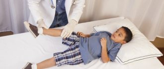
Massage for hip dysplasia Massage for hip dysplasia is included in complex treatment to stabilize the joint and restore full range of motion. Massage strengthens muscles, ensures motor activity and physical development of the child.
Newborns are often born with a dislocated hip - hip dysplasia. Dislocation also develops with improper treatment or lack of it in the very first months after the birth of the child. Therefore, early diagnosis and prompt treatment, including massage for hip dysplasia, will allow the child to develop correctly anatomically and functionally.
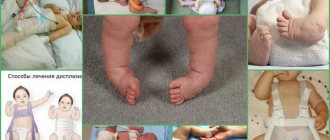
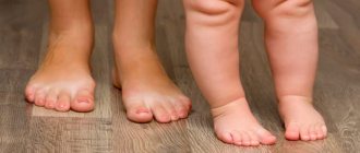
At the birth of a baby, every mother examines his body daily and may notice the presence of some discrepancy in the hip joint:
- different lengths of legs;
- asymmetry of the gluteal and crural folds;
- asymmetry when the leg is abducted to the side;
- incomplete abduction of the leg bent at the knee;
- shortened thigh;
- symptom of slipping or clicking.
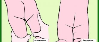
Parents may not immediately notice asymmetric skin folds on the thighs, but only by 2-3 months if there is a bilateral pathology. The depth and shape of the folds, their location in the presence of congenital dislocation will be different. For diagnosis, you need to pay attention to the location of the gluteal, popliteal and inguinal folds.
When the head of the femur is displaced posteriorly relative to the acetabulum, shortening of the femur will be noticeable: the child is placed on his back, the knees and hip joint are bent. One knee will be lower when the hip is shortened.
Massage for hip dysplasia
Forms of hip dysplasia
Hip dysplasia comes in three forms:
The first form , “pre-dislocation,” occurs when the joint is immature and unstable. Timely treatment and massage for dysplasia will ensure normal development of the joint in the future. If left untreated, subluxation may develop due to a stretched capsule, slight dislocation and reduction of the head into the socket. This is called a positive slip sign.
The second form , subluxation, is a morphological change in the joint: the femoral head moves upward and to the side relative to the acetabulum. The contact between the head and the socket will be maintained, since the head will not go beyond the limbus, but will only push it upward. Proper treatment and massage will help form a full-fledged joint. Otherwise, a defective joint will form and a complete dislocation may develop.
The third form is congenital dislocation with complete displacement of the femoral head, which is the most severe form of joint dysplasia.
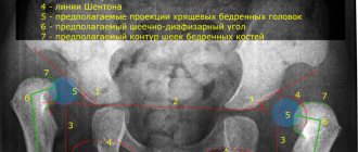
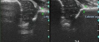
The diagnosis of hip dysplasia is confirmed after an ultrasound examination and radiographs, after which immediate treatment begins. Parents need to know the form of dysplasia of the child’s joints and have an idea of the anatomical location of the joints, as this is necessary in order to correctly perform massage on their own and to prevent dysplasia.
Many orthopedists and related specialists call dysplasia a congenital pathology of the joint when it is underdeveloped, which leads to dislocation or subluxation of the femoral head. Other surgeons and orthopedists use the diagnosis “dysplasia” as a collective concept, which includes all anomalies associated with the hip joint: the slightest underdevelopment of the roof of the joint without displacement and true dislocation.
Parents should be aware of and differentiate on radiographs between hip dislocation and complete loss of contact between the head and acetabulum. As a result of subluxation, contact is only partially lost. With preluxation (dysplasia), the development of the joints of the pelvis and hips is disrupted without displacement of the elements articulating the joint.
Massage for hip dysplasia
Efficacy and contraindications for massage
Massage for dysplasia is prescribed only by an orthopedic doctor and is part of a comprehensive treatment of pathology. As a result of the massage course:
- the condition of the hip joints is stabilized;
- the process of joint reduction improves;
- the condition of muscle tissue improves;
- motor function is normalized, incl. the number of movements increases;
- muscle tone improves;
- the movement of biological fluids improves;
- child development improves.
Contraindications for the procedures are:
- epilepsy;
- exacerbation of hepatitis or diathesis;
- hernia;
- inflamed lymph nodes;
- light weight;
- violation of the integrity of the skin;
- presence of congenital heart disease;
- diseases of the nervous system;
- diseases of the kidneys, liver, blood;
- inflammation of muscle and bone tissue.
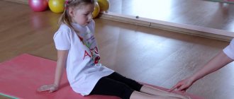
Massage technique for the treatment of dysplasia
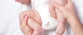
Stroking. The child is placed on his back to perform general stroking movements along the front surface, relaxing the arms, legs (hips, legs, feet, soles), chest and abdomen. You can cover the entire hip joint or just the front surface. Light straight and spiral movements are directed from the lower leg to the thigh. Massage is not performed on the inner thighs near the genitals - damage to the lymph nodes is possible. Children perceive this massage (2-3 minutes) for dysplasia as a game and affection.
Trituration. With some reinforcement, deep-lying muscles, tendons and ligaments must be manipulated with the pads of the fingers in circular and spiral movements. In this case, it is necessary to feel the joint and not press hard and forcefully on it.
- When performing a massage for dysplasia from the back, after stroking, rub the back surface of the thighs, gradually directing the hands to the buttocks and performing a local massage on the dysplastic joint with rubbing, pinching, tapping, and circular movements. Then fix the hip joint with one hand.
- With the second hand, gently and measuredly spread the leg, easily grasping it by the knee, lightly pressing on it and rotating the thigh inward.
- Stroking is performed again, pressing your hands tightly to the skin without moving it.
- Using the thumb and forefinger and the entire palm, massage for dysplasia using spiral movements, excluding the surface of the thighs near the genitals, for 3-5 minutes, then begin rubbing using the same technique, but displacing the skin. This technique is especially important for affected joints with dysplasia. The fingertips should penetrate deep into the muscles for 10 minutes.
- With the legs apart, carefully and without sudden movements, bend and spread them to the side. Perform inward circular movements with the legs in the area of the hip joint.
- Wallow. Cover the baby's thigh with cupped palms and roll the leg between the palms like a cutlet for a minute.
- Perform the “bicycle” movement - with the legs bent at the knees and hip joint, simulating riding a bicycle.
- Bend and straighten your legs together and in turn, alternating with light kneading (pressure) of the calf muscles with four fingers.
- When the child is next turned onto his stomach, stroking is performed again, turning into rubbing. Then they move their legs to the side one by one, imitating crawling.
- They switch to massaging the back and lumbar area with stroking and rubbing movements. Going down to the buttocks, massage for dysplasia with stroking, rubbing and pinching movements, lightly tapping the fingertips and patting.
- Along the spine, spiral kneading movements are performed with the phalanges of the fingers, sawing in a herringbone pattern from the spine to the periphery. Rub the lumbar area with your palm.
- Stroke and rub the area of the hip joint and the outer surfaces of the thighs, alternating with light kneading, moving the legs to the sides (“crawling”) and “hovering” - lifting the child above the table, supporting it under the chest and pelvic area.
- Rub and knead the buttocks with spiral movements. When performing a massage, you must remember that you cannot roughly examine the hip joint and look for congenital dislocation. This can damage the growth plate and delay the development of the femoral neck, deform the head (plow vara) and lead to early coxarthrosis. Improve blood circulation with foot massage.
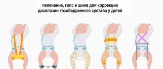
Parents need to remember that massage and exercises in the “standing” or “squatting” position can only be performed with the permission of an orthopedist, since vertical load may worsen the deformity of the hip joint.
Pathology treatment methods
Fibrous dysplasia, or Braitsov's disease, usually manifests itself in pediatric patients and is very rarely first diagnosed in elderly patients. The pathology was first described about a hundred years ago, and over the ensuing years many studies have been carried out that have not provided specialists with information to determine the exact causes of its occurrence. Today, it is believed that hereditary predisposition plays a major role in the abnormal development of tissue that gives rise to bone in the prenatal period.
There is a monoosal form of dysplasia, in which only one bone is affected; painful symptoms debut in patients of any age; areas of skin pigmentation and endocrine disorders are not detected. The polyostotic form developing in children manifests itself in damage to several bones, accompanied by endocrine disorders and melanosis (the appearance of dark spots on the skin).
According to another classification, intraosseous, tumor, fibrocartilaginous and some other forms of fibrous dysplasia are distinguished. In some cases, total bone damage is observed.
Characteristic signs of the disease are the appearance in the patient of intense pain, pathological fractures and gradually progressive bone deformations. If the pathological focus is localized in the tubular bones of the legs, they become bent, shortened, and acquire a pathological shape. The patient also complains of lameness. The appearance of an area of fibrous dysplasia in the spine is accompanied by the development of scoliosis or kyphosis, and postural disorders.
In many patients, the pathology, especially in the initial stages, occurs without clinical symptoms, so Israeli clinics tend not to prescribe specific treatment unless absolutely necessary. If there are no signs of disease progression, the patient is observed by the attending physician, and he must attend a preventive examination at least once every six months and undergo prescribed examinations.
The only effective treatment method is surgical removal of the pathological focus, for which progressive gentle surgical methods are used.
Surgery
Most often, to eliminate the affected area of bone tissue, segmental resection is used, carried out within the boundaries of normal tissue. During this operation, the continuity of the bone is disrupted, as a result of which the defect is subsequently restored using a bone grafting procedure. The endoprosthesis replacement procedure involves the installation of endoprostheses, which are individually manufactured for each patient. An autograft can also replace the articular surface. Pathological fractures require placement of an Ilizarov apparatus on the patient.
Drug therapy
If, during the examination, multiple lesions are revealed in the patient, he is prescribed medications that prevent deficiency of bone tissue density, which is one of the main causes of pathological fractures, and its deformation. The prescribed drugs help strengthen bone tissue and prevent bone atrophy. In addition, the patient is prescribed vitamin D and calcium-containing medications. To eliminate severe pain, analgesics and other painkillers are indicated.
- Oncological operations
Advantages of treatment in Israel
- International level of specialist training.
- Modern material and technical base of clinics
- Performing surgical interventions using progressive methods.
- Comfortable conditions for treatment.
- Loyal prices.
Carrying out therapy at the initial stages of the disease allows most patients to forget about the disease and quickly restore lost functions. Do not hesitate, contact an Israeli clinic and undergo a full course of treatment and rehabilitation.
- 5
- 4
- 3
- 2
- 1
(0 votes, average: 5 out of 5)
Publications in the media
A huge number (hundreds!) of nosological units with the generic word “Dysplasia” are known. This article lists in alphabetical order those nosological units that could not be placed in other articles in the reference book characterizing dysplasia (Craniofacial dysplasia, Ectodermal dysplasia, Epiphyseal dysplasia, Dental development disorders, Chondrodysplasia, Achondrogenesis). Many dysplasias, like the vast majority of genetic diseases and phenotypes, are also difficult to identify using the ICD-10 system.
Acromicric dysplasia (102370, В), congenital acromicria. Clinically: moderate facial anomalies, shortening of the hands and feet, severe growth retardation, short metacarpal and phalangeal bones. Laboratory findings: disorganized cartilage growth. ICD-10. Q87.1 Syndromes of congenital anomalies manifesting predominantly as dwarfism
Arterial fibromuscular dysplasia, see Fibromuscular dysplasia.
Diastrophic dysplasia - skeletal dysplasia with severe curvature of bones • Diastrophic dysplasia (222600, 5q31–5q34 5q32–5q33.1, mutations of the transmembrane sulfate transporter gene DTD, r). Clinically: congenital dwarfism with short limbs, impaired ossification and congenital epiphyseal cysts, hypertrophy of ear cartilage, cleft hard palate, kyphosis, scoliosis, abducted thumb, fusion of proximal interphalangeal joints, brachydactyly, bilateral clubfoot, calcification of rib cartilage • Pseudodiastrophic dysplasia ical (264180 ). Clinically: rhizomelic shortening of the limbs, interphalangeal and metacarpophalangeal dislocations, elbow dislocations, severe clubfoot, increased distance between the coronal sutures of the skull, hypoplasia of the middle third of the face, hyperthermia, platyspondyly, tongue-like deformities of the lumbar vertebrae, scoliosis, hypoplasia of the 2nd vertebra, pronounced lumbar lordosis • Congenital bone dysplasia de la Chapelle (#256050, r). Clinically: Lethal at birth, severe micromelia, kyphosis of the cervical spine, clubfoot equinovarus, abducted hallux, abducted toes, duplication of the middle phalanges, cleft palate, patent foramen ovale, respiratory failure, laryngeal stenosis, softening of the cartilages of the larynx and trachea, hypoplasia lungs, shortness of breath, small chest, congenital bone dysplasia, triangular fibula and ulna, platyspondyly, pathological metaphyses and epiphyses, sacral anomalies, additional pelvic ossification points. Laboratory: lacunar halos around chondrocytes in skeletal cartilage. ICD-10. Q77.5 Diastrophic dysplasia.
Oculomaxilloosseous dysplasia (*164900, Â). Corneal opacity and multiple anomalies of the lower jaw and limbs. Synonym: OMM syndrome (from: o phthalmo m andibulo m elic). ICD-10. Q78.8 Other specified osteochondrodysplasias.
Greenberg dysplasia (215140, r) is congenital lethal dwarfism. Clinical picture: dwarfism with short limbs, prenatal death, severe fetal hydrops, noticeably shortened, “moth-eaten” long tubular bones, unusual ectopic ossification points, pronounced platyspondyly, pronounced extramedullary hematopoiesis. Synonym: hydropic chondrodystrophy. ICD-10. Q77.1 Small stature incompatible with life.
de Morsier dysplasia (septo-optic dysplasia, 182230, Â?). Hypoplastic optic discs with a double edge, absence of the septum pellucidum, GH deficiency, pathology of the corpus callosum and cerebellum. ICD-10. Q04.4 Septooptic dysplasia.
Diaphyseal dysplasia (Engelmann's disease) is a progressive symmetrical hyperostosis of the diaphysis of long tubular bones from the periosteum and endosteum with sclerosis of the newly formed bone tissue. Clinically: asthenic physique, severe pain in the bones of the legs, fusiform swelling of the lower leg, multiple subungual hemorrhages, myopathy, waddling gait, compression of cranial nerves, weakness, muscle fatigue, scoliosis, lumbar hyperlordosis, hypogonadism, anemia, leukopenia, increased ESR, hepatosplenomegaly, onset aged 10 to 30 years, sensitivity to GC, dysplasia, osteosclerosis and hyperostosis of the diaphysis. Synonyms •• Camurati-Engelmann disease •• Ribbing's disease •• generalized hyperostosis •• systemic diaphyseal congenital hyperostosis •• progressive diaphyseal dysplasia •• systemic hereditary osteosclerosis with myopathy. ICD-10. Q78.3 Progressive diaphyseal dysplasia.
Dissegmental dysplasia is a group of hereditary skeletal dysplasias manifested by dwarfism, damage to the brain and internal organs. At least 2 forms, differing in clinical, radiological and morphological signs • Handmaker-Silvermann dissegmental dysplasia (224410, r) - lethal form. Clinically: vertebral bodies of various sizes and shapes, early death, the clinical picture resembles Kniest syndrome • Dissegmental Rolland–Debuquois dysplasia (224400, r) is a milder form. Clinically: congenital chondrodystrophy, dwarfism, abnormal segmentation of the vertebrae, limited joint mobility, micromelia, curvature of the limbs, high palate, cleft hard palate, hydrocephalus, hydronephrosis, hypertrichosis. Synonyms: dissegmental dwarfism •• anisospondylic campomicromelic dwarfism •• Rolland-Debuquois syndrome • Dissegmental dysplasia with glaucoma (601561) - the phenotype resembles Kniest dysplasia (156550), and dissegmental dysplasia (224400, 224410), combined with severe glaucoma oh. ICD-10 • Q77.1 Small stature, incompatible with life • Q77.3 Point chondrodysplasia • Q77.5 Diastrophic dysplasia.
Campomelic dysplasia (114290, Â, more often *211970, 17q24.3–q25.1, SOX9 gene [CMD1, SRA1, SRY], r) - congenital lethal dwarfism with short limbs, small cartilaginous skull size, platybasia, hypertelorism, depressed nasal bridge , micrognathia, cleft palate, recessed tongue, pulmonary hypoplasia, tracheal hypoplasia, narrow pelvis, hip abnormalities, platyspondyly, kyphoscoliosis, hypotonia, absence of olfactory nerves, small hypoplastic scapulae, 11 pairs of ribs, short phalanges of the hands and feet, moderate curvature of the femoral and tibial bones, leg equinovarus deformity • Grant family syndrome (138930, Â) is one of the forms of skeletal dysplasias of the campomelic type. Clinically: blue sclera, hypoplasia of the jaws, campomelia, curvature of the clavicles, femurs and tibias, sloping shoulders, additional bones in the sutures of the skull. ICD-10. • Q77.1 Small stature incompatible with life.
Bone dysplasia with medullary fibrosarcoma (112250, BDMF gene, 9p22–p21, r). Clinically: skeletal dysplasia, malignant fibrous histiocytoma, bone fractures with minimal trauma, multiple necrosis of the bone diaphysis, compaction of the cortical layer of the diaphysis. ICD-10. C41 Malignant neoplasm of bones and articular cartilage of other and unspecified locations; C41.8 Damage to bones and articular cartilage, extending beyond one or more of the above locations.
Craniocarpotarsal dysplasia (*193700, Freeman–Sheldon syndrome, Â, r). Clinically: hypoplasia of the nose, mouth, deep-set eyes, ocular hypertelorism, camptodactyly; scoliosis. ICD-10. Q78.8 Other specified osteochondrodysplasias.
Cranio-metaphyseal dysplasia - dysplasia of the metaphyses of long bones in combination with severe sclerosis and thickening of the skull bones (leontiasis ossea), hypertelorism. ICD-10. Q78.8 Other specified osteochondrodysplasias.
Mesomelic Nivergelt dysplasia (*163400, Nivergelt syndrome). Clinically: short limb, dwarfism recognized at birth, radioulnar synostosis, rhomboid tibia and fibula, synostosis of the tarsal and metatarsal bones. ICD-10. Q77.8 Other osteochondrodysplasia with growth defects of long bones and spinal column.
Mesomelic Reinhardt-Pfeiffer dysplasia (191400, Â). Congenital dwarfism, hypoplasia of the bones of the forearm and lower leg. ICD-10. Q78.8 Other specified osteochondrodysplasias.
Metatropic dysplasia (dysplasia) - congenital dwarfism with damage to the metaphyseal cartilages • Non-lethal form (156530, Â) • Lethal form (*250600, r): death in utero or shortly after birth. Clinically: intrauterine growth retardation, relatively short spine, severe scoliosis, kyphosis, anisospondyly, pelvic anomalies, hyperplasia of the femoral epicondyles, abnormal shape of the metaphyses, respiratory failure. Laboratory examination: violation of the formation of cartilage of the trachea and bronchi, absence of spongy substance of the metaphyses. ICD-10. Q78.5 Metaphyseal dysplasia.
Metatropic Knistic dysplasia is a group of hereditary skeletal diseases manifested by rhizomelic dwarfism, probably due to collagen defects (#156550, collagen gene COL2A1 [120140], Â): metatropic dwarfism, macrocephaly, flat face, myopia, retinal detachment, cataract, hearing loss, cleft palate, platyspondyly, inability to clench the hand into a fist. Laboratory examination: pathological collagen of cartilage under electron microscopy, excretion of keratan sulfate in the urine. ICD-10. Q78.5. Metaphyseal dysplasia. OMIM. Metatropic dysplasia: •• type I (*250600) •• type 2 Cnista (#156550) •• with protruding lips and ectopic lens (245160) •• lethal (245190).
Metaphyseal dysplasia. Impaired transformation of the metaphyses of long bones into a normal tubular structure; at the same time, the ends of the long tubular bones become thickened and porous, the cortical layer becomes thinner. ICD-10. Q78.5 Metaphyseal dysplasia.
Metaphyseal multiple dysplasia is a congenital disease characterized by thickening of long tubular bones, valgus deformation of the knee joints, flexion ankylosis of the elbow joints, enlargement and deformation of the skull cranial metaphyseal dysplasia. ICD-10. Q78.5 Metaphyseal dysplasia.
Mondini dysplasia is a congenital anomaly of the bones and membranous ear labyrinth, characterized by aplasia of the cochlea of the inner ear and deformation of the vestibule and semicircular canals with partial or complete loss of auditory and vestibular functions. ICD-10. Q16.5 Congenital anomaly of the inner ear.
Oculoauriculovertebral dysplasia (*257700) is a syndrome characterized by epibulbar dermoid, anomaly of the auricle, micrognathia, vertebral and other anomalies “Goldenhar syndrome. Q18.8 Other specified malformations of the face and neck.
Oculovertebral dysplasia - microphthalmia, coloboma or anophthalmia with a small orbit, unilateral dysplasia of the upper jaw, macrostomia with underdeveloped teeth and malocclusion, malformations of the spine, cleft and underdeveloped ribs. ICD-10. Q87.8 Other specified congenital anomaly syndromes not elsewhere classified.
Otodental dysplasia (*166750, Â) - sensorineural hearing loss, dental anomalies (ball-shaped teeth, absence of small molars, molars with two pulp chambers, taurodontia, pulp stones). ICD-10. Q87.8 Other specified congenital anomaly syndromes not elsewhere classified.
Spondylometaphyseal dysplasia is a heterogeneous group of skeletal diseases with impaired growth and formation of the spine and long tubular bones; it differs from spondyloepimetaphyseal and spondyloepiphyseal dysplasias by involving only the metaphyses of the tubular bones. All three groups of dysplasia have spinal abnormalities. Spondylometaphyseal dysplasias are often observed as isolated cases, but various inherited forms with dominant, X-linked and recessive modes of inheritance have been described. ICD-10. Q77.8 Other osteochondrodysplasia with growth defects of long bones and spinal column. OMIM: Spondylometaphyseal dysplasia: • Goldblatt (184260) •• with angular fractures (184255) •• Algerian type (184253) •• with enchondromatosis (271550) •• Richmond type (313420).
Spondyloepimetaphyseal dysplasia (SEMD) is a heterogeneous group of skeletal diseases with impaired growth and formation of the spine and long bones. SEMD differs from spondylometaphyseal dysplasia (SMD) and spondyloepiphyseal dysplasia (SED) by involving both the metaphyses and the epiphyses. In all three groups of dysplasias (SEMD, EDS and SMD), there are spinal anomalies. SEMD is often observed as isolated cases, but various inherited forms with dominant, X-linked and recessive modes of inheritance have been described • Kozlovsky spondyloepimetaphyseal dysplasia (*184252, Â): short stature, usually manifests between 1 and 4 years of age, short trunk, pathological femoral necks and their trochanter, general platyspondyly • Spondyloepimetaphyseal dysplasia with White's hypotrichosis (183849, Â): congenital hypotrichosis, rhizomelic short stature, limited abduction of the hips, enlarged metaphyses, delayed ossification of the epiphyses, areas of decay in the metaphyses, vertebral bodies in the thoracic and lumbar regions pear-shaped spine • Strudwick spondyloepimetaphyseal dysplasia (#184250, 12q13.11–q13.2, type II collagen a1 chain gene COL2A1 [120140], Â, the eponym “Strudwick” comes from the surname of one of the patients): severe dwarfism, “chicken” chest", scoliosis, cleft hard palate, retinal detachment, facial hemangioma, inguinal hernia, clubfoot, disproportionately short limbs, normal mental development, sclerotic changes in the metaphyses of long bones, the lesion is greater in the ulna than in the radius and in the fibula more than tibia, delayed maturation of the epiphyses • Spondyloepimetaphyseal dysplasia with joint laxity (*271640, r) • Spondyloepimetaphyseal dysplasia with short limbs (271665, r). ICD-10. Q77.8 Other osteochondrodysplasia with growth defects of long bones and spinal column. OMIM: Spondyloepimetaphyseal dysplasia • Kozlovsky (184252) • White (183849) • Strudwick (184250) • with joint laxity (271640) • with short limbs (271665) • X-linked (300106) • with abnormal dentin development (601668) • type Missouri (*602111) • micromelic (601096).
Spondyloepiphyseal dysplasia is a group of hereditary skeletal diseases that differs from spondyloepiphyseal dysplasias in the absence of damage to the metaphyses of long tubular bones • Congenital spondyloepiphyseal dysplasia (#183900, collagen gene COL2A1 [120140], Â). Клинически: врождённая карликовость с коротким туловищем, нормоцефалия, плоское лицо, миопия, отслойка сетчатки, расщелина твёрдого нёба, платиспондилия, короткая шея, подвывих шейных позвонков, гипоплазия зубовидного отростка, кифоз, сколиоз, поясничный лордоз, цервикальная миелопатия, гипотония, умственная отсталость, barrel-shaped chest, sensorineural hearing loss, hypoplasia of the abdominal muscles, abdominal and inguinal hernias, insufficient ossification of the pubic bones, distal epiphyses of the femur and proximal tibia, talus and calcaneus, flattening of the vertebral bodies • Dysplasia spondyloepiphyseal Maroto (184095, Â): platyspondyly, normal nal intelligence, shortening of the extremities, x-shaped legs deformation, abnormal shape of the input opening of the pelvis • Spondyloepyphysular dystrophy with retinal dystrophy (183850, â) • Spondyloepyphysular dysplasia, myopia and neurosenory hearing loss (184000, â), possibly allel with sticker syndrome • Display of spondyloepyphyphyphyphyphypically Shimke (*242900, r) • Spondyloepiphyseal dysplasia, Irapa type (*271650, r), common among the Irapa Indians in Venezuela and Mexico. Clinically: shortening of the spine, platyspondyly, short bones of the metacarpus and metatarsus, pathological proximal femoral epiphyses and distal humerus • Spondyloepiphyseal dysplasia with atlantoaxial instability (600561, Â) • Spondyloepiphyseal dysplasia pseudoachondroplastic (3 types: 177150, Â; 2641 50, r; #177170 [19p13.1, gene for oligomeric cartilage matrix protein COMP, Â]) is one of the most common skeletal dysplasias. Patients appear normal at birth, and growth retardation is rarely recognized until the second year of life or later. Unlike achondroplasia, the head and face are normal. The fingers are short but do not have the trident shape typical of achondroplasia. There are various deformities of the lower extremities, and ligament weakness is noted. Clinically: short-limb dwarfism, recognized in childhood; lumbar lordosis, kyphosis, scoliosis, dislocations in the atlantoaxial joint, brachydactyly, ulnar deviation of the wrists, limited straightening in the elbow and hip joints, ligament weakness, X-shaped deformity of the legs, chronic myelopathy of the cervical spinal cord, platyspondyly, deformation of the vertebral bodies, shortening of the tubular bones, expansion of metaphyses, abnormal epiphyses • Late dominant spondyloepiphyseal dysplasia (*184100, Â): dwarfism with shortening of the trunk, recognized in childhood, wide face, platyspondyly, short neck, subluxation of the cervical vertebrae, hypoplasia of the odontoid process, kyphoscoliosis, lumbar lordosis, barrel-shaped chest, pathology of the femoral heads with degenerative changes • Late spondyloepiphyseal dysplasia with a characteristic face (600093, r): microcephaly, developmental delay, wide root and tip of the nose, short wide filter (philtrum), thick lips, progressive narrowing of the intervertebral distances, smoothed knee epiphyses • Late spondyloepiphyseal dysplasia with progressive arthropathy (*208230, 6q, PPAC gene, r). Synonym: progressive pseudorheumatoid arthropathy. Clinically: arthropathy, progressive morning stiffness, swelling of the finger joints; histologically: normal synovial membrane, age of onset - about 3 years, reduced mobility of the cervical spine, smoothed vertebral bodies, ossification defects, widened proximal and middle phalanges of the fingers. Laboratory: normal ESR, negative rheumatoid tests, bone dysplasia, pathological acetabulum, short stature in adults (140–150 cm) • Late spondyloepiphyseal dysplasia (*313400, À): congenital dwarfism with short limbs, normal skull shape, flat face, short neck, platyspondyly, subluxation of the cervical vertebrae, hypoplasia of the odontoid process, kyphoscoliosis, lumbar lordosis, barrel chest, degenerative arthritis of the hip joints, diagnosis cannot be made before 4–6 years of age • Late recessive spondyloepiphyseal dysplasia (*271600, r) • Late spondyloepiphyseal dysplasia with mental retardation (271620, r). Clinically: mild or moderate mental retardation, tongue-like shape of the lumbar vertebral bodies, platyspondyly, expansion of the iliac bones, deformity of the acetabulum with hip subluxation and varus deformity in the joint, thin femoral necks. ICD-10. Q77.7 Spondyloepiphyseal dysplasia.
Trichodental dysplasia (601453, Â) - hypodontia and abnormal hair growth. ICD-10. • Q84.2 Other congenital hair abnormalities • K00.8 Other disorders of dental development.
Fibrous bone dysplasia is a violation of the structure of the tubular bone in the form of replacement with fibrous tissue, which leads to its symmetrical curvature and thickening; the process may be limited to one bone or involve many bones (multiple fibrous osteodysplasia) “fibrous osteodysplasia” Lichtenstein–Braitz disease “fibrous osteoma” osteofibroma “local fibrous osteitis. ICD-10. • D48 Neoplasm of undetermined or unknown nature in other and unspecified localizations • D48.0 Bones and articular cartilage.
Frontofacial dysplasia (*229400, frontofacial dysostosis, r) - brachycephaly, cerebral hernia, hypoplasia of the frontal bone, blepharophimosis, ptosis, hare's eye, coloboma of the eyelid and iris, hypertelorism, cataracts, microphthalmos, microcornea, hypoplasia of nasal structures, cleft lip/ palate. ICD-10. Q87.0 Syndromes of congenital anomalies affecting primarily the appearance of the face.
Cranioclavical dysplasia (#119600, 6p21, defect in the CBFA1 transcription factor gene [600211, AML3], Â; 216330, r, severe form). Clinically: moderate growth retardation, brachycephaly, hypoplasia of the middle third of the face, delayed eruption of primary and permanent teeth, supernumerary teeth, spina bifida occulta, widened sacroiliac joints, hypoplasia or aplasia of the clavicles, abnormal position of the shoulder blades, narrow chest, shortened ribs, hypoplasia pubic bones, widening of the symphysis, hypoplasia of the hip joint with dislocation of the hip, brachydactyly, acroosteolysis, joint laxity, syringomyelia, permanently open sutures of the skull with protrusion of the fontanelles, shortening of the middle phalanx of the fifth finger, thin diaphyses of the phalanges and metacarpal bones of the fingers, cone-shaped epiphyses, moderate delay bone age in childhood • Younis-Varon syndrome (*216340, r): large skull with dehiscence, micrognathia, poorly defined lips, absence of clavicles, thumb, distal phalanges of the fingers, hypoplasia of the proximal phalanx of the big toes, pelvic dysplasia, bilateral hip subluxation. ICD-10. Q87.5 Other congenital anomaly syndromes with other skeletal changes.
Epithelial dysplasia of the mucous membranes (*158310, Â). Clinically: damage to the red border of the lips, photophobia, follicular keratosis, nystagmus, keratoconjunctivitis, cataracts, moderate baldness, chronic nail infections, repeated pneumonia, cystic fibrosis lung disease, cor pulmonale, candidiasis of the skin and mucous membranes, diarrhea in infancy, T disorders - and B cellular immunity. Laboratory: in smears from the vagina, oral cavity, urinary tract - large immature cells containing vacuoles and strip-like inclusions, histology of the mucous membranes - dyskeratosis and lack of keratinization, ultrastructure of epithelial cells - lack of keratohyalin, a decrease in the number of desmosomes. ICD-10: coded according to the clinically most significant syndrome for a given treatment.