Impingement syndrome in medicine is interpreted as a mechanical conflict that occurs between the femoral head or neck and the edge of the acetabulum of the hip joint. This phenomenon in the joint is scientifically called femoroacetabular (FAI) and femoroacetabular (FA) impingement. Its source is a unilateral or bilateral defect in the anatomy of the hip bones of congenital or acquired etiology. In another way, this process can be characterized as follows: a pathological collision of the bone components of the joint during movement due to the discrepancy between their shapes.
Femoroacetabular impingement syndrome, which should not normally occur, causes abnormal bone friction. Osteophytes form around the femoral head or along the periphery of the acetabulum (AC). Bone protrusions prevent the sliding of interacting segments, and at a certain point of movement they collide. As a result, the acetabular lip, which borders the pelvic cavity at the top, constantly experiences chronic trauma. Over time, this problem leads to its rupture, as well as abrasion and destruction of the cartilaginous covers of the articulating bones of the hip joint. As a result, to progressive motor-support disorders and increasing pain.
Impingement syndrome very often acts as a stimulator of pain in the hip joint and a provocateur of the early appearance of coxarthrosis in young people.
Causes of impingement syndrome
Predisposing factors to the development of impingement between the femur and pelvic bone are congenital and acquired anomalies of the structure of the hip joint.
Common birth defects that can cause FAI include:
- ellipsoidal head of the femur;
- protrusion (tubercle) of the bone body in the area of the neck-head junction;
- shortened femur bone;
- defective configuration of the acetabulum.
Acquired pathogenesis often results from injuries and diseases, such as:
- epiphysiolysis (Salter-Harris fracture);
- osteochondropathy of the femoral head (Perters disease);
- infarction of the bone tissue of the femoral head (avascular necrosis);
- local fractures, bruises, sprains, tendon ruptures, other types of injuries;
- inflammatory pathologies of the synovial bursa (bursitis, etc.);
- atrophy of the muscles surrounding the joint;
- unsuccessful surgical interventions on the joint.
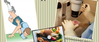
Note that severe endocrine diseases can cause a vicious ratio of articular bones and, as a result, impingement syndrome. People suffering from diabetes are at high risk, since poor metabolism and problems with blood circulation negatively affect the morphology of bone and cartilage tissue. The risk category includes people who regularly experience heavy physical stress on the pelvic girdle and lower limbs. Possible causes of FAI also include primary and secondary diseases of the musculoskeletal system and connective tissue, for example, arthritis of the hip joint, gout, scoliosis, lumbar hernia, etc.
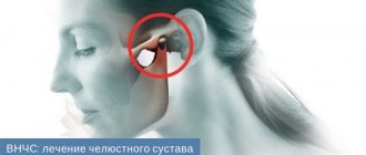
Impingement syndrome of the right shoulder joint: symptoms, treatment and prevention
Impingement syndrome of the shoulder joint is a serious disease, but if treatment is started on time, the disease does not pose a threat.
In the article you will find how the disease develops, causes and treatment, prevention and diagnosis of shoulder impingement syndrome. Also in the article you will find traditional medicine treatment and exercises that should be performed regularly. And I think you will also be interested in learning about the symptoms of shoulder impingement syndrome.
This information will be useful to anyone who is faced with this disease. The article also contains videos in which the doctor will give you the advice you need and, I hope, in which you will find answers to your questions.
Impingement syndrome of the shoulder joint - characteristics
The beautiful name “impingement syndrome” is associated with acute pain that significantly reduces the quality of life in a joint: the shoulder, and sometimes in others, for example, in the hip, ankle. This disease is especially common among athletes and those engaged in heavy physical labor.
The shoulder joint is a complex and no less unique system of components that mechanically interact with each other. The movable base of the scapula, humerus, and clavicle allows the human shoulder to perform a variety of mechanical movements.
With the help of tendons, the minor, subscapularis, supraclavicular and subclavian round muscles are attached to the scapula and bone of the shoulder, which interact only with the help of a rotator cuff formed by other tendons. Sometimes it happens that friction occurs between the acromion (scapular process) and these tendons of the rotator cuff, resulting in a pain syndrome that is commonly called impingement syndrome of the shoulder joint.
This process, called impingement syndrome, occurs in a large number of people due to compression of the tendons and joint capsule between the head of the humerus and the acromion when raising the arm in a vertical position.
Impingement syndrome, which is clinical in nature with the appearance of a painful syndrome, is characteristic of people whose professional activities are directly related to long-term static postures. These are positions that imply a fixed holding of the hand in a raised position.
Among the most common professions in which this disease can occur are: painters, plasterers, athletes, carpenters. In general, a similar shoulder joint syndrome can be caused by various conditions in which the gap between the coraco-acromial arch (rotator cuff tendons and acromion) decreases.
Shoulder impingement syndrome is painful. Sometimes it is tolerable, less often it is strong and exhausting, but almost always it is sudden and inexorable. It is quite difficult to call such a pathology rare, but, nevertheless, it is practically unknown to most domestic doctors. And the point here is not so much the reluctance of many doctors to spend time learning something new, but rather a certain ossification of thinking characteristic of many doctors.
Formally, impingement syndrome has been known in medicine for more than 100 years (the first reports of it date back to 1872), but in those days it was called “scapulohumeral periarthritis.” However, due to the imperfections and inaccuracy of instrumental diagnostic methods of the 19th century, a clear explanation of the mechanisms of pain was not found then.
A century later (1972), the pathology received its modern name (in Russian-language literature you can find variants like “impeachment syndrome” or even “impeachment syndrome”), but most practicing doctors still use the old name.
The essence of the problem
The shoulder joint is one of the most complex mechanical devices created by nature. It consists of many elements, precisely adjusted to one another. Unfortunately, over time, the ideal interaction between them is disrupted, and the system begins to malfunction, which we perceive through pain attacks. One of the most likely “problem” places is the subacromial space.
This is a narrow (no more than 7 mm) gap formed above by the acromion process, and below by the head of the humerus. With age, due to the accumulation of salts, a decrease in the amount of lubrication and a general decrease in joint mobility, the subacromial space decreases, strong friction occurs and a distinct pain attack occurs, which is called impingement syndrome.
The only point that needs further clarification concerns the age range of the pathology. Logic dictates that such changes are more common in older people, but in practice, impingement syndrome occurs even in 30-40 year old patients.
Therefore, the common misconception that joint problems are the lot of old people must be recognized as false. Therefore, when the first symptoms appear (when the problem can be effectively dealt with using conservative methods), you need to consult a doctor, and not postpone treatment until better times. Which, as practice shows, can come much faster than you think.
Risk factors
Impingement syndrome (IS) can be caused by a variety of diseases. Some of them are widespread (especially in developed countries), others, with some reservations, can be called rare:
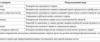
- adhesive capsulitis (“frozen shoulder”);
- suprascapular nerve neuropathy;
- arthrosis of the shoulder and acromioclavicular joints;
- cholecystitis;
- calcification of the supraspinatus tendon;
- osteochondrosis of the spine, localized in the cervical region.
But if we discard the theory and focus on the practical side of the issue, it turns out that the incidence of IS depends to a greater extent on the characteristics of professional activity (painters, carpenters, plasterers, athletes) than on the age of the patients. In other words, impingement syndrome is more of an occupational disease than an aging disease.
There is no generally accepted classification of IP, especially if we leave out the professional, highly specialized language. Most practicing doctors divide “impingement syndrome” (after all, such a spelling also has a right to life) into two conditional groups.
Primary IS. Occurs due to mechanical irritation of the periosteal muscle due to:
- post-traumatic deformity (acromial or clavicular processes, greater tubercle of the humerus);
- rotator cuff tendon injuries;
- congenital changes in the shape of the acromion process.
Secondary IC. Caused by a narrowing of the subacromial space caused by:
- tear of the rotator cuff or biceps tendon;
- violation of the integrity of the ligaments of the acromioclavicular joint;
- congenital ligament weakness;
- thickening of the rotator cuff or bursa (a consequence of ossification or chronic bursitis);
- paralysis or muscle weakness;
- displacement of the greater tubercle of the humerus (usually as a result of trauma).
Causes
When you raise your arm, the tendons and joint capsule between the head of the humerus and the acromial process of the clavicle (acromion) are always slightly compressed. This regularly recurring condition is called impingement (in English impingement and actually means “blow”, “collision”).
Under certain circumstances, soft tissues can become pinched, irritated, inflamed, damaged, and here we are talking about painful impingement syndrome. Due to the narrowing of the space under the acromion, there is not enough space for the normal functioning of all the structures located there. The situation becomes even more complicated in cases of non-standard shoulder shape and the presence of bone processes.
The supraspinatus tendon, which is located directly under the acromion and is part of the rotator cuff, which covers the head of the humerus, especially often suffers from compression. Therefore, impingement syndrome is often directly associated with damage to the important rotator (or rotator) cuff.
This disease is the most common cause of pain in the shoulder joint, which occurs depending on movements or body position, including at night. Those suffering from the syndrome are often unable to move their arm upward (the most painful sensation is at an angle of 60 to 120° - the so-called “painful arc”), and can hardly put on a shirt or wash their back. Ultimately, impingement syndrome, that is, the severe pain associated with it, significantly limits the functioning and mobility of the entire affected joint.
There are two types of subacromial impingement syndrome, that is, affecting the shoulder joint: primary (outlet) and secondary (non-outlet). In the first case, the sub-shoulder space is narrowed due to mechanical reasons, such as the presence of a bony process in the joint or a strongly sloping shoulder.
In the second case, possible causes of impingement syndrome include chronic inflammation of the periarticular bursa (bursitis), damage to the long biceps tendon, rupture of the rotator cuff, or biomechanical disorders such as muscle stiffness or imbalance. With muscle imbalance, we are talking about functional impingement syndrome of the shoulder.
Thus, the occurrence of problems is based on various reasons, among which the most common are pinching, damage, wear, cracks, inflammation of muscles and tendons. If the causes are not hereditary (the special structure of the shoulder joint), then, as a rule, they have their origins in the type of human activity, which is characterized by constant physical overloads placed on the upper part of the body.
This risk group includes athletes, especially tennis players, handball players, volleyball players, basketball players, as well as representatives of certain working professions - painters, plasterers and others.
Symptoms
The shoulder is made up of a complex system of interconnected tendons, and crowding under the acromion can cause pain when moving the arm in certain ways. The pain occurs when the tendons are pinched or ruptured due to heavy loads and intensifies when raising the arm above the head. If inflammation persists, other symptoms may appear. Constant irritation of the synovial bursa between the rotator cuff and the acromion leads to night pain, which is quite severe and painful for the patient, which disturbs sleep and the person cannot sleep on the inflamed shoulder.
In addition to pain, there is a restriction in movement in the shoulder joints as long as the symptoms persist. This condition is referred to as secondary shoulder stiffness or frozen shoulder. Inflammation of the tendons leads to excruciating and long-term chronic pain.
Signs of the presence of the disease in its early stages are aching pain in the shoulder, which can appear even during sleep. Then the painful sensations intensify, and limited movements gradually appear.
When you raise or abduct your arm at the shoulder, you may feel unpleasant clicking, snapping, or crunching sounds. As a result, the muscles in this area weaken and lose tone. With further stress, the tendons may rupture, the pain will become unstable, and the joint will become almost immobile.
Impingement syndrome of the shoulder at the very beginning develops as inflammation of the tendons, then dense scar connective tissue appears in the area of inflammation - fibrosis, and as the disease progresses, fibrosis is replaced by calcium deposits.
In a healthy person, this gap is wide enough for the supraspinatus muscle to move freely, but with impingement syndrome, the gap narrows. This narrowing makes it difficult for the muscle to work and creates conditions for its microtrauma.
In a sense, anatomy itself created the prerequisites for the emergence of this pathology. Indeed, oddly enough, the development of impingement syndrome depends on the shape of the acromion, and the shape is different for all people.
However, if you think about it, the question arises: if the cause of impingement syndrome of the shoulder joint is a bent acromion, then compression of the muscle should have occurred already in infancy, why then does the disease appear only over the years?
Indeed, the shape of the acromion is an important, but not the only factor, and curvature alone is not enough for the occurrence of impingement syndrome of the shoulder joint. In general, it is worth recalling here that our body is quite resistant to the occurrence of any pathology, including this one. And in order for impingement syndrome to occur, the action of not one, but several factors is necessary.
So, the second such factor is the natural ability of bones to grow. As you know, bone can grow throughout your life. Only in childhood and adolescence does this growth ensure the “growing up” of a person.
And although an adult has grown, the ability of bones to grow has not completely disappeared - it has been preserved. Why is this necessary? Everything is very simple. For example, so that if something happens, fractures heal. Or in order to strengthen the place of increased load, just as it happens with the skin on the feet.
After all, it’s no secret that if we walk barefoot for a long time, our feet will become rough. And from a medical point of view, such coarsening means that the skin has thickened due to the growth of additional layers to protect the feet. The same thickening, in response to overload, occurs in the bones.
By the way, the growth of bone tissue is considered by people far from medicine to be salt deposition. However, science has clearly established that this is not salt at all, but a protective reaction to overload. Depending on the area of the congested area, bone growth can be extensive or localized.
It is precisely this point growth at the end of the acromion that creates something like a spike. And if with a flat acromion this is not critical, then with a hook-shaped acromion it significantly narrows the already small opening. In general, the ability of bones to grow in response to physical activity is the second factor and cause of shoulder impingement syndrome.
Thus, for the occurrence of shoulder impingement syndrome, at least two conditions are necessary: an anatomical predisposition in the form of a hooked acromion and overload of the shoulder joint.
Moreover, you don’t need to think that overload means hard physical work or sports. For example, among our patients with this diagnosis there are many people from creative professions: violinists, cellists, artists, cameramen, especially those who often shoot “from the shoulder,” etc.
In addition, you should not delude yourself into thinking that impingement syndrome occurs only in the case of a bent acromion. Long-term overload of the shoulder and intense growth of bone tissue can lead to disease even with an initially flat acromion.
In addition, it is necessary to take into account the direct connection between shoulder impingement syndrome and diseases of the cervical spine. After all, the stimulus for the formation of impingement syndrome is very often disc pathology: protrusion, disc herniation, osteochondrosis, etc.
In general, remember the golden rule more often: “a disease is easier to prevent than to treat,” and try to always follow this rule. And for this, at the slightest suspicion of impingement syndrome, contact a chiropractor.
The onset of the disease is manifested by discomfort, which is gradually replaced by episodic pain. Then the pain becomes more frequent and constant. In general, everything is increasing: from slight discomfort to constant pain.
And if the disease is not stopped in time, everything can end in surgery. Of course, an operation is not a death sentence; on the contrary, it allows one to get out of the crisis in which a person finds himself. But any operation is a large-scale undertaking and high costs.
Moreover, even the most successful operation entails restorative treatment, which can be even more difficult and more expensive than the operation itself, and without which all the results of the operation may come to nothing. That is why you should not neglect the disease, and if you or your loved ones are faced with this problem, you must do everything possible to avoid surgical treatment of shoulder impingement syndrome.
Modern approaches to gentle manual therapy, most often, make it possible to overcome the disease and treat impingement syndrome without surgery.
Diagnostics
The symptoms of impingement syndrome of the shoulder joint are clear - these are acute pain in this area when raising the arms and other movements, in a lying position on the affected side of the body. The pain can spread only to the shoulder or further, to the forearm and arm.
Often, due to the transparency of symptoms, the doctor can make a diagnosis based on the results of a routine examination of the patient and simple clinical functional tests, in which he must perform certain movements. Orthopedists encounter complaints typical of impingement syndrome very often and, as a rule, have enough experience in this area.
During the examination, the doctor often asks the patient about his daily work, since impingement syndrome is mainly an occupational disease (plasterers, installers), which also affects athletes (swimmers, shot putters, volleyball players).
To determine the source of pain, x-rays are prescribed. Additional examination using magnetic resonance imaging may be necessary. This technique can visualize bone and soft tissue and determine whether the rotator cuff is torn.
If it is not possible to determine the cause of the pain, which, in addition to inflammation in the joint, can be cervical osteochondrosis, an anesthetic is administered to help determine the source.
Treatment of shoulder impingement syndrome occurs in two stages: conservative and surgical. Conservative treatment is prescribed at an early stage of the disease, and involves taking drugs such as Voltaren or Xefocam. Sometimes, in case of severe pain, the doctor prescribes diprospan, the purpose of which is to reduce inflammation and swelling.
All necessary injections are made in the acromion area. At the same time, to improve mobility, a number of physical exercises are performed. Traditionally, during this pathological process it is customary to distinguish three stages. This classification was developed by Dr. Neer CS at the end of the twentieth century, but even now it is actively used by orthopedic traumatologists:
- The first stage usually occurs at a young age, between 20 and 40 years of age. It manifests itself as moderate pain after physical activity. Swelling and hemorrhages can be detected in the rotator cuff at this stage.
- As the disease progresses, a transition to the second stage is noted. The rotator cuff thickens due to constant mechanical injuries. Fibrosis develops in it, which is accompanied by inflammatory processes in the tendons - tendonitis. Typically, the second stage occurs in young and middle age - from 30 to 50 years. The best effect in this situation is demonstrated by surgical treatment.
- In the absence of treatment, the disease enters the third stage. It is characterized by tears of both the rotator cuff and the biceps tendon. A formed bone spur can also be found in the joint. Since the shoulder muscles - short rotators - are degeneratively changed during this period, they become unable to provide dynamic stability of the joint.
Constant injuries lead to inflammation and degeneration of not only the joint, but also the adjacent soft tissues.
This clinical and radiological picture can be seen in patients of the middle and older age groups - 30–70 years old. Treatment of shoulder joint impingement syndrome
The disease requires individually selected therapy, depending on the underlying cause, stage of development, and duration of pain. First, conservative treatment is usually carried out, including medication and physiotherapeutic procedures. A certain effect may occur only after several weeks, and sometimes even months, so you need to be patient.
The goals of therapy are to restore joint function, return the muscles to their former strength and stability, and, of course, eliminate pain. For this purpose, both painkillers and anti-inflammatory non-steroidal drugs are used in combination with therapeutic exercises, physical therapy (treatment with cold, heat, electrical impulses, ultrasound) and sometimes manual therapy. In addition, the doctor may include glucocorticoid injections into the shoulder into the treatment plan, however, due to the risk of additional tendon damage, the number of injections should not exceed three.
In no case should you underestimate the importance of physiotherapy, especially special gymnastics, which allows you to strengthen the muscles and change the position of the head of the forearm, freeing up more space for the tendons. It is better to carry out such training daily for several months, remembering that gymnastics should not cause pain and without overdoing the duration (no more than 15-30 minutes) and intensity of exercises.
Throughout the entire treatment period, you should refrain from overloading the shoulder joint. In some acute cases, the doctor may even resort to fixing it. Is therapy possible when such a diagnosis is made? How is shoulder impingement syndrome treated? If the disease is detected at an early stage, traumatologists recommend starting with conservative methods. Progress of treatment:
- First of all, it is necessary to deal with pain. For this purpose, non-steroidal anti-inflammatory drugs are used in the form of ointments, gels, tablets, suppositories or injections. If their effect turns out to be insufficient or short-lived, doctors resort to heavy artillery - hormonal drugs. This therapy gives good results with a long-lasting effect, but the risk of side effects is significantly higher.
- Among the auxiliary methods, physiotherapy is widely used - treatment with a magnet, ultrasound, local administration of drugs by electrophoresis.
- Regardless of which conservative method is chosen as the main one, it is necessarily complemented by physical therapy. It is aimed at strengthening the muscles of the shoulder and arm.
- If conservative therapy does not produce the expected results within 3–5 months, traumatologists recommend surgery. As a rule, we are talking about diagnostic arthroscopy, during which the correction of disorders is simultaneously carried out - elimination of osteophytes, restoration of tendons, changing the shape of the acromion.
But not all patients trust traditional medicine. Some are wary of major interventions due to the risk of side effects. Is alternative therapy possible?
If conservative therapy does not bring the expected result, especially if the pain only intensifies, there is talk of surgery. Professional athletes are also often inclined to the surgical method of solving problems.
This operation is called subacromial decompression of the shoulder joint, or acromioplasty, and is designed to expand the subacromial space to restore the normal functioning of all elements in this area. However, surgery is indicated mainly for the primary (outlet) form of the syndrome, which is caused by mechanical reasons. In the secondary form, that is, in the presence of bursitis or other inflammation, acromioplasty is generally not recommended, except in some cases of rotator cuff injury.
Today, such operations are almost always performed using endoscopic, minimally invasive methods, through very small incisions. During the day, the shoulder joint is then fixed in a special way. The first two weeks after surgery require special movement exercises with the participation of a physiotherapist, starting from the third week - self-performed gymnastics.
At the same time, the rotator cuff muscles are strengthened and built up step by step. Movement is very important, especially given the risk of developing a post-operative complication called frozen shoulder. Preventing the development of shoulder impingement syndrome is very difficult. In any case, balanced physical activity contributes to the healthy functioning of all joints and muscles. When doing fitness and gymnastics, it is recommended to pay special attention to training the rotator cuff.
Usually they start with conservative treatment. You may be prescribed non-steroidal anti-inflammatory drugs such as diclofenac or ibuprofen. Rest and ice can relieve pain and inflammation. If the pain persists, cortisone injections into the joint may help.
Cortisone is a powerful drug that reduces inflammation and pain. The effects of cortisone are temporary, it effectively relieves pain and inflammation, but negatively affects articular cartilage. Your doctor may also prescribe physical therapy and exercise therapy. If there are no contraindications, then various procedures can be used to relieve inflammation, including cold and heat. Lessons with an instructor may be required.
Gradually increasing the strength and coordination of the rotator cuff and scapula muscles allows the humeral head to move directly into the center of the scapula without impinging on the tendons or bursa under the acromion. It may take four to six weeks for your shoulder to regain range of motion and function. If conservative treatment is unsuccessful, then surgery is necessary.
The operation is called acromioplasty. The goal of the surgery is to increase the space between the acromion and the rotator cuff tendons. The greater the space between the above structures, the less chance there is of impingement of the cuff tendons between the shoulder and acromion.
The surgeon first removes any bone spurs that are irritating the cuff tendons and bursa. It often becomes necessary to remove a small part of the acromion and even the acromial end of the clavicle. In patients who have a downward slope of the acromion, more bone tissue needs to be removed.
Treatment of shoulder impingement syndrome can be surgical or non-surgical. And, as we said, surgery is a last resort, it is done only when all other types of treatment have been tried and they have not worked. But this rarely happens.
Non-surgical treatment of impingement syndrome requires drug and non-drug methods. The main and main non-drug method is gentle manual therapy. Additional treatments include physiotherapy and exercise therapy.
For acute pain and severe swelling of deep tissues, treatment begins with medications. For this purpose, anti-inflammatory and decongestant medications are used: movalis, voltaren, ortofen, and in case of persistent pain - diprospan. When swelling and pain decrease, begin gentle manual therapy. This is the most effective non-surgical treatment for shoulder impingement syndrome. Manual therapy has various manual methods in its arsenal.
Each of them is aimed at a specific structure: muscles, joints, tendons, ligaments, spine. It is best to use a combination of these methods. This approach gives the highest effect and the most lasting treatment result.
As the patient recovers, special therapeutic exercises are prescribed. Usually they start with simple and gentle movements, and then gradually increase the amplitude, volume and load. And so on until the normal functioning of the shoulder joint is completely restored.
ethnoscience
Impingement syndrome of the shoulder joint can be treated using traditional medicine. But you need to understand that we are talking exclusively about symptomatic therapy - in order to reduce inflammation and pain. When treating subacromial conflict, the following are most often used:
- Anti-inflammatory teas. Infusions of chamomile and yarrow, tea made from lingonberries and rose hips, and currant leaves have a good effect.
- Applying cabbage or plantain leaves directly to the affected area.
- Various compresses. You should be careful with this method, as it can aggravate the inflammatory process. The composition of the compress must be agreed upon with the attending physician.
- Treatment with honey. It can be applied as a thin film to the affected area.
But still, traditional medicine methods are not able to replace traditional medicine and save the patient from the disease forever. They can only be used as an additional treatment.
Prevention and prognosis
With timely diagnosis and comprehensive treatment, impingement syndrome cannot be classified as an incurable disease. But due to the neglectful attitude of both patients and medical personnel towards joint diseases, some patients may develop irreversible changes in the body and permanent disability.
Therefore, we repeat, the sooner you seek help, the greater the chances of a complete cure. Also, do not neglect simple prevention methods, compliance with which will significantly reduce the likelihood of developing pathology:
- minimizing the risk of shoulder injury;
- during prolonged physical activity, special protective and supportive bandages should be used;
- mandatory treatment of even minor shoulder wounds with antiseptics, followed by application of a bactericidal dressing.
VERY IMPORTANT INFORMATION:
If you are reading this, it means only one thing - you have serious problems and you really need help. We understand you like no one else!
97.4% of our readers have already encountered similar problems!
You need to answer honestly 3 simple questions:
- Are you familiar with pain and discomfort in your joints, back, knees or arms?
- Do you trust doctors without recommendations?
- Do you think it is right to treat the symptoms of a disease or eliminate the cause itself?
Make an appointment with a doctor at Veteranov 16, St. Petersburg
To make an appointment with a doctor, please call: +7 (812) 756-92-92 , +7 (952) 204-59-83 , or fill out the ONLINE appointment form.
[contact-form-7 404 “Not Found”]
Types of femoroacetabular impingement
The so-called “impact conflict” during the motor act of the TB joint is classified according to localization into 3 main types of FAI:
- acetabular, or pincer type;
- femoral, or cam type (cam);
- mixed look, or mixed.
- Pinser type
. The cause of the impact is an anatomical failure in the form of an increase in the coverage of the acetabulum with a normal proximal femur. Occurs 3 times more often in women than in men. The age range of patients is 40-57 years. On radiographs in anteroposterior and lateral projections, the following can be seen:
- increasing the depth of the cavity, its protrusion;
- signs of cross-over;
- central-edge angle G. Wiberg more than 39°;
- VP roof inclination angle is less than 0°;
- symptom of the posterior edge of the pelvic cavity;
- linear depression of the bone in the neck area.
- Cam-type
. The primary source of impingement is the abnormal shape of the femur in the proximal part with loss of sphericity with an unchanged acetabular socket. This problem occurs 14 times more often in male patients. The age group of patients is 21-50 years. X-ray images in the direct and lateral planes show:
- deformation of the metaepiphysis of the femur like a “gun handle”;
- neck-shaft angle less than 125°;
- horizontal epiphyseal plate symptom;
- ∠α (alpha angle) from 50° or more;
- abbreviated cervical offset – below 8 mm, offset index less than 0.18;
- posterior deviation of the femoral neck (retrotorsion).
- Mixed type.
The clinical picture is observed simultaneously on two bone components of the articulation: the acetabular cavity and the femur. In 90% of cases this type is diagnosed.
Symptoms of subacromial syndrome
Clinical manifestations of the disease occur with a gradual increase. Pain in the shoulder area and along the muscles is the main symptom. At the initial stage, pain occurs after exercise, then gradually becomes permanent. Movement in the joint is limited. Raising your hand becomes sharply painful. Local swelling of the shoulder may be observed. At a later stage, atrophy of the muscles of the shoulder girdle becomes visually noticeable. A clicking sound may be heard when you lower your hand. If the tendon has not ruptured, then patients, as a rule, do not consult a doctor for a long time, making a mistake. After all, in case of advanced disease, only surgical treatment is effective.
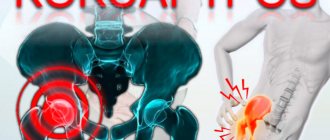
Symptoms of hip impingement syndrome
Characteristic clinical signs are pain and limited mobility. These criteria are relative, as there are exceptions when a person does not notice any discomfort at all. He lives in peace and is not even aware of the existing problem. Therefore, everything is individual, but in the majority of people, the painful condition still makes itself felt with the following symptoms:
- pain, its frequent localization is the groin area, buttock area, lower back;
- sudden onset of pain in the hip joint during its extreme positions of internal rotation, ghosting, flexion;
- persistent limited range of motion in the joint;
- the lesion is usually localized in one of the hip joints;
- increased pain after a long stay in the “sitting” position;
- increasing discomfort after prolonged or intense physical activity;
- pain relief at rest.
We would like to point out that similar symptoms manifest themselves in many diseases not only of the hip joint, but also of other components of the musculoskeletal system. Therefore, with complaints of this nature, it is important to reliably determine whether the symptoms originate from the hip joint. In addition, whether they are provoked by impingement, what type and origin it is, how strong the femoroacetabular conflict is and whether it is combined with any other diseases of the joint. A competent differentiated approach to diagnosis will help you avoid making mistakes in choosing treatment tactics.
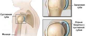
Tears of the labrum.
Injuries to the labrum (lat. labrum) cause severe pain and can often cause tissue entrapment during movements and blockade of the hip joint. The cause of damage to the labrum may be trauma or chronic femoroacetabular conflict.
These injuries do not respond to conservative therapy, such as taking medications, reducing physical activity, physical therapy, and even injections. Over time, mobility restrictions increase and begin to make it difficult to perform professional duties, interfere with housework and the usual way to spend free time. In this case, surgical treatment becomes inevitable.
Clinical examination, diagnosis
For patients, diagnostic measures begin with functional tests of the hip. They consist of assessing the functions of flexion, extension, adduction, abduction, and rotation of the problem joint. The doctor tests the patient's limb in different physiological trajectories of movement.
A specialist can make a preliminary conclusion about the diagnosis, for example, based on the “log rolling” test. To do this, the patient lies on his back, after which the doctor rolls his leg from the outside in and back. If during such an experiment a peculiar crunch occurs locally, then this is highly likely to indicate damage to the labrum.
To identify a bone conflict, the tactics of bending the limb at the hip joint to a right angle are used, then the leg is brought in and rotated inward, then outward. If pain occurs at the end point, the test is considered positive. More often, such pain indicates a collision of the anterosuperior acetabular area and the surface of the femoral neck.
The region of interest can also be tested for Drehmann's sign and the C sign. In the first case, the diagnostic criterion is the ability to bend the leg at the joint exclusively from the position of external rotation, which indicates torsion of the femur according to the retroversion type. In the second, pain occurs when grasping and compressing the supratrochanteric part with the thumb and forefinger, forming the letter “C”. A certain C-symptom indicates a distorted morphology of the acetabular element.
No good specialist will make a diagnosis or prescribe treatment based solely on functional tests. The next stage, which allows us to confirm or reject initial assumptions about the problem and extract maximum information about the anatomical disharmony of the joint, is instrumental diagnostics. It is based on the implementation:
- radiography;
- magnetic resonance imaging;
- computed tomography.
First there was impingement, and then arthrosis developed.
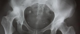
Despite the existence of highly promising methods for obtaining cross-sectional images of the joint, standard radiography remains the first and mandatory diagnostic method in this diagnostic chain. The diagnostic information value of classical radiography for determining BVI is quite high: 90% and above. It is possible to make a correct diagnosis based on X-ray results only if images are taken in all of the following projections:
- anteroposterior (standard);
- according to Launstein (laying in the “frog” position);
- lateral;
- in the position of hip flexion at angles of 90°, 45°;
- “false” profile (oblique projection technique, Lequesne’s false profile).
Tomography methods (CT, MRI) are used when a complicated generalized form of pathology is suspected, requiring a more in-depth assessment of the structural components of the hip joint. The best picture of the condition of soft tissues will be provided by magnetic resonance imaging using a high-power MRI machine. This type of diagnosis determines pathologies of articular cartilage, synovial membrane and lip, well visualizes cysts, muscle tendinosis, joint synovitis, bone marrow edema.
Types and degrees of impingement
There are two main types of impingement syndrome: anterior and posterior.
Anterior impingement is mainly a consequence of traumatic injury to the ligaments of the ankle. Any instability of the joint, even if it is very weakly expressed, contributes to its injury in the position of extreme extension.
According to statistics, athletes mostly suffer from anterior impingement. In them, the disease is provoked by frequent high load on the front part of the joint, which causes permanent damage. First of all, with this disease, the amplitude of extension of the joint decreases.
Posterior impingement is often associated with the anatomical features of the ankle structure and injuries. The posterior type of impingement is characteristic mainly of ballet dancers. Indeed, during this dance, a person is forced to walk a lot on his toes, which leads to strong flexion of the ankle joint in the back and, as a result, to injury.
The existence of posterior impingement is often forgotten, although in fact the disease can lead to serious impairment of joint function.
The disease can also be divided into several degrees:
- I degree – characterized by the presence of a spur up to 3 mm in size on the tibia, called the synovial degree;
- II degree – osteochondral, the size of the spur exceeds 3 mm;
- III degree – exostoses with or without fragmentation can be identified, a spur also appears on the talus;
- IV degree – changes characterizing arthrosis develop.
Basic principles of treatment for FAI
In the treatment of patients with hip impingement, depending on the severity of the clinical case, either conservative or surgical treatment is used. Today, due to incomplete knowledge of the natural course of the femoral-acetabular conflict, it is impossible to predict the success of non-surgical therapy.
After operation.
Regarding prognosis after surgical interventions, we note that in patients with severe secondary arthritis, treatment results are worse. Surgery has a good effect if the joint space is narrowed by no more than 1/2 of the normal values, that is, the width of the gap is not less than 2 mm when the norm is 4 mm. The patient's young age and the short period of time from the onset of FAI to the patient seeking medical attention significantly increase the chances of success of the operation. help.
Diagnostics and preoperative preparation.
Before the operation, you need to agree on a meeting in our consultation department with a leading specialist (telephone 8 812 559 9783).
You will undergo an intensive examination by our medical team. Laboratory tests necessary for general anesthesia must be performed. These include ECG, fluorography, blood and urine tests. These tests can be done on an outpatient basis to reduce hospital stay. If you have completed test results (for example, x-rays, CT scans, nuclear magnetic resonance imaging scans), bring them with you to your consultation. Based on the examination data, we will be able to answer all your questions about the intervention plan, the proposed anesthesia and the subsequent recovery period.
Conservative treatment tactics
A conservative approach involves prescribing complex treatment, including:
- limiting physical activity that causes pain;
- temporary immobilization of the joint (with severe inflammation, swelling)
- physical therapy aimed at increasing the range of movements and strengthening the muscles that stabilize the joint apparatus;
- physiotherapy procedures (ultrasound, laser, magnet, electrophoresis);
- drug treatment - the use of painkillers from the NSAID series; for unbearable pain, blockades with intra-articular administration of corticosteroids (diprospan, hydrocortisone, etc.) are used.
Injections of steroids and hyaluron are justified if there is a proven fact of the presence of a lesion in the cartilaginous formation attached to the edge of the acetabular bed along the circumference. The pathogenesis associated with lip damage can only be confirmed by MRI. Purely on the basis of x-rays that simply prove the presence of a nosocomial infection, injections of hormones and hyaluronic acid are not prescribed.
If the symptoms of a pathological phenomenon cannot be stopped conservatively, it is advisable to perform surgical intervention to eliminate the causes of pathogenesis. Remember, an unfavorable pathogenetic mechanism can progress, causing a serious disease of the entire hip joint - deforming arthrosis. Often such complications require total joint replacement with an endoprosthesis.
If non-surgical treatment fails within 3-4 months, it is important for people of working age, athletes and adherents of an active lifestyle to undergo surgical intervention as soon as possible.
Surgery and hospital stay.
Patients are usually admitted to the hospital on the day of surgery. If you need to make a long journey to the hospital, you may be admitted to the hospital the evening of the previous day. Our friendly nursing staff will help you find accommodation in a single or double room. During the day, the operation will be performed, and dinner will be delivered to you in bed. Typically, observation in a hospital is necessary for 3-5 days from the date of surgery, after which discharge to an outpatient stage is possible. You can
leave the clinic independently, on two crutches, or accompanied. We will be happy to help you arrange transportation if you wish.
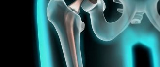
Surgery is performed arthroscopically under general anesthesia. This means that a video camera with a light source will be inserted into the joint cavity through a skin incision about a centimeter long. All important structures of the joint will be examined, and findings can be recorded in the form of photographs and HD video.
Reconstruction of damaged structures is carried out through the same incision.
Surgery for hip impingement syndrome
We emphasize that impingement is not always a reason for operating on a patient. The first thing that draws attention is how much FAI interferes with a person in everyday life (at work, in sports, at home). So, surgical treatment is carried out using open or arthroscopic approach technology. The surgical session is aimed at removing exostoses (bone growths), which will eliminate the pathomechanical factor of the components of the joint hitting each other.
An open operation is performed through a standard incision, about 8-10 cm. Interventions with a wide opening of the articulation are prescribed, for example, for the posterior cam type, a generalized increase in the area of the acetabular covering, acetabular retroversion of idiopathic etiology. Open intervention is accompanied by cutting off the greater trochanter and dislocation of the hip. Next, taking into account the indications, the necessary manipulations are performed (resection, plastic reorientation interventions, etc.) that will help restore the shape of the proximal metaepiphysis of the femur and/or the glenoid cavity.
Reducing buildup.
Arthroscopic procedures are performed using the optical instrument of an arthroscope, but with impingement not clearly visible on radiographs. The arthroscope tube is inserted into the joint through a small skin incision, the size of which is approximately 1 cm. During arthroscopy under video control, the surgeon performs a simulating resection: clears the acetabular and femoral components of excess bone growths. If lip injuries are discovered during the session, the defects are corrected by anchor refixation, autoplasty or mechanical (sometimes plasma) debridement.
Recovery period
After almost all hip joint surgeries, the person is allowed to get out of bed the very next day. At first, he moves with crutches and loads the operated limb only 50%. After arthroscopy this period lasts 1.5-2 weeks, after open surgery - 3-5 weeks.
The duration of rehabilitation after arthroscopic surgery is 3-4 months, after open surgery – 5-6 months. During the recovery period, the patient engages in physical therapy and undergoes physical procedures. This allows him to quickly return to his usual lifestyle.
Rehabilitation is an important part of surgical treatment. Lack of medical care during the recovery period increases the risk of complications.
Rehabilitation is the most important part of treatment.
How much does the operation cost?
The cost of surgical treatment of hip impingement in Russia starts from approximately 15 thousand rubles, the maximum can reach 100 thousand rubles. The estimated price for arthroscopy of a simple level of complexity in Moscow is 20-25 thousand rubles. Orthopedic clinics in Israel and Germany offer promising prospects for complete, safe, low-traumatic removal of impingement. The price of an operation for this problem in medical institutions in Israel is from 14 thousand dollars or more, in clinics in Germany - at least 10 thousand euros.