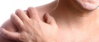
The disease capsulitis or frozen shoulder is an inflammation of the shoulder tissues and the development of adhesions in the joint cavity. Capsulitis is accompanied by severe pain and difficulty raising the arm; these symptoms are similar to other diseases of the shoulder joint, but the treatment is very different, so before starting treatment, it is necessary to undergo an examination to make an accurate diagnosis.
A medical diagnostic specialist successfully treats shoulder capsulitis ; in addition to treatment, you can undergo a comprehensive examination with us using modern equipment to clarify the diagnosis.
Why does adhesive capsulitis occur? What happens to the shoulder joint?
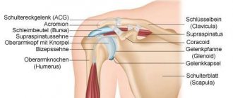
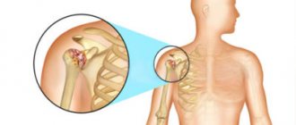
The articular processes of the bones, tendons and ligaments that form the shoulder joint are enclosed in a capsule of connective tissue. It provides stability to the joint. With adhesive capsulitis, the joint capsule hardens and tightens, thereby making movement difficult.
The reasons for the development of the disease are not fully known. Some factors increase risks:
- Diabetes. Adhesive capsulitis of the shoulder occurs in 20–30% of diabetics.
- Parkinson's disease is a neurological disease in which, as a result of the death of special nerve cells in the brain, the regulation of movement and muscle tone occurs.
- Pathologies of the thyroid gland, in which there is an increase (hyperthyroidism) or decrease (hypothyroidism) in its activity.
- A stroke or surgery are conditions when a person is immobilized for a long time.
- Suffered shoulder fractures.
Reasons for the development of capsulitis
Capsulitis is most often observed in people from 40 to 60 years of age who have suffered shoulder injuries, work regularly with increased physical activity and poor treatment of musculoskeletal diseases.
The main reasons for the development of capsulitis:
- Age-related pathological changes.
- Professional sports.
- Weakened immunity.
- Infectious diseases.
- Congenital pathologies of joint development.
- Injuries, bruises and shoulder dislocations.
- Some diseases of the cardiovascular system.
- Previous heart attacks and strokes.
Symptoms of adhesive capsulitis of the shoulder
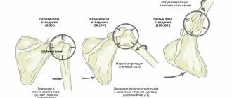
Symptoms usually develop slowly and the disease occurs in three stages. At first, you experience dull, aching pain in the shoulder joint, which intensifies with every movement. For some people, they occur at night and prevent them from falling asleep. A person cannot find a comfortable position in bed in which the joint would stop “aching.” At the same time, the deterioration of mobility in the shoulder joint increases.
Gradually the pain goes away, but mobility impairment increases. This condition is called frozen shoulder syndrome.
After some time, gradual “unfreezing” begins.
On average, each of the three stages lasts 3–4 months. Relapses are rare. In right-handed people, adhesive capsulitis of the shoulder most often develops on the left, in left-handed people - on the right.
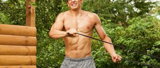
Physiotherapy
Exercise therapy is widely used as the initial treatment for many shoulder conditions, including AK. Physical therapy should include an exercise program that can restore shoulder movement. The patient should be included in an exercise program to restore and maintain range of motion. Patients receiving exercise therapy should begin an active exercise program for shoulder range of motion, as well as gentle passive stretching exercises, including forward elevation, internal and external rotation, and cross adduction. These exercises should be performed five to six times a day. And it is important to perform several sessions of 5-10 minutes a day, since during the time between sessions the shoulder becomes stiff and stiff again.
Improvement in range of motion was significantly better in the physical therapy groups than in the exercise-only groups. It was emphasized that, compared with exercise, physiotherapy interventions lead to significant reductions in anxiety and worry, which are closely related to symptoms. It was reported that 90% of 75 patients treated with a specific four-directional shoulder stretching program had satisfactory results at a mean follow-up of 22 months.
A systematic review and meta-analysis of randomized controlled trials evaluating the effectiveness of steroid injections and exercise therapy found that both interventions had similar effects on improving shoulder function, increasing passive range of motion, and reducing pain in AK.
Many studies have shown that physical therapy is a supportive intervention that produces good results. NSAIDs have been shown to be more effective when used in combination with physical therapy compared to NSAIDs alone. Similarly, steroid injections used in combination with physical therapy resulted in better results compared to injections alone.
Where to contact? What studies and tests can a doctor prescribe for adhesive capsulitis of the shoulder?
A rheumatologist diagnoses and treats frozen shoulder. During the examination, the doctor will ask you to raise, abduct, and flex your arm to evaluate movement disorders. Then he will ask you to relax your hand, take you by the shoulder and try to move it himself (in doctors' language this is called testing passive movements - with adhesive capsulitis they are also difficult).
Usually, a doctor can make an accurate diagnosis after an examination, without additional examination. If there are suspicions of other pathologies of the shoulder joint, you will be prescribed an X-ray, computed tomography or magnetic resonance imaging.
Take care of yourself, book a consultation now
Message sent!
expect a call, we will contact you shortly
Classification
Scapulohumeral periarthritis includes the following types of lesions:
- supraspinatus muscle (rupture, tear, tenomyositis , entensopathy - periarthritis of the shoulder joint is associated with this pathology in most cases);
- rotator cuff muscles;
- subscapularis muscle (its distal parts);
- inflammation of the biceps tendon ( tendinitis );
- teres minor and infraspinatus muscles;
- acromioclavicular joint;
- enthesopathy of the subscapularis muscle;
- subacromial bursitis;
- shoulder-hand syndrome;
- capsulitis of the shoulder joint.
Forms of periarthritis:
- simple;
- acute;
- chronic (“locked or frozen shoulder”).
Periarthritis is divided into:
- primary;
- secondary.
Primary periarthritis of the shoulder develops with prolonged muscle tension or disturbances in the motor pattern. Secondary occurs in various diseases: arthrosis , arthritis , developmental abnormalities, spondyloarthritis , joint hypermobility , connective tissue dysplasia , diabetes mellitus , hypothyroidism , calcium metabolism disorders, oncopathology or vascular disorders.
Why are the periarticular tissues of the shoulder joint often affected? This is one of the complex joints and the high incidence of damage to periarticular tissues is due to the anatomy, biomechanics of the joint and high loads. The shoulder joint does not have strong intra-articular ligaments that strengthen the joint, and the joint capsule is thin. Joint stability depends on the muscles that provide movement. The appearance of pain during degenerative processes of the tendons is associated with the addition of inflammation (tendinitis).
The duration of glenohumeral periarthritis depends on the course. The duration can be several weeks or several years (recurrent course). The disease can begin slowly and progress gradually, or it can progress quickly, leading early to dysfunction, tendon dystrophy and muscle wasting. A long-term course is observed with bilateral damage to the joints - the second joint is affected due to mechanical overload when performing the function of the first joint.
Cervicoscapular periarthritis is considered a complication of osteochondrosis . Vertebrogenic radiculopathy of the C4-C6 roots is characterized by pain not only in the neck, but also in the shoulder girdle, as well as in the scapula. In this case, the pain is associated with damage to the cervical spine. MRI of the cervical spine, which reveals osteophytes, arthrosis of the facet joints, calcification of the ligaments, and intervertebral hernias, allows for a correct diagnosis.
As stated above, there may be inflammation of the tissues surrounding any joint. The tissues of the knee and hip joint are also affected, but somewhat less frequently. This is due to the fact that the hip and knee joints have strong external ligaments and intra-articular ligaments that support the joints well.
Periarthritis of the knee and hip is most often secondary, that is, it develops against the background of arthrosis and arthritis . Thus, with osteoarthritis, the pathological process involves all structures of the joint - ligaments, capsules, synovial bursa and periarticular muscles. Also, mechanical pain of the knee joint develops with excessive load, intensifies in the evening and significantly decreases or disappears after resting at night.
In the area of the knee joint, tissue damage is represented by bursitis, enthesopathies (inflammation at the site of attachment of the tendon to the bone) and tenosynovitis (inflammation of the tendon and tendon sheath). Prepatellar bursitis (the term “parquet floorer’s knee” is often used) is caused by repeated trauma or stress (prolonged kneeling).
It is manifested by edema, swelling, pain, redness of the skin and a local increase in temperature. If the skin is damaged, infection can occur. When the prepatellar bursa becomes infected, there is a sharp, twitching pain and swelling, and redness of the skin. The contents of the bag contain pus. Recurrence of bursitis is avoided by eliminating the causative factor and protecting the joint with an orthosis.
The patellar ligament, which bears a large load, is often affected (it connects the quadriceps and lower leg and is involved in the extension of the joint). Damage to the patellar ligament occurs during stress and trauma in the form of enthesopathy . The ligament is damaged at the junction with the tibia (the term “footballer’s knee” is used) and the edge of the patella (the so-called “jumper’s knee”). “Jumper's knee” (patellar ligament ligamentitis) occurs when the joint is mechanically overloaded. The pain may be sudden or chronic. “Jumper’s knee” is observed in tennis players, track and field athletes, basketball players, and volleyball players and is associated with long jumps. Symptoms include pain below the patella, worse when sitting, swelling, and limited mobility.
In the hip area, pain syndromes are associated with damage to the femoral tendons, ligamentous structures and muscles.
Iliopsoas tendonitis causes pain in the upper thigh that makes walking difficult, as well as limited extension associated with back and groin pain.
When the hip joint is fully extended, painful clicking sounds occur and sometimes there is pain in the abdominal cavity. With this lesion, there is pinching of the femoral nerve ( Bernhardt-Roth neuralgia ), manifested by numbness of the outer thigh and paresthesia.
A common cause of hip pain is enthesitis of the greater trochanter, which complicates osteoarthritis at 40-60 years of age, but enthesitis can occur without coxarthrosis . Patients experience pain radiating along the outer thigh.
A typical complaint is the inability to lie and sleep on the side where the ligament is affected and pain when abducting the hip. enthesopathy of the abductor muscles occurs , and the presence of constant pain in the hip joint indicates trochanteric bursitis .
How to treat adhesive capsulitis of the shoulder?
To combat pain, the doctor prescribes non-steroidal anti-inflammatory drugs, for example, aspirin, ibuprofen. For severe pain, injections of corticosteroids - hormones of the adrenal cortex - are given into the shoulder joint. They are especially effective in the early stages of the disease.
To restore movement in the joint, they resort to physiotherapy and therapeutic exercises.
Sometimes they resort to more radical methods of treatment. For example, a doctor can inject sterile water into the cavity of the shoulder joint - it will stretch the capsule and improve mobility. Sometimes, under anesthesia, the compacted tissues are manually stretched, moving the shoulder in different directions.
If the above methods do not help, surgical intervention is performed, during which scar tissue and adhesions are removed.
In most cases, symptoms of adhesive capsulitis of the shoulder disappear within 1-3 years. But sometimes the disease leads to permanent limitation of movements. Timely treatment will help improve the condition and outcome. Make an appointment with a rheumatologist by phone: +7 (495) 230-00-01
The material was prepared by Natalya Yuryevna, a neurologist at the International Clinic Medica24, Candidate of Medical Sciences Lasch.
Medicines
In the initial stages of painful freezing, the treatment strategy is aimed at relieving pain. Although it is common to give patients nonsteroidal anti-inflammatory drugs (NSAIDs), NSAIDs alone do not affect the natural course of AK. There are no randomized controlled trials confirming the effectiveness of NSAIDs for specific AK conditions.
Oral corticosteroids are also used in the treatment of AK. The study reported that oral glucocorticoids (0.5 mg/kg/day methylprednisolone) in 33 patients with AK improved clinical outcomes: mean visual analogue scale (VAS) score; average score on the Constant scale; mean score of American shoulder and elbow surgeons at first-year follow-up. In another randomized clinical trial, 40 patients with idiopathic AK were treated with an oral corticosteroid regimen (20 patients) or an intra-articular corticosteroid injection (20 patients). Patients receiving oral medications showed significant improvements in pain and functional outcomes at 4-week follow-up. However, patients receiving intra-articular injection showed superior results in objective measures of shoulder function, range of motion, and patient satisfaction compared with the oral steroid group. As described in previous studies, oral steroid treatment appears to provide early benefit in terms of both pain relief and functional outcome; however, long-term benefits have not yet been established.
Description of the disease
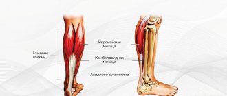
Rotator cuff
– anterior outer part of the capsule of the shoulder joint. It combines the tendons of the supraspinatus, infraspinatus, and teres minor muscles. Despite the difference in the functions they perform, such an anatomically close location of fixation of the muscles allowed traumatologists to identify them in a common group (the rotator cuff).
Damage to the rotator cuff can be considered a rupture of one or a group of tendons that make up its composition. Most often this is caused by injury, dislocation or previous chronic inflammatory process.
Treatment options for rotator cuff tears
The very first action of the doctor will be measures to relieve pain: usually, these are anti-inflammatory painkillers and ointments. It is recommended to completely rest the injured arm and fix it using a bandage or abduction splint. Applying cold, such as an ice pack, will help relieve swelling.
Surgery
A complete rupture of the cuff cannot heal on its own, and therefore immediate surgical intervention is required so as not to lose the motor function of the joint. In this case, it is important to perform the operation as early as possible, because an old injury leads to shortening of the muscle and the inability to stretch it to its original length. In such cases, it is very difficult to return the tendon to its place and will require a lot of effort from the surgeon. The optimal period for performing the operation is several months from the moment of rupture.
During surgery, the damaged tendon is stretched, attaching it to its original position, and also sutured, if necessary. All lifeless tissue that has undergone degeneration is removed so that the tendon can better adhere to the site of artificial attachment. Anchors are mainly used to attach the tendon. The anchor is screwed into the area of the bone where the soft tissue will subsequently be connected. The sutures attached to the anchor are passed through the rotator cuff and pulled to the bone using interrupted sutures. Thus, the sutures hold the tissue until the tear sites heal completely. Surgery to restore the function of the rotator cuff can be called quite complex and is performed through an incision.
Arthroscopic treatment
Arthroscopy is the most progressive method of surgical treatment of rotator cuff tears. It is performed without an incision by creating a special puncture with a diameter of 1-2 centimeters. A camera, an arthroscope, is inserted into the joint through the puncture cavity, allowing the surgeon to see a clear picture of the internal space of the joint. The image obtained from the surface of the arthroscope is transmitted to a screen, looking at which the surgeon carries out all the necessary manipulations and simultaneously controls them.
An operation performed in this way is most preferable, since healing occurs much faster, and the tissues surrounding the joint are practically not damaged during the intervention. After surgery, the patient's arm is immobilized for several weeks by applying a splint. This will protect against the possibility of re-rupture and allow the tissues to heal after surgery.
Symptoms of a rotator cuff tear
The rupture is always accompanied by a sharp attack of pain localized in and around the shoulder joint. The pain often radiates to the hand, neck and forearms. A characteristic symptom is increased pain when trying to make a certain movement with the arm, for example, lift it or move it to the side. In some cases, patients are completely unable to move their arm. The individuality of symptoms and the degree of their severity depends on whether the rotator cuff tear was complete or partial. Patients also very often complain of the inability to sleep on the side where the joint is damaged.
The location where the center of pain is located directly depends on the location of the damaged tendon. The most common rupture in clinical practice is the rupture of the supraspinatus tendon. This case can be diagnosed by asking the patient to move his arm to the side. If we are dealing with just such damage, the patient will not be able to complete this task. If abduction of the arm is possible, but pronounced pain is felt, it is most likely that the tendon is not completely torn, but only severely damaged.