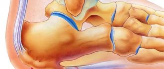
Open reduction and osteosynthesis of a calcaneal fracture
It includes open reduction with external osteosynthesis of the calcaneus with plates of various configurations or less invasive osteosynthesis using a pin for the calcaneus, however, the indications for the use of this technique are more limited.
Most often, surgeons use the standard extended lateral (external) approach, as it is technically the simplest and provides excellent visualization, but we should not forget about the risk of ischemic necrosis of the flap and infectious complications that often occur when using it. With sufficient experience and skill, the surgeon can perform reduction and osteosynthesis of the fracture from a less invasive approach to the subtalar joint.
A detailed description of the technique of bone osteosynthesis from an extended lateral approach using plates of various configurations can be viewed at https://www2.aofoundation.org.
Localization of cracks
These areas of the foot are most susceptible to cracks in the skin:
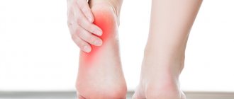
- heel - cracks on it cause pain when walking and are usually a consequence of non-compliance with hygiene rules; they also often occur in those who walk barefoot a lot;
- on the big toe - can signal the presence of a fungal infection, metabolic disorders, and a number of diseases;
- between the fingers - a consequence of a fungal infection, diabetes, thyroid diseases, etc.
Closed reduction and minimally invasive osteosynthesis of a calcaneal fracture
Given the peculiarities of the blood supply to soft tissues and the high risk of infectious and ischemic complications, minimally invasive methods of surgical treatment of calcaneal fractures are gaining popularity all over the world. The basic principles of minimally invasive osteosynthesis are: restoration of the height and length of the heel bone, elimination of valgus/varus displacement of the heel tubercle, elimination of displacement of the posterior facet, restoration of the width of the calcaneal tubercle, without the use of large skin incisions and exposure of the bone. Elimination of displacement is carried out taking into account the principle of ligamentotaxis - due to traction in one, two or three directions, followed by fixation of fragments with screws or knitting needles.
Ligamentotaxis – due to traction in one, two or three directions, reposition of calcaneal bone fragments is achieved.
Elimination of displacement of calcaneal bone fragments due to ligamentotaxis
Depression of the area of the posterior facet of the calcaneus is eliminated with the help of an impactor from the approach to the subtalar sinus or from access along the plantar surface.
Elimination of displacement of the posterior facet of the calcaneus using an impactor
Percutaneous fixation of fracture fragments using screws.
Also, thick Kirschner wires or Steinman or Shants pins can be used for reposition; after installation in a bone fragment, such a “joystick” allows you to easily manipulate the fragment under the control of the image intensifier.
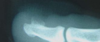
Intra-articular fracture of the calcaneus with avulsion of the tuberosity
Installation of a thick Kirschner wire into the calcaneal tuberosity
Reposition of fragments under x-ray control
Temporary fixation of fragments with a knitting needle
Sequential fixation of a calcaneal fracture with three cannulated screws
Restoring the Gissan angle
Cracked heels
Fungus
Iron deficiency
Diabetes
16054 November 18
IMPORTANT!
The information in this section cannot be used for self-diagnosis and self-treatment.
In case of pain or other exacerbation of the disease, diagnostic tests should be prescribed only by the attending physician. To make a diagnosis and properly prescribe treatment, you should contact your doctor. Cracked heels: causes of occurrence, what diseases cause them, diagnosis and treatment methods.
Description
Cracks in the heels are linear tears in the skin that occur as a result of loss of elasticity and thickening of individual areas. Most often, cracks form in places where the skin is naturally folded and stretched and can be of varying depths. A person often complains of itching, burning, pain, and discomfort while walking.
For most people, cracked heels are a problem related to the appearance of the feet. However, in some cases they can cause considerable discomfort - they can be painful, lead to bleeding, and as a result there is a risk of infection.
In addition, cracked heels are one of the symptoms of certain diseases.
Types of cracked heels
The following types of heel cracks can be distinguished:
- superficial and deep;
- dry (arising from excessively dry skin) and wet (arising from excessively wet skin - this phenomenon is called maceration);
- uninfected and infected;
- resulting from:
- endogenous causes (dermatological diseases, hormonal disorders, diseases of the gastrointestinal tract; associated with a lack of vitamins, microelements);
- external factors.
Possible causes of cracked heels
The most common causes of cracked heels include conditions associated with local physical effects on the skin of the foot: exposure to the sun, wind and cold, temperature changes, chlorinated water, dry air, poor hygiene, excessive care (frequent peeling - removal of the upper stratum corneum of the skin), use of alkaline soap, improperly selected shoes.
However, in some cases we may be talking about general conditions that affect the skin of the heels: smoking and alcohol abuse, fasting, unbalanced diets, small amounts of fluid to drink, congenital skin characteristics (dryness, sensitivity), pregnancy, menopause, aging of the body.
Pathologies that provoke the appearance of cracked heels include fungal infections, dermatological diseases (psoriasis, ichthyosis), diseases of the gastrointestinal tract, helminthiases, conditions of deficiency of vitamins and microelements (iron deficiency anemia, hypovitaminosis), hormonal disorders (thyroid diseases, diabetes mellitus). diabetes), as well as diseases leading to impaired trophism (nutrition) of the lower extremities (impaired innervation and blood supply).
As a result of negative environmental influences (sun, wind, etc.), the skin loses its elasticity, becomes thinner, and the function of the sebaceous and sweat glands decreases. As a result, growths of thick, rough, dry skin appear on the heels. Against the background of increased pressure on the foot (prolonged standing, wearing tight shoes, obesity), cracks may appear. Failure to comply with the drinking regime and lack of vitamins aggravates the problem.
Cracked skin is not only an aesthetic problem, over time it can cause pain when walking, cracks can bleed and become infected. Poor hygiene and the presence of concomitant diseases complicate the problem.
Insufficient levels of thyroid hormones (hypothyroidism) leads to a decrease in metabolism in the body, blood supply to tissues, and the function of sweat and sebaceous glands. Dry and rough skin on the feet becomes prone to excessive keratinization and cracking.
Psoriasis is a chronic systemic disease affecting the skin, nails, and joints. It is characterized by the appearance of small red spots with scales (psioriatic plaques) rising above the skin, which tend to enlarge, merge, and thicken. Thick and hard skin on the feet itches and cracks. Since psoriasis of the feet is associated with a high risk of trauma to the elements of the rash, they often become infected.
Psoriatic plaques on the skin of the feet Diabetes mellitus is a chronic endocrine disease, which is based on metabolic disorders due to insulin deficiency and the resulting high level of glucose in the blood. The long course of the disease leads to damage to nerves and blood vessels with impaired innervation and blood supply to tissues. If the feet are not properly cared for, ulcers and cracks form on their surface, and patients with diabetes need more time to heal.
Foot fungus can be another cause of cracked heels. The disease is characterized by redness, itching, peeling and thickening of the skin, and the appearance of cracks of varying depths and sizes. An unpleasant odor may be present. Based on the picture of the disease and the patient’s complaints, the doctor suggests a fungal infection of the feet, but to confirm the diagnosis, a laboratory test is necessary to determine the presence of the pathogen in the affected area of the skin.
Foot fungus
Keratoderma is a group of skin diseases characterized by disruption of the keratinization process. With this disease, keratosis of the palms and soles is observed. Keratoderma can be diffuse (continuous lesion of the skin of the palms and soles) and focal (the lesion is located in the form of islands, linearly, pointwise).
Keratoderma of the skin of the feet
In children with Unna-Tost keratoderma (congenital ichthyosis of the palms and soles), excessive keratinization of the skin is observed at 1-2 years of age. The disease begins with mild thickening of the skin of the palms and soles. The horny layers are smooth, yellowish in color. Deep cracks may form.
Acquired ichthyosis develops at a later age and is manifested by dry and rough skin with a large number of scales. The skin is affected not only on the torso and limbs, but also on the palms and soles. Acquired ichthyosis is a sign of other diseases (hypovitaminosis A, malignant neoplasms of internal organs, mycosis fungoides, etc.).
Which doctors should I consult for cracked heels?
If changing tight shoes and performing hygiene procedures do not help get rid of cracked feet, you should consult a podiatrist (a doctor who diagnoses and treats foot diseases).
To exclude diseases that provoke the occurrence of cracked feet, you may need to consult a dermatologist or rheumatologist.
Diagnostics and examinations for cracked heels
To determine the causes of cracked heels, the doctor may prescribe a set of laboratory and instrumental diagnostic methods.
- Tests for fungal infections of the skin of the feet.
Closed reduction of a calcaneal fracture with fixation in an external fixation device.
It is often used as an intermediate method of fracture fixation in cases of significant soft tissue damage at the planned surgical site.
It can also be used as a final method of fixation, especially in cases where the surgeon knows the technique of installing the device with Ilizarov apparatus modules.
External fixation often allows one to achieve good reposition without significant damage to soft tissues; after swelling subsides and wounds heal, a transition to internal fixation is possible.
Preventive measures
To avoid the problem of cracks in your feet, just follow a few simple rules:
- Choose comfortable shoes that fit your size. It must be made from natural materials.
- Watch your diet - your diet should contain a sufficient amount of vitamins and microelements necessary for the body.
- Observe the rules of personal hygiene - do not neglect the care of the skin of your feet and apply for a pedicure only to specialized institutions. Do not wear (or even try on) someone else's shoes.
- Seek qualified help if you experience any, even the most minor, problems with the skin of your feet.