What is exostosis
Exostosis is a bone or bone and cartilaginous growth of a non-tumor type on the surface of bones (a type of linear, spherical and other formation).
Exostosis in its structure consists of cartilaginous tissue (ossified in its similarity to normal cartilage tissue) and therefore the name “cartilaginous” exostosis does not accurately indicate the essence of the entire process. The process of ossification during exostosis is usually accompanied by transformation into spongy bone, enclosed on the outside in a thin and dense bone shell.
The surface of the bone exostosis is a layer covered with hyaline cartilage, the thickness of which is only a few millimeters. From such a cartilaginous head the growth of the entire exostosis subsequently results.
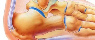
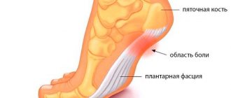
Marginal bony exostoses of the calcaneus
Bone exostosis of the calcaneus is better known as a heel spur. This is a neoplasm that begins to develop from tendon and fascial tissue. The primary factor in the inflammatory reaction is a violation of the integrity of individual fibers of the plantar fascia. It begins to gradually thicken. Plantar fasciitis develops.
This pathology entails constant injury to the synovial cartilage tissue in the heel bone area. The cartilage begins to grow and marginal bone exostoses form, causing severe suffering to the patient when walking.
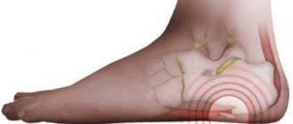
It is possible to cure bone exostoses on the heels without surgery only in the initial stages. Therefore, if you experience pain in the heels that develops after physical activity (for example, after walking long distances), you should immediately consult an orthopedist.
The doctor will order an x-ray of the heel bone. It will show the formation of marginal bone exostoses along the surface of the heel bone. These formations injure the surrounding soft tissues when walking. An inflammatory process occurs, which stimulates further growth of cartilage and bone tissue.
Causes of exostosis
The causes of the formation of exostosis can be an inflammatory process, bruise, pinching, abnormalities of the periosteum and cartilage, infectious diseases such as syphilis, insufficiency of the functions of the endocrine system or its individual glands. Exostosis is presented, in general, as a persistent formation, however, there are cases when the process of formation of exostosis decreases over time and the exostosis disappears forever.
Often, slowly increasing and not causing pain, exostosis is not marked by clinical symptoms, remaining invisible to both the patient and the doctor. Exostosis is detected by X-ray examination, or by palpation of seals that are already visible during examination.
A large number of scientific works are devoted to elucidating the causes of exostosis; their attention is directed to the study of heredity in this disease . However, even the presence in certain cases of family exostoses, which are inherited, does not yet provide any basis for explaining the occurrence of this disease.
Symptoms
Exostosis of the tooth appears in the form of a convex growth that appears for no apparent reason. Main symptoms:
- Sensation of a foreign body in the mouth;
- discomfort when eating, talking (with large osteophytes);
- pain when pressing on the tumor;
- redness, thinning of the mucous membrane in the pathological area.
A small anomaly can only be detected during a dental examination, since visually it does not manifest itself in any way.
Osteochondral exostosis
Osteochondral exostosis can continue to go unnoticed for a long time, since the growth of osteochondral exostosis is very often not accompanied by symptoms. Exostosis can be detected randomly, for example, during an X-ray examination or when growths or indurations are identified.
Often bone growths do not appear until the age of 8, however, during active skeletal growth in the period from 8 to 16 years, activation may occur and exostosis may develop. Accelerated development of osteochondral exostosis is observed during puberty and is found on the fibula and tibia, as well as in the lower part of the thigh, on the scapula and clavicle.
Osteochondral exostosis affects the hands and feet much less frequently and never affects the skull area. The number of growths with osteochondral exostosis can vary - from a few to dozens; the situation is similar with sizes - from a pea to a large orange. It is not always possible to palpate exostoses during research, so radiography is used to accurately determine their number. This is the only way to obtain data on the size, shape and structure of osteochondral exostosis.
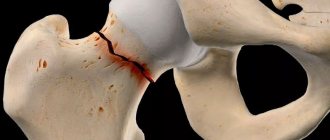
There are two types of osteochondral exostosis: solitary osteochondral exostosis and multiple exostotic chondrodysplasia. Both types of exostoses can affect any bone. The favorite localization is the metaphyses of the long tubular bone. 50% of all osteochondral exostoses affect the femur, proximal metaphysis of the shoulder joint and tibia. Osteochondral exostosis usually manifests itself in adolescence and childhood.
The clinical picture of osteochondral exostosis depends on the form of the disease , its location, the size of the exostosis, shape and connection with nearby tissues and organs.
Exostoses of enormous size can affect nerve trunks and blood vessels, causing pain. Osteochondral exostosis in the spinal region, with further growth into the spinal canal region, can lead to compression of the spinal cord.
Indications
- Rapid growth of osteophyte;
- exostosis after wisdom tooth removal;
- discomfort, pain;
- the appearance of cosmetic defects on the lower or upper jaw (they look like white balls on the jaw, noticeable when smiling or talking);
- the need for implantation, removable or fixed prosthetics;
- the risk of tumor transformation from benign to malignant.
If you need to install prostheses or implants, exostoses will become an obstacle to the procedure. Dentures will injure the bone growth on the gum, and implants will not be able to take root normally in the bone due to the pressure of osteophytes.
Treatment of exostosis with surgery
Treatment of exostoses is only surgical . In the case of the formation of multiple exostoses, the first step is to remove overgrown areas of bone tissue that compress nerves and blood vessels. Treatment of exostosis with surgery is carried out by orthopedic traumatologists under general or local anesthesia, depending on the size of the growths on the bone surface and their location. During the operation, overgrown areas of bone tissue are removed and then smoothed.
When treating exostosis, our traumatology and orthopedics center performs surgery with minimal tissue trauma and the use of modern technology, as well as the application of internal cosmetic sutures, which allows you to return to an active lifestyle in the shortest possible time. Timely methods for diagnosing exostosis with further effective treatment (if necessary) help to avoid subsequent complications of this disease.
Bone exostoses of the spine and ribs
Cartilaginous and bone exostoses of the spine are a consequence of a long-term degenerative dystrophic process in the area of the intervertebral discs and endplates of the vertebral bodies. The degeneration process begins with a violation of the diffuse nutrition of cartilage tissue. It is able to receive nutrition only when the muscular frame of the back is actively working. If a person leads a sedentary lifestyle, muscles lose elasticity and performance. They cannot transport blood and nutrients dissolved in it to the cartilage tissue of the intervertebral discs.
As a result, the process of dehydration of the fibrous ring begins. It loses its shock-absorbing ability and becomes covered with a network of cracks. Calcium salts are deposited in them.
As a compensatory reaction, chondrosis and sclerosis of the endplates of the vertebral bodies begin. They increase in volume and become deformed. As a result of strengthening the blood supply point, the process of nutrition of bone tissue is enhanced. Deformation of the vertebral bodies and bases begins.
Bone exostoses of the ribs are formed as a result of traumatic exposure and curvature of the spinal column. These tight junctions fix the deformation of the chest, limit the vital capacity of the lungs and can lead to severe oxygen deficiency in the structures of the brain and vital internal organs (heart, liver, spleen).
Diagnosis is made using x-rays. For treatment, conservative methods of influence can be used, such as manual therapy, physiotherapy, shock wave therapy, etc. Surgical intervention for this localization of exostoses is used relatively rarely.
Why Pacient.Club?
We have been working in the field of medical tourism for a long time and know how to quickly and efficiently organize treatment for our clients. You should trust us because:
- The medical institution is selected taking into account the nature of the disease, individual wishes and financial capabilities of the client;
- We help you prepare documents quickly and without hassle;
- We provide a full package of services upon arrival;
- We select a personal translator and accompanying person who will help you quickly adapt to your stay in another country.
The best clinics for the treatment of exostosis
This disease is effectively treated in the following leading clinics and medical centers:
- Assaf Harofeh;
- Assuta Express Medical;
- Hisar Intercontinental Hospital;
Countries where exostosis is treated
To get rid of growths on the bones, you can go for treatment to countries such as:
- Switzerland;
- Greece;
- South Korea;
- Latvia;
- Germany;
- Finland;
- Israel;
- Türkiye.
List of sources
- Zatsepin, S.T. Bone pathology of adults / S.T. Zatsepin. – M.: Medicine, 2001. – 640 p.
- Neustadt, E.L. Tumors and tumor-like bone diseases / E.L. Neustadt, A.B. Markochev. – St. Petersburg: Foliant, 2007. – 344 p.
- Epidemiology of skeletal tumors: educational manual / Chernyakova Yu.M., Ivanov S.A., Yadchenko V.N. – 2012.
- Volkov, M.V. Bone diseases in children / M.V. Volkov. – M: Medicine, 1985. – 511 p.
- Lanzman Yu.I. Benign tumors / Bone tumors. – Tomsk: Tomsk University Publishing House, 1990. – P. 88158.
Diagnostics
If signs of subungual exostosis are detected, you should consult an orthopedist. The doctor will examine the fingers and foot as a whole, interview the patient about the nature of the existing complaints and cases of nail injury in the past. The presence of an osteochondral formation at the end of the phalanx of the first finger can be established by palpation, although it is impossible to reliably determine its nature in this way.
X-ray
The fact is that other diseases, in particular benign and malignant tumors, can manifest themselves in a similar way. Therefore, to accurately establish the nature of the formed protrusion, instrumental diagnostic methods are prescribed. Typically, x-rays are used. This method provides information about the state of bone structures and will be informative in case of long-term existence of subungual exostosis, when it has already had time to ossify. If you conduct a study when the protrusion has just appeared, it may be inconclusive, since the cartilaginous structure will not be visible on the pictures.
Other instrumental diagnostic methods are rarely used. They are mainly required in controversial cases when x-rays turned out to be uninformative. In such situations, patients may be prescribed MRI and CT.