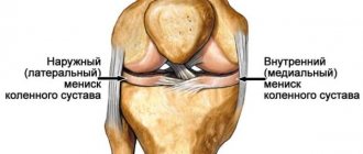
Meniscal injury is the most common knee injury. Its independent restoration is impossible. Fortunately, modern medicine makes it possible to restore (or clear damage from) damaged cartilage surgically.
An important role in the postoperative period is played by rehabilitation, namely the selection of correct and effective exercises that in a short time will help strengthen tendons, ligaments and restore normal function of the meniscus.
Clothes should be loose, it is advisable to remove shoes. Perform all exercises (especially the first days) smoothly and gradually. Remember the important principle: “Endure mild pain, do not allow severe pain.”
You can do the first 7 exercises right away. The rest only after your doctor allows axial loading.
Half squats with resistance
Stand facing the door. Fasten one end of the expander under the knee of the leg being worked out, and the other end to a door or other stationary object at the level of the knee joint. Raise your free leg off the floor; you can hold on to a chair or armchair for balance. Bend the knee you are working on slightly (do a half squat on one leg), then slowly straighten your leg. Repeat 15 times. You can simplify the exercise by not lifting your free leg.
Exercises on a balancing platform
Stand on the balancing platform. Optimal position: feet shoulder-width apart.
Meniscus injury
Meniscus injury
Early rehabilitation is considered to be the period when soft tissue healing occurs. They are damaged during surgery. The patient is in the hospital for an average of 2 weeks. This is how long early recovery after a meniscus injury and its surgical treatment continues.
The main goals that are achieved through rehabilitation measures:
1. Elimination of pain and inflammation. They always occur in the postoperative period. Moreover, it does not matter what kind of operation was performed. Swelling and release of inflammatory mediators are a standard response to traumatic tissue damage.
2. Stimulation of contractile activity of the thigh muscles. To do this, isometric exercises are performed.
3. Combating knee contracture. This term refers to the limitation of passive (not carried out by the patient himself, but with the help of external influence) movements. The knee is designed to ensure normal mobility.
4. Improvement of joint trophism. That is, access to blood, and with it oxygen and nutrients. The recovery time of damaged tissue directly depends on the intensity of blood circulation. This is evidenced by many facts. For example, wounds on the face heal twice as quickly as on the leg. With diabetes, blood supply deteriorates, and any damage takes a very long time to heal. Areas of the meniscus that are not supplied with blood are not capable of regeneration at all. And fragments of the pink zone heal much more slowly than parts of the red zone, which is located near the joint capsule and is better vascularized.
5. Maintaining the patient's body. A person must be able to take care of himself and not experience severe discomfort.
Platform rotation
- Rotate the balancing platform clockwise until its edge is in constant contact with the floor , then counterclockwise. Repeat 30 times in each direction.
- Rotate the balancing platform clockwise until its edge touches the floor , then counterclockwise. Repeat 30 times in each direction.
magnetotherapy
Magnetic therapy
1. In the first days after surgery, a person studies in the ward. He spends about 20 minutes doing the exercises. Movements can be performed on the operated limb only in the ankle and hip joint.
2. No movement during exercise should cause pain. There should be no knee swelling after training. Its formation indicates the development of synovitis. There should be no load on the articular cartilage after removal or suturing of the meniscus. For this purpose, unloading body positions are used when the patient is lying down or sitting. Smooth polished surfaces are used to facilitate movement.
3. The most important feature of early rehabilitation after a meniscus injury remains a gentle attitude towards the extensor apparatus of the knee. This is important for the restoration of patellar cartilage.
4. The duration of training gradually increases. Over the course of 14 days, they increase from 20 minutes to 2 hours a day.
5. From the third day of rehabilitation, drainage massage is indicated. It reduces swelling and improves knee function. The massage effect should not affect the knee joint itself. It is carried out in the thigh area.
6. The main purpose of exposure to cold in the first days after surgery is to eliminate pain, as well as exudation (fluid accumulation in the joint cavity). Due to the low temperature, vasospasm occurs and the release of fluid from the vascular bed into the tissue decreases. Due to this, the swelling goes away.
7. Each patient requires 1 or 2 types of physiotherapy. A total of about 10 sessions are required.
8. On the first day after arthroscopy, you can walk on crutches without leaning on the problematic leg. The total walking duration should not exceed 15 minutes. This takes into account the fact that the patient visits the restroom from time to time and goes to change dressings.
9. Dosed loads on the operated limb are allowed from 3-5 days (depending on the characteristics of the operation performed and the course of the rehabilitation period). The exception is when a person’s articular cartilage is injured along with the menisci. Then loads are prohibited for 4 weeks.
10. If only the menisci are damaged, a person can exercise on an exercise bike within a week after arthroscopy. In case of additional cartilage damage, no earlier than after 4 weeks.
Late rehabilitation
Home care
Any home therapy should be discussed with your doctor, because it is only an auxiliary component of drug treatment. Traditional medicine can only be indicated for relieving pain and swelling:
- Compresses of a hypertonic solution draw out excess fluid, thereby relieving swelling. A solution is prepared from a tablespoon of salt dissolved in 250 ml of water. The cloth is soaked in salt water and applied to the injured knee.
- Peppermint rubs. For this, essential oils of clove, mint, camphor and eucalyptus are taken. After mixing the ingredients, take a few drops of the product and rub it into the injury site with slow and careful movements. Menthol will create a feeling of freshness and coldness, which will alleviate the patient's condition.
- Pine baths. They can improve blood flow to the injured knee and relieve pain. In addition, pine needles have a restorative effect on the body. To prepare baths, you can use pharmaceutical pine mixtures, which just need to be brewed immediately before preparing the bath. If it is not possible to find pine needles, you can use pine salt.
Treatment
Treatment options depend on the severity of the injury. The choice of treatment strategy is also influenced by which meniscus is damaged. The medial one is much less well supplied with blood vessels, so its rupture requires longer treatment and the use of auxiliary drugs.
Conservative treatment methods include drug therapy, exercise therapy, and physiotherapeutic procedures. If they are ineffective, or if there are extensive injuries, surgical intervention is required.
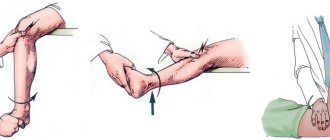
Removing the blockade of the knee joint
To restore knee function, perform a series of sequential actions:
- puncture of the joint to remove effusion and blood from its cavity using a special puncture needle. Then anesthetics are administered to reduce pain during further manipulations;
- After the anesthetic has taken effect, the doctor gradually pulls the foot of the affected leg down using his hands or a loop of rolled up bandage. Moves the tibia outward to widen the joint space. Next, he turns the shin inward to return the cartilage to its normal position;
- the doctor checks the movements in the knee: if they are fully restored and do not require any effort, the procedure is considered successful. The leg is fixed in a plaster cast for 5-6 weeks.
The blockade cannot always be eliminated the first time; 3 attempts are allowed, and if they are ineffective, surgical treatment is indicated.
Drug therapy for knee meniscus tears
Involves prescribing different groups of drugs:
- non-steroidal anti-inflammatory drugs - to relieve inflammation and reduce pain. Prescribed in the form of tablets, injections in combination with preparations for external use: ointments, creams;
- glucocorticosteroids - hormonal agents to relieve severe inflammation;
- chondroprotectors – a group of agents for stimulating regenerative processes in damaged cartilage;
- vitamins - to provide cartilage tissue with necessary substances during the recovery period.
Symptomatic therapy includes taking painkillers and applying cold to the affected area.
Exercise therapy and physiotherapy
Physiotherapeutic procedures involve the use of various physical factors that reduce the severity of pain, inflammatory, and degenerative processes. The required type of physiotherapy is selected taking into account the severity of the condition; more often they use:
- electrophoresis with various medications;
- ozonokerite;
- cryotherapy, thermotherapy;
- electromyostimulation;
- magnetic therapy;
- UHF, etc.
Exercise therapy classes are indicated from the first day of conservative therapy. A set of exercises is selected individually, taking into account the severity of the injury. At the beginning of treatment, general strengthening exercises are used, which are more designed for the healthy limb, but there are movements aimed at tensing the muscles of the affected leg. To reduce the likelihood of stagnation of venous blood in the limb, it is recommended to lower the leg down and raise it to an elevated position. After the cast is removed, classes continue and gradually become more difficult.
Surgery
If a complete meniscus tear occurs and conservative therapy does not produce results, the only treatment option is surgery. It is also indicated in the following cases:
- crushing of the meniscus;
- inability to eliminate the knee block manually or its reoccurrence;
- hemarthrosis;
- meniscopathy.
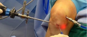
The volume and technique of surgical intervention are selected taking into account the severity and location of the damage. The most effective and safe method is joint arthroscopy, which is mainly performed under spinal anesthesia. This minimally invasive technique allows you to carry out all the necessary manipulations without incisions.
A camera and the necessary instruments are inserted through the punctures. The arthroscope includes a lighting system and a set of lenses; through a camera, the image is transmitted to a screen on which the doctor controls all movements. The main advantage of the method is minimal tissue trauma and a short rehabilitation period. After the operation, there are practically no marks left on the body.
Arthroscopy has a low risk of complications such as bleeding and wound infection. The duration of the operation is significantly shorter, in contrast to open techniques, which also reduces the drug load on the body associated with the introduction of anesthesia. Open surgical treatment techniques involve wide incisions. They are more traumatic, but allow for more extensive manipulations, which is required in case of severe injuries.
The extent of surgical treatment depends on the area in which the injury is located, age, level of physical activity, and how long ago the injury was. More often, partial removal of the affected meniscus is performed. If it is impossible to restore the cartilage plate, the biomechanics of the joint changes. The pressure of the femur on the tibia will increase, which over time can lead to damage to their joints. Therefore, the goal of arthroscopic surgery is to preserve tissue as much as possible and remove it only as a last resort.
Conservative therapy
Shock wave therapy is an effective method of conservative treatment for minor disorders.
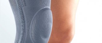
Conservative treatment is aimed at reducing inflammation and accelerating healing when the rupture is incomplete and surgery can be avoided. If there is pathological fluid in the joint - blood due to vascular damage or effusion due to inflammation - it is removed using a puncture. The limb is partially immobilized with an orthosis in order to avoid excessive stress when moving. A course of non-steroidal anti-inflammatory drugs is prescribed. Sometimes intra-articular corticosteroids are required. Injections of hyaluronate, platelet-rich plasma, help cartilage quickly recover. For this purpose, oral chondroprotectors are prescribed. You need to start developing the joint under the supervision of a rehabilitation specialist. Rehabilitation is aimed at restoring all functions and strengthening the thigh muscles. In addition to kinesiotherapy, mechanotherapy and massage are used. Physiotherapeutic methods (UHF, electrophoresis, electromyostimulation) accelerate the healing process.
Diagnosis of ruptures
To make a diagnosis, a thorough examination and instrumental diagnostics are required. At the first appointment, the doctor clarifies the circumstances that led to the injury, determines the pain and its severity, and compares the affected and healthy joints. During examination, assesses gait, position of the affected limb, range of motion. To recognize injury, there are a number of tests and tests, for example, McMurray, Apley, etc.
To visualize the rupture, the following is prescribed:
- radiography;
- ultrasonography;
- MRI.
X-ray data provide information about bone structures. The lesion of the meniscus itself cannot be examined, but the examination is necessary to exclude fractures inside the joint and to detect loose bodies.
Ultrasound is the safest diagnostic method that can be repeated an unlimited number of times. Helps identify any meniscus tears, post-traumatic cysts. MRI is an additional diagnostic method required to clarify information in complex clinical cases.
menisci
menisci
The goals of the recovery period are as follows:
- restoration of normal gait;
- adaptation to long or fast walking;
- training the muscles of the lower extremities, restoring their strength and tone;
- adaptation to slow running;
- psychological recovery.
To achieve these goals, training in the gym is used. They should be daily, lasting up to 2 hours. These include walking, running, pool and exercise bike exercises. The total duration of physical activity per day can exceed 3 hours (including those carried out outside the gym).
To increase the volume of the muscles of the lower extremities after a meniscus injury, the following exercises are used:
- leg press – can be used a month after arthroscopy, and the operated leg can be used only after a month and a half;
- climbing stairs – from 7 weeks;
- exercise on an exercise bike begins on the 7th day of recovery, but then the load is increased;
- Half squats are allowed after 2 months.
Running can be started only in the absence of inflammatory changes in the knee, after the complete elimination of the contracture, as well as restoration of the muscle volume of the thigh. In the first 1-2 workouts, the reaction to the load is assessed. If there are no complaints, you can continue running using different techniques.
Types of breaks
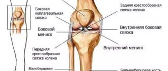
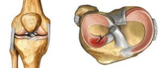
There are many classifications of damage, one of the significant criteria is the degree of cartilage rupture: incomplete or complete. In the first case, the cartilage is partially damaged; no changes in its structure occur. In a complete tear, a fragment of the meniscus is torn away from the main part. Taking into account the blood supply and the possibility of independent recovery, the following types of ruptures are distinguished:
- in the red zone - the wound can heal on its own without surgery;
- at the border of the red and white zones - spontaneous healing, as a rule, does not occur, but after surgical treatment the tissues can recover;
- in the white zone - this area does not have a blood supply, so the tears do not heal and require surgical intervention.
Taking into account the nature of the damage, ruptures are distinguished:
- “watering can handle” type – longitudinal injury with movement of the fragment into the intercondylar eminence;
- “parrot's beak” type – transverse tear, which most often occurs at the junction of the posterior horn and the body of the meniscus;
- patchwork – horizontal tear with displacement;
- complex - a combination of various discontinuities.
Transverse injuries to the body of the internal meniscus are most rarely observed in the area under the tibial collateral ligament. More often, injuries along the longitudinal axis are diagnosed with a rupture of the middle part of the meniscus.
