Modern diagnostic techniques allow a detailed and painless assessment of the condition of the knee joints. It may not be easy for a patient to figure out on his own whether to choose CT or MRI.
Despite the similarity in names, the surveys have different operating principles.
Computed tomography is based on x-rays, essentially an improved x-ray that allows you to take several pictures at once in different projections, which can then be used to create three-dimensional models.
Magnetic resonance imaging works by using a strong magnetic field that affects hydrogen atoms in the body. The equipment records vibrations, and the result is a black-and-white series of cross-sectional images.
What is hardware diagnostics?
The era of hardware diagnostics began with the discovery of X-rays at the end of the 19th century. This is how an X-ray machine appeared in the arsenal of doctors. A little later, fluorography and mammography appeared on its basis. In the early 30s of the 20th century, the diagnostic capabilities of ultrasound were discovered, and medical centers began to perform ultrasound diagnostics of various internal organs and evaluate fetal development during pregnancy. In the 70s of the 20th century, the first attempts were made to use the power of magnetic resonance for diagnostic purposes. Since the 80s of the last century, medical institutions in Russia began to be equipped with magnetic resonance imaging scanners. Medicine never stands still, and with the development of X-ray technology, an improved version of radiography appeared, which was called computed tomography. It was widely used in clinical practice in the 80s of the 20th century. First-generation computed tomographs have been replaced by spiral and multispiral tomographs - MSCT. Now most medical institutions in St. Petersburg are equipped with them.
- MRI
- Ultrasound
MRI tomograph:
Siemens Magnetom C
Type:
Open (expert class)
What's included in the price:
Diagnostics, interpretation of images, written report from a radiologist, recording of tomograms on CD + free consultation with a neurologist or orthopedist after an MRI of the spine or joint
Ultrasound machine
HITACHI HI VISION Avius
Class:
Expert (installation year 2019)
What's included in the price:
Diagnostics, interpretation of images, written diagnostic report
Preparing the premises before putting equipment into operation
The room for equipment in a medical institution must be prepared and comply with building codes and regulations: provide sufficient ventilation and air conditioning for the equipment in use, the presence of a water source and drainage system, as well as additional grounding, uninterruptible power supplies, etc. It is necessary to focus on the requirements of the manufacturer of the purchased medical equipment for the premises, the standards of sanitary and hygienic requirements necessary for operation. If it is necessary to re-equip the premises, you need to worry about coordinating the design of the converted premises with the relevant authorities. At this stage, the Balt Medical service department supervises the preparation of the room for putting the device into operation.
Attention!
All preparatory and repair work for the preparation and re-equipment of the premises must be carried out before the start of operation of the medical equipment, otherwise the equipment may lose part of the warranty period!
What is a CT scan of the knee?
A CT scan of the knee is a non-invasive and painless examination of the knee joint area.
A CT scan uses X-rays and a computer to create numerous images of the knee at different levels and from different angles. A multislice computed tomograph is an X-ray machine with a gantry ring in the center. During the examination, the scanner table with the patient lying on it moves through the installation tunnel, and sensors located in the walls of the ring emit beams of X-rays. They pass through the patient's body at different speeds and are caught by the detector. Data from the detector is sent to the computer. It processes all residual X-ray strength signals and creates volumetric images from all sections of the knee joint. As a result, on tomographic images, the diagnostician can see the exact anatomical structure of all the bones and cartilage of the knee and identify the exact location and nature of any bone pathologies. Initial appointment with an ORTHOPEDIST
ONLY 1800 rubles!
(more about prices below)
Installation of medical equipment and commissioning works
Carrying out installation and commissioning work of newly delivered medical equipment before the start of operation is the responsibility of the supplier. But you need to take into account that these works are subject to licensing. If the supplier of your device does not have such a license, then he is obliged to enter into an agreement with a company that has the right to install and commission new equipment. has been on the market for many years, has the right to carry out this type of work and has all the necessary licenses.
When installing equipment, the following factors are taken into account:
- property location;
- design features that may ultimately affect the operation of the equipment;
- specialization of the medical institution;
- regulations requirements for putting equipment into operation.
Attention!
A medical clinic must have an employee whose responsibilities include interaction with equipment suppliers and service departments, including signing documents on the acceptance of goods and the direct organization of all stages of acceptance of new equipment.
What is MRI of the knee?
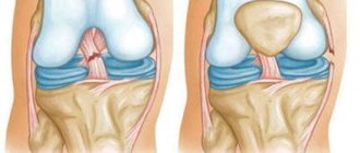
Magnetic resonance imaging is also a non-invasive and painless method for diagnostic imaging of soft and bone tissues of the body. It allows doctors to see joint structures and their changes. Unlike a CT scan, an MRI of the knee does not use radiation, but rather a magnetic field and radiofrequency waves comparable to those found in a cell phone or microwave oven. As with CT, the goal of the MR method is to obtain volumetric images of the area under study. However, the principle of creating tomograms is based on the physics of nuclear magnetic resonance, and not on X-rays. There are hydrogen atoms in the tissue cells of our body. These charged particles can release energy when they change their positions. When a patient is placed inside an MRI machine, a strong magnetic field is created around the area being examined. It causes hydrogen atoms to change their positions for a short time, lining up along the wave. They then return to their original position, releasing energy in the process. This is what the detector in the device catches, registers and transmits to the computer. It digitizes the information received and displays three-dimensional images of the patient's knee anatomy. There are more hydrogen atoms in those tissues where there is a lot of water. Therefore, magnetic resonance imaging better shows cartilage, muscles, tendons, and nerve endings.
| Service | Price according to Price | Discount Price at Night | Discount Price During the Day |
| from 23.00 to 8.00 | from 8.00 to 23.00 | ||
| MRI of the knee joint | 4000 rub. | 3190 rub. | 3690 rub. |
| Appointment with an orthopedist | 1800 rub. | free after MRI | free after MRI |
| First aid program for joints (8 studies + appointment with an orthopedist + MRI of the joint) | 13000 rub. | 7500 rub. | 7500 rub. |
Acceptance of new equipment by the clinic
When accepting goods, when the equipment is delivered directly to the end user, for various reasons it may happen that the equipment in the supplied form does not fit into the door openings and this will require partially dismantling the device. Under no circumstances should you do this yourself without some confirmation. It could be another similar problem. Service engineers will help you accept oversized goods, document the need to disassemble this or that element, without in any way violating the warranty terms. It is necessary to understand that after signing the transfer acceptance certificate, only the medical institution is responsible for the technical condition of the equipment.
Advantages and disadvantages of CT and MRI of the knee
Each of the tomographic methods has its own advantages and disadvantages. Compared to CT, MRI of the knee joint has the following advantages:
- absence of radiation and ionizing radiation;
- accessibility to any category of patients (pregnant women, children of any age);
- unlimited number of studies. Due to radiation exposure, CT scans should not be done more often than once every six months;
- significantly larger coverage area. Using MRI, doctors can examine both bone and soft tissue.
However, the quality of bone structure is difficult to assess on MRI images. There is little water in the bones, so on an MRI of the knee joint they will not be visible very accurately and clearly. For qualitative examination of bone tissue, X-ray methods remain more significant. Bones are much better visible on CT scans and x-rays.
CT scan of the knee, in comparison with MRI, will benefit in the following parameters:
- speed of examination. MSCT of the knee will take 5-7 minutes, unlike an MRI examination, which will take from 20 to 40 minutes.
- the examination step on a computed tomograph is 1 mm. To achieve the same level of scanning on a magnetic resonance imaging scanner, you need to use powerful high-field machines.
Both MRI and CT of the knee joint are convenient for the patient in terms of the speed of obtaining diagnostic results. Immediately after the procedure (30-40 minutes), the examinee receives a written report from the radiologist, the images themselves and a detailed description of the identified pathologies and abnormalities. Such operational data allows doctors to get closer to an accurate diagnosis and prescribe treatment as quickly as possible.
MRI and CT of the knee joint with contrast
Sometimes in medical centers you can observe the following picture. Radiologists begin to ask the patient the question: “Should you do an MRI or CT scan with or without contrast?”, and the person being examined is lost and cannot answer. To avoid such confusion, let's clarify when CT and MRI of the knee should be done with contrast, and when the basic protocol will be enough.
CT and MRI of the knee joint in most cases are performed without contrast. Diagnosticians use contrast enhancement in those rare cases when a tumor is suspected. To identify a tumor and diagnose its benign or malignant nature, radiologists inject a special contrast agent through a vein into the patient. It helps to better visualize the location of the space-occupying formation, and by the nature of the accumulation of the drug, doctors can guess what kind of tumor it is and at what stage of development it is.
Different chemical compositions are used as a contrast agent in magnetic and computer studies. Computed tomography uses iodine-containing contrast agents. Contrast for MR imaging is based on gadolinium salts. The gadolinium-containing composition is less toxic than iodine and almost never causes allergies. An allergic reaction to iodine-based contrast agents occurs in 1% of cases.
Medical staff training
Training of medical staff of institutions that will directly use the equipment in their work is one of the important stages of commissioning medical equipment, be it a tomograph, ultrasound machine or something smaller, according to GOST 12.0.004-2015 “Organization of occupational safety training.” A detailed briefing is required, the fact of which is recorded in the contract for work on commissioning the equipment. However, one should not be confused that the equipment supplier provides instructions, and the actual training is carried out by companies that have a license for this. After instructions and training have been completed by the personnel regarding the technical use of the equipment, you can sign the act of putting the device into operation. From this moment the warranty period of the new equipment begins.
What does the user get when contacting?
The undoubted benefits of cooperation with us are positively assessed by all medical institutions; each client receives:
- assembly, installation and commissioning work by experienced qualified engineers;
- quality of work ensuring reliable, uninterrupted and accurate operation of medical equipment;
- full compliance with the requirements of the regulations when installing equipment;
- full reporting and preparation of documentation and necessary regulations on the work done;
- competent instruction and training of medical personnel in handling the installed equipment;
- the possibility of concluding a contract for the maintenance of your equipment by our service engineers.
MRI or CT of the knee joint - which is better?
In answering this question, it is best to trust the judgment of the attending physician. He will be able, based on diagnostic purposes, medical history, primary diagnosis, to select the most effective diagnostic method. If you find it difficult to choose between magnetic and computed tomography on your own, we will be happy to offer you a consultation with an experienced orthopedist. He will be able to make a primary diagnosis and, if further examination is necessary, give a referral for the necessary type of diagnosis
Author: Usenko Nikita Sergeevich
Orthopedist-traumatologist with 8 years of experience