Joint arthroscopy > Knee joint > Injuries to the knee joint
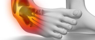
Knee injuries
– rupture or sprain of tendons, bone fractures (patella, tibia, fibula, femur), damage to the meniscus. Problems like these are widespread. Injuries are often differentiated by severity - from mild tissue damage to bone fracture. Knee joint bruises usually occur due to falls. A knee injury may be indicated by pain, tissue swelling, and limited mobility. Usually the damage is accompanied by hemorrhage into the joint cavity. To make a diagnosis, the following methods are used: x-ray, ultrasound, arthroscopy, computed tomography, MRI. Treatment depends on the location and extent of the damage.
General information
Knee injuries
– occupies a leading place among other injuries. This position is associated with the features of the normal anatomy (structure) and physiology (load) of the knee joint. Most injuries occur from falls and impacts. The resulting injuries are usually minor bruises, so treatment is carried out by a traumatologist on an outpatient basis. Amateur and professional athletes also often injure the knee. Their knee joint injuries are much more serious.
Less frequently reported are cases of knee injuries in road accidents, falls from height, and at work. For patients who are involved in such an incident, damage inside the joint is typical. They are characterized by the fact that the integrity of the anatomical structure is violated. Relief of symptoms and restoration of the structure occurs in the trauma department of a medical institution. In addition to a knee joint injury, there may also be a TBI (traumatic brain injury), fractures of other bones, and organ ruptures.
First aid
A victim with a knee fracture needs qualified medical care. Therefore, the first task of the surrounding people is to urgently deliver the patient to the nearest emergency room.
To reduce the risk of possible complications, the patient requires high-quality first aid at the scene of the incident:
- In case of an open fracture, bleeding is controlled using an aseptic bandage and tourniquet. If it is necessary to transport the victim for a long time, the tourniquet is removed every 40 minutes in the winter and after 90 minutes in the summer.
- When closed: the injured limb is immobilized, a cold compress is applied and it is fixed with a splint.
Any type of fracture requires high-quality pain relief.
Self-reduction of fragments is strictly prohibited. This action will further injure the patient and worsen his situation.
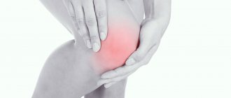
Knee contusion
Knee bruise
- this is damage to surrounding tissues (skin, subcutaneous fatty tissue), which is not accompanied by a violation of the anatomical structure. Against the background of this injury, inflammation of the elements that are located inside the joint occurs, as well as hemorrhages. Symptoms are often not as pronounced with other injuries. Because of this, the diagnosis is made when other injuries have been ruled out. Signs that the victim has a knee joint injury may include: pain, moderate swelling of the soft tissues, and slight bruising. At the same time, support is maintained, but the person may begin to limp and be slightly limited in some movements.
To determine pain, palpation is performed in the area of the bruise. Feeling the bones and ligaments should not cause attacks of pain, as well as pathological mobility. There may be an accumulation of fluid, at first it is blood, and after two or three weeks it is exudate. Injury to the knee structures is excluded using x-rays. Research methods such as MRI, computed tomography, ultrasound and arthroscopy are also used. Care for a knee injury is provided at the emergency room. If there is synovitis or blood inside the joint, a puncture is performed. If the patient has a slight bruise, then the main actions are aimed at maintaining peace. But for severe cases, the traumatologist applies a plaster cast for 2 - 3 weeks. For the first two days, it is recommended to apply something cold to the site of the bruise, and from the third day, UHF therapy is prescribed. The patient must visit the doctor at the appointed time and follow his instructions. If indicated, repeat arthrocentesis is performed. If the knee joint is bruised, the disability lasts 2–4 weeks.
Diagnostics
It is possible to accurately determine the presence or absence of a fracture after an x-ray. At the same time, to see the damage, they often take pictures from different angles. Using it, the doctor determines the severity of the injury and what treatment will be optimal in a particular case. Only on an x-ray will a specialist see how the bone is splitting, whether it is displaced and how many fragments have formed. To obtain accurate data, the picture is taken in three projections.
- Straight. Makes it possible to see damage to the intraarticular condyle.
- Axial. Necessary to determine the presence or absence of a vertical osteochondral fracture or subluxation of the knee.
- Axial. This projection involves a lateral view of the damaged area. It allows you to see the transverse fracture.
Meniscus injuries are visible on any photograph. If they are suspected, an ultrasound of the knee or an MRI may also be indicated to clarify the information, which depends on the equipment of the medical institution. Many doctors prefer ultrasound, since in this case it is also possible to assess the condition of the muscles in the area of injury.
Important! If a lobular fracture of the patella is assumed, a photograph of the healthy knee must be taken to compare external changes in the injured area.
In the case of an open penetrating fracture in the knee joint, a “salt test” is also performed. This method is combined with a procedure for removing blood from the joint cavity. After anesthesia, the blood accumulated inside the knee is released through a needle that is inserted into the wound. Then saline solution is poured into the cavity through it. Then, if it actively flows from the wound, then an open penetrating fracture is diagnosed. If the fluid does not flow out, then this is an indicator of simply a minor traumatic injury to the knee without a fracture.
It is impossible to diagnose a knee fracture independently based on external signs. Symptoms of a fracture of the knee joint, with the exception of cases of an open fracture, when bone fragments are visible from the wound, are similar to the manifestations of severe bruises, as well as its subluxation. If you have the slightest suspicion of such an injury, you should seek medical help. The sooner proper treatment is started, the lower the risk of complications.
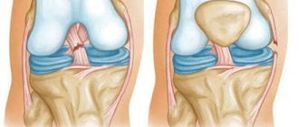
Ligament damage
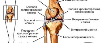
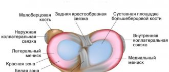
The medial and lateral collateral ligaments, the anterior and posterior cruciate ligaments (ACL, PCL) take part in the formation of the knee joint. All injuries to these structures can be divided into partial and complete. A preliminary diagnosis can be made by studying biomechanical processes. Pain during supination (turning the leg outward) indicates damage to the medial collateral ligament. If the patient has problems with pronation (turning in on the inside), then the lateral collateral ligament may be injured.
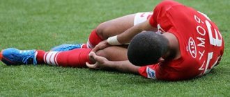
Injuries to the ACL and PCL usually occur after direct impacts, hyperextension (overextension), and rotation of the hip with a fixed shin. Such injuries are typical for athletes (basketball players, wrestlers, track and field athletes). Other reasons that may cause a sprain or rupture of ligaments include carelessness in everyday life, road accidents, falls from a height. When an injury occurs, severe pain occurs.
A rupture of the ACL is accompanied by a click, which is not the case with the PVD. With such injuries, instability of the knee joint and a feeling of displacement when walking are observed. When visually assessing the patient's condition, swelling of the soft tissues in the knee area and hemorrhage inside the joint are observed. The last symptom may be absent if the posterior cruciate ligament is torn. The reason is that damage to adjacent structures - the posterior part of the knee joint capsule - is possible. Because of this, blood enters first into the popliteal fossa, and then into the space between the fascia. On palpation, the patient has a pronounced pain syndrome.
Injury to the medial and lateral collateral ligaments is accompanied by abnormal lateral mobility of the tibia, and the ACL and PCL are characterized by “drawer” displacement. During the acute phase, the patient is prescribed local anesthesia. When the sensation of pronounced pain disappears, instability of the knee joint remains. In order to avoid pathological displacements while walking, the leg should be wrapped with an elastic bandage. Over time, there is a gradual decrease in muscle mass due to atrophy, as well as symptoms of post-traumatic arthrosis.
When performing an X-ray of the knee, a narrowing of the joint space is observed. Using MRI, you can find out about the condition of the ligaments. Diagnosis is best done through a procedure such as arthroscopy. This is a minimally invasive diagnostic method for visualizing structures on a monitor screen. In addition to diagnostics, arthroscopy is also used to restore the integrity of ligaments. Tears are eliminated through conservative treatment. Arthrocentesis is also performed and a cast is applied for four weeks. Afterwards, the doctor may prescribe physical therapy and massage. If a ligament rupture occurs, it is repaired through surgery (stitching, plastic surgery). During the rehabilitation period, a number of physiotherapeutic procedures are prescribed for a speedy recovery.
Rupture of the connective tissue of the quadripses and part of the patella (the ligament itself) can occur with a strong blow, as well as with simultaneous overstrain of the thigh muscles and sharp flexion of the lower leg. When injured, sharp and intense pain manifests itself, and gait is disturbed (the leg bends reflexively). Blood does not enter the joint cavity. Palpation of the damaged area is accompanied by increased pain, there is no displacement and pain in the bones.
Kinds
The effectiveness of the prescribed therapy depends on the correct diagnosis and determination of the type of injury. Fractures happen:
- Open. Accompanied by damage to the integrity of the skin.
- Closed. The skin is not injured.
Open knee fractures carry a high risk of infection and severe blood loss. Lack of timely first aid can lead to the death of the victim.
© Photographee.eu - stock.adobe.com
Intra-articular knee fractures are:
- with displacement of fragments;
- without displacement.
Depending on the position of the parts of the patella, damage is classified into:
- Osteochondral. A small part of the patella is torn off.
- Horizontal. Breaking the bone into two parts.
- Multi-fragmented. The bone is crushed into several fragments.
- Vertical. The calyx breaks lengthwise.
Based on the location of bone fragments along the axis, fractures are:
- With displacement of fragments. Surgery is required.
- No offset.
- Compression. The bone is pressed in.
The severity of a displacement injury largely depends on the sprain. If the victim does not have problems with the tendons, shrapnel displacement can be avoided.
Old fractures require longer treatment and rehabilitation in contrast to primary ones.
Meniscal injuries
Meniscus injury
– one of the popular problems with the knee joint, which occurs with greater frequency in athletes (football players, basketball players, track and field athletes, skiers). This injury is no less common in people who dance, do heavy work, and are artists. Without preliminary mechanical action, damage to the menisci can also occur with arthrosis of the knee joint. The nature of the injuries ranges from small tears to complete ruptures. Often, damage to the meniscus is combined with injury to other structures of the knee.
The acute period is characterized by the following symptoms: pain, swelling, swelling, stiffness of movement. There may be exudate inside the joint. Within three weeks, the symptoms of the inflammatory process disappear, and the signs of meniscus injury become more pronounced. This is expressed in the appearance of a painful cushion at the level of the joint space. The following clinical picture is also recorded: pain when going down the stairs, making circular movements with a bent shin, or the “Turkish” position.
X-rays are not used for diagnosis (only to exclude other damage). Arthroscopy or MRI is usually prescribed. Ultrasound diagnostics of the knee joint is rarely performed. Treatment is prescribed based on the examination results. Small tears can be repaired with therapy. More serious damage can only be eliminated through surgery. Possible operations may include suturing, resection, or complete removal of the menisci. The latter is rarely performed due to the increased risk of arthrosis.
Contraindications
Performing operations of this type is a fairly common practice in medicine, but the inserted plate for a knee fracture may have contraindications. Their list includes:
- large area of affected tissue;
- the presence of torn, crushed, infected surfaces;
- manifestation of acute infection;
- exacerbation of a chronic pathological condition;
- local inflammatory process with suppuration;
- state of shock;
- severe combined injuries;
- great blood loss.
With a large number of torn tissues, cleaning, treatment and healing of wounds is performed. The operation is prescribed after the patient’s condition improves.
Fractures in the knee joint
Violation of the integrity of the patella is possible if the anterior part of the knee is damaged. In this case, acute pain syndrome occurs, problems with bending the leg, swelling of the soft tissues, and hemorrhage in the middle of the joint. Diagnosis is made using an X-ray of the knee. If the patella fracture is not displaced, then treatment is conservative. The leg is immobilized for 6 to 8 weeks. If there is displacement, surgical intervention is performed. During the rehabilitation period, physical therapy, physiotherapeutic procedures, massages, and pain relief are prescribed. Full recovery is observed within 2 - 3 months.
list
Rehabilitation
When choosing a rehabilitation system, the doctor must take into account for each patient the characteristics of his general condition and the injury itself. Based on general recommendations, the specialist selects the best option for knee restoration. Prescriptions for rehabilitation after a knee fracture of various locations include the following:
- massage – carried out within 10 days after removal of the plaster. It must be warming up. This is required in order to improve blood circulation in the damaged area and restore normal muscle tone. And also improved blood circulation allows you to increase the rate of regeneration of damaged tissues, which continues even after the plaster is removed;
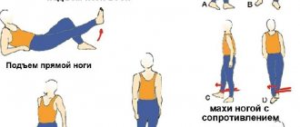
- moderate physical activity - they should be selected by the doctor taking into account the general condition of the patient and the degree of damage. At first, the loads are minimal. Their gradual increase occurs as the tissues are restored. For this, a complex of physical therapy is used. It differs for those whose fracture is treated conservatively or surgically. Performing physical therapy exercises requires special care and a clear understanding by the patient of how to do it correctly. Often classes begin 2-3 days after surgery and 5-7 days with conservative therapy. This is necessary for the development of the limb;
- physiotherapy - carried out only according to the indications of the attending physician and strictly taking into account the changes that are visible on the x-ray. If necessary, the doctor makes adjustments to prescriptions based on the results of the x-ray;
- diet – proper nutrition can significantly speed up the process of tissue repair. The diet should be enriched with foods high in calcium and phosphorus, which are necessary to speed up the fusion process. It is also mandatory to consume foods rich in collagen, which is necessary for the formation of cartilage tissue.
You should not change your doctor’s recommendations for rehabilitation after a knee fracture on your own. This can cause a significant deterioration in the condition and prolong the period for the injured knee to return to normal mobility and the ability to fully walk.