Clinic of Dr. Glazkov. Treatment of the knee and shoulder joint.
Clinic of Dr. Glazkov.
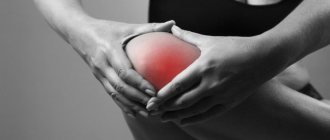
Treatment of the knee and shoulder joint. Cartilage tissue covering the articular surfaces of bones performs an important biological function. It reduces the load on the joint by providing shock absorption and also promotes less friction on the joint surfaces during active and passive movements.
Cartilage does not contain blood vessels; its cells (chondrocytes) are nourished by the diffusion of nutrients and oxygen from the synovial fluid filling the joint cavity. Tissue destruction occurs due to trophic disturbances against the background of changes in the composition and properties of the synovial fluid.
The degenerative-dystrophic process leads to disruption of the functional state of cartilage. Their surface becomes less smooth, which during movements contributes to even greater damage. Next, the tissues become inflamed and swell. The pathology is manifested by stagnation of blood with progression of pain.
Orthopedics and traumatology services at CELT
The administration of CELT JSC regularly updates the price list posted on the clinic’s website. However, in order to avoid possible misunderstandings, we ask you to clarify the cost of services by phone: +7
| Service name | Price in rubles |
| Appointment with a surgical doctor (primary, for complex programs) | 3 000 |
| MRI of the knee joint (1 joint) | 7 000 |
| MRI of the shoulder joint (1 joint) | 7 000 |
All services
Make an appointment through the application or by calling +7 +7 We work every day:
- Monday—Friday: 8.00—20.00
- Saturday: 8.00–18.00
- Sunday is a day off
The nearest metro and MCC stations to the clinic:
- Highway of Enthusiasts or Perovo
- Partisan
- Enthusiast Highway
Driving directions
Causes
Chondromalacia of the patella of the knee joint is a pathological process, the development of which can be provoked by the influence of several etiological (causal) factors. Among them:
- Genetic predisposition.
- Congenital change in the anatomical relationship of the patella in relation to other structures.
- Knee injury, in which the impact of the damaging factor is directed directly to the kneecap (fall on the knee, mechanical shock).
- Increased loads on the knee that occur in athletes during systematic running, jumping, and squats.
- Reduced tone and strength of the quadriceps femoris muscle, which makes normal movement of the cup difficult.
The simultaneous impact of several factors leads to the development of more severe chondromalacia of the patella. How many cartilaginous structures are damaged and what degree of their destruction are determined by the type and strength of the damaging factor.
Radiation diagnostics[edit | edit code]
The initial examination includes radiographs in frontal, lateral, tunnel and axial projections. X-rays in frontal and lateral projections allow us to assess the position of the patella and identify its high or low position. An axial radiograph shows minor changes, such as narrowing of the joint space of the lateral femoral-patellar joint. This is often necessary to identify patellar tilting, either with or without obvious subluxation. For examination in the axial projection, the knees are bent at an angle of 45°, and the X-ray tube is directed caudally at an angle of 30° to the plane of the femur. Often, especially in the early stages of the disease, the x-ray picture of the joint is normal. An additional examination method for tilting and subluxation of the patella is CT. With its help, you can take a series of horizontal slices at any flexion of the knee joint and determine at what stage of the movement symptoms occur. In difficult cases, CT can also show changes in subchondral bone structure and cystic changes that often occur in the lateral femorotibial joint. MRI is best suited to clarify the extent and location of damage. Cartilage and soft tissue are very clearly visible on it, but disturbances in the relative position of the structures of the femoral-patellar joint are often better visible on conventional radiographs and CT scans.
Chondromalacia of the patella - degrees and types
Chondromalacia of the patella - degrees and types
- Stage 1 – small local thickenings of cartilage develop in the form of swellings.
- Stage 2 – cracks and depressions appear on the surface, the dimensions of which do not exceed 1 cm.
- Stage 3 – cracks and depressions exceed 1 cm in size, they can penetrate through the entire thickness of the cartilage to the surface of the bone.
- Stage 4 is the most severe degree of damage, in which the surface of the bone is almost completely exposed.
Treatment[edit | edit code]
Conservative treatment[edit | edit code]
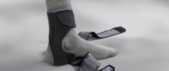
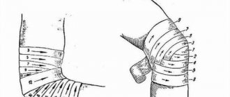
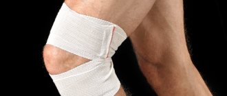
Treatment usually begins with conservative methods: rest, NSAIDs, changes in training regimen. After this, a rehabilitation program is prescribed, which is based on stretching the extensor muscles, the iliotibial band, the ligaments that support the patella, and the posterior thigh muscles. It is also important to increase the strength of the quadriceps muscle, especially the oblique part of the vastus medialis muscle, as the main limiter of patellar mobility. It is believed that due to the weakness of this muscle compared to the vastus lateralis muscle, the patella is subject to external subluxation. Low-range quadriceps strengthening exercises and straight leg raises reduce the patellofemoral joint's response to this imbalance. In addition to exercises, you can prescribe elastic bandaging of the knee joint, fixation of the patella with a bandage or orthopedic device, and the patient should be explained the essence of the disease and encouraged. For most patients with isolated arthrosis of the patellofemoral joint, such treatment is sufficient to relieve symptoms. However, persistent pain, effusion, and crepitus combined with patellar subluxation indicate progressive degeneration; in this case, you should move on to other methods of treatment.
Surgical treatment[edit | edit code]
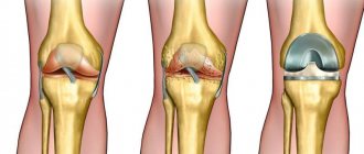
There are many options for surgical treatment of arthrosis of the femoral-patellar joint. Most of them are aimed at restoring the correct anatomical position of the femoral-patellar joint, a small part - at the regeneration of the cartilaginous lining. In severe cases, contouring of the articular surfaces and patellectomy are indicated.
Knee arthroscopy is not only an important diagnostic, but also a therapeutic procedure. Although the advisability of irrigating the joint cavity and removing pathological tissue during arthroscopy remains a matter of debate, the value of this examination in determining the stage of the disease and planning surgical treatment is obvious. Arthroscopic lavage temporarily reduces pain and improves function by removing dead tissue and proteoglycans produced by inflammation. But since the cause of the disease is not eliminated with this manipulation, the symptoms usually reappear.
When arthroscopically staging the disease, the Outerbridge system is widely used because of its simplicity and reproducibility of results. The system is based on determining the location, shape, size and depth of the defect. Defects of the first degree are soft thickenings, sometimes swellings. Grade II is characterized by depressions and cracks with a diameter of less than 1 cm. Grade III damage looks like deep cracks with a diameter of more than 1 cm, reaching the bone. Finally, grade IV is characterized by exposure of the subchondral bone.
Lavage and removal of dead tissue is more appropriate for injuries without evidence of patellar instability than for degenerative lesions of a nontraumatic nature. If the patella is tilted and there is minimal damage to the articular surfaces, especially the lateral facet, the lateral edge of the patella can be mobilized with arthroscopy. This intervention is only appropriate for clinically obvious patellar tilt and non-severe joint injuries. In general, arthroscopic lavage and removal of pathologically changed tissues with or without mobilization of the lateral edge of the patella are justified for lesions of grades I-II; in grades III-IV, long-term results are usually poor. For severe cartilage degeneration, arthroscopic chondroplasty is used. Methods of abrasive and microgap chondroplasty include mechanical penetration into the underlying bone with the introduction of bone marrow mesenchymal stem cells into the defects, stimulating the regeneration of fibrocartilaginous tissue. Arthroscopic chondroplasty is usually performed on people under 30 years of age with well-demarcated grade III lesions; for more severe lesions it is contraindicated.
Additional methods are aimed at restoring hyaline (articular) cartilage. To regenerate cartilage, implantation of one's own chondrocytes, transplantation of an osteochondral auto- or allograft, and plastic surgery with a fragmented osteochondral graft (mosaic plastic) are performed. A detailed description of these methods is beyond the scope of this publication, but some idea of them should still be given. Implantation of native chondrocytes is carried out in case of significant through defects in the cartilaginous lining of the femoral condyle, which manifest themselves clinically. First, the patient's chondrocytes are harvested, then they are cultured and placed under a periosteal flap, on the articular surface defect cleared of pathological tissue. According to long-term cooperative studies, good and excellent results were obtained in 79% of cases. The method is indicated for young (20-50 years old) active patients with isolated (2-4 cm2) traumatic defects of the cartilaginous lining of the femoral condyles. The results of filling defects in the patellar surface of the femur or patella are much worse. Contraindications include extensive osteoarthritis, instability or tilting of the patella with subluxation, and previous meniscectomy.
Transplantation of an osteochondral autograft and mosaic plastic surgery are interesting because they use their own intact cartilage to fill deep defects. However, to accurately restore the relief of the articular surface requires considerable technical skill. In addition, the number of donor areas is limited and complications at the sites of cartilage collection cannot be excluded. Osteochondral allografts are usually used for large (10 cm2 or more) defects of the femoral condyles and often after failure of other methods. In fresh allografts, chondrocytes are more viable, but at the same time they are more immunogenic and increase the risk of infection transmission. Moreover, fresh allografts are difficult to handle and require the surgeon and the patient to clearly plan the timing of the intervention in a relatively short time. Fresh frozen allografts are less likely to cause immune reactions and provide greater freedom in choosing the timing of surgery, but both chondrocyte viability and long-term graft viability are lower. Patellectomy and patellar contouring are used for extensive injuries to the patella that cause significant impairment of function due to pain, and also when other treatment methods have not produced the desired effect. The results of the methods are ambiguous. Surgeries that require movement of large amounts of tissue, such as osteotomy, transfer and elevation of the tibial tuberosity, and others, will be discussed in the section on patellar tilt and subluxation.
Classification by localization and prevalence
Depending on the prevalence of the pathological process, chondromalacia of the patellar facet is distinguished (thickening of the cartilaginous surface, repeating the shape of the knee joint), which is divided into 2 types:
- Chondromalacia of the medial facet of the patella - damage primarily affects the thickening of the cartilage of the patella, localized on the inner (medial) side.
- Chondromalacia of the lateral facet of the patella - the cartilaginous thickening located on the outer (lateral) surface of the patella is predominantly damaged.
- Combined lesion affecting all structures of the cartilaginous surface of the cup.
Also, pathological destruction of cartilage tissue can be isolated or affect other structures of the knee. The most common sites are chondromalacia of the patella and tibial condyle.
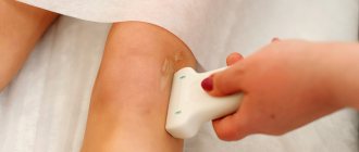
Diagnostics
When making a diagnosis, the data of the anamnesis and physical examination are taken into account. To make a final diagnosis, functional tests and instrumental visualization methods are used:
- radiography, in a standing position on the affected leg (determining the angle of deviation of the limb and the central axis)
- computed tomography (assessment of the condition of the underlying bone, detection of cystic cavities)
- MRI (obtaining informative images of all soft tissues of the patellofemoral joint, identifying subchondral changes, incongruence of articular surfaces, pathological proliferation of blood vessels in cartilage tissue)
- diagnostic arthroscopy (quantitative characterization of degenerative lesions)
Magnetic resonance imaging is recognized as the most highly sensitive diagnostic method. Safe non-invasive imaging technology allows you to accurately determine the degree of chondromalacia of the patella, assess the syndrome of imbalance and develop further treatment tactics.
Chondromalacia patella - symptoms
Chondromalacia patella - symptoms
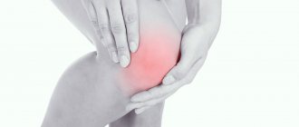
- Acute, severe pain that intensifies with increasing load on the knee (climbing or descending stairs, running, squats, kneeling).
- Chronic constant pain at rest. It is less pronounced than when performing movements in the knee.
- Characteristic clicks and crunches that occur when performing active or passive movements in the joint.
- Limitation of range of motion in the knee.
- Swelling and redness of the skin in the area of the kneecap, indicating the development of an inflammatory reaction.
Chondromalacia of the medial condyles and patella has pronounced clinical symptoms. In general, clinical manifestations tend to gradually progress over a fairly long period of time. As degenerative-dystrophic processes progress, as well as against the background of an inflammatory reaction, the intensity of symptoms increases significantly.
Forecast[edit | edit code]
The goal of rehabilitation for lesions of the femoral-patellar joint is to return the patient to the level of physical activity at which he was before the disease. To do this, you must first relieve inflammation, restore range of motion, strength, endurance and muscle flexibility while maintaining a good functional state of the cardiovascular system. The final stages of rehabilitation restore the agility, speed and skills needed in a particular sport. In each case, the program should be tailored individually, taking into account age, the initial state of the muscles and cardiovascular system, the patient’s motivation and how well he is familiar with the training equipment.
chondromalacia patella
Chondromalacia of the patella
After the diagnosis of chondromalacia of the patella has been established, a comprehensive treatment is prescribed by the doctor. It includes conservative drug therapy (use of anti-inflammatory drugs and chondroprotectors), physiotherapy and surgery. The choice of volume and direction of therapeutic measures depends on the severity and localization of the pathological process.
Return to sports[edit | edit code]
Regardless of the treatment method, resumption of sports activities is possible only after restoration of range of motion and stability and strength of the affected lower limb. All four phases of rehabilitation must be completed, and sport-specific exercises must not cause pain, functional limitations, or accumulation of effusion in the patient. In addition, it should be emphasized the need to continue training the last stage of rehabilitation and avoid movements involving tight contact of the femur and patella.
Read also[edit | edit code]
- Supplements for ligaments and joints Glucosamine sulfate
- Chondroitin sulfate
- Collagen
- Anterior cruciate ligament rupture
- Subluxation and dislocation of the patella