A joint is an articulation of bones of a certain type that provides the ability to perform various movements. The joint of the bones has its own cartilaginous capsule, inside of which there is synovial fluid. The heads of the bones entering the joint are covered with a dense layer of cartilage tissue, which is capable of absorbing and releasing synovial fluid. There is no bare bone tissue in the joint in normal condition.
But it can appear if the cartilaginous synovial layer is destroyed. This provokes the appearance of growths on the joints - bone calluses, with the help of which the body strives to restore the damaged integrity of the heads of the bones.
Very often, hard growths on the joints of the fingers and toes are a consequence of developing deforming osteoarthritis. But no less rarely, such a symptom signals another dangerous disease - rheumatoid polyarthritis. This pathology is systemic in nature and can provoke the destruction of heart valves and form their defects.
Bone growths on joints can be osteophytes or exostoses, have a dense structure consisting of calcium and phosphorus salts, or contain fibrin and cartilaginous fibers. The structure of the growth can be determined using an x-ray image. If in the image the neoplasm looks the same as the surrounding bone tissue, then it is an osteophyte or callus. If the neoplasm tissue is sparse and less dense in structure, then most likely it is osteochondral exostosis. At an early stage, it is quite easy to treat conservatively.
If any growths appear on the bones, it is necessary to exclude the possibility of developing oncological tumors (sarcoma, osteoma, blastoma, etc.). To exclude such diseases, a consultation with an oncologist is always scheduled. If tumor diseases are excluded, then further diagnosis and treatment is carried out by an orthopedist or osteopath. In Moscow, you can make an appointment for a free appointment with an orthopedist at our manual therapy clinic. The doctor will conduct a full examination and make a preliminary diagnosis. He will recommend additional examination methods if necessary. Then, after clarifying the diagnosis, an individual course of treatment will be developed.
Potential causes of growths on joints can be different, ranging from the consequences of traumatic exposure to the formation of tumor tumors. Let's look at the most basic and common risk factors for the appearance of bone growths:
- injuries (cracks, fractures, damage to the periosteum with the formation of a hematoma);
- excess amounts of calcium and phosphorus salts in the human body, which provokes deposition on bone and cartilage surfaces;
- excess amount of uric acid in the body, which provokes the development of gout and the formation of tophi nodules in the area of large and small joints of the upper and lower extremities;
- rheumatoid polyarthritis and its consequences in the form of joint deformation (most often it affects the small interphalangeal joints of the hands);
- deforming osteoarthritis (causes destruction of the cartilage protective layer and exposure of bones, which in the future leads to the formation of thorns and growths);
- genetic abnormalities, poor heredity.
Bone destruction can be caused by such common pathologies as osteomalacia or osteoporosis. These diseases have several identified causes. One of them is associated with diseases of the large and small intestines (Crohn's disease, ulcerative colitis, long-term carriage of Shigella or pathogenic E. coli). A constant inflammatory process in the thickness of the intestinal mucosa does not allow the body to absorb calcium and phosphorus. A deficiency of these microelements begins. They are necessary not only for the construction of bone tissue. First of all, calcium and phosphorus are used in the form of blood electrolytes (basic elements that transmit electrical impulses and are involved in the destruction of myofibrils).
In conditions of calcium deficiency, seizures may occur and disruption of the cardiovascular system may occur. Therefore, the body begins the process of leaching this microelement from bone tissue. It becomes deformed, and rough cartilaginous exostoses form on the articular surfaces. There is an increased risk of fractures and cracks, which is also fraught with the subsequent formation of rough callus.
The second important factor is vitamin D deficiency. It is produced in the human body, but only if the skin is exposed to sufficient ultraviolet rays from the sun. Therefore, if a person rarely goes out into direct sunlight or constantly uses protective UF creams, then there is a high probability of vitamin D deficiency. And without it, calcium is not absorbed at all. Therefore, rickets occurs in children, leading to total deformation of bone tissue, and in adults this can provoke deformation in the form of bone growths on large and small joints of bones.
A third potential cause of osteomalacia or osteoporosis is hormonal imbalance. Women during menopause are more susceptible to it. Therefore, if you have problems with the formation of hard growths on the joints, you should first visit an endocrinologist and do a series of tests.
Other potential causes include ankylosing spondylitis, aseptic necrosis of bone tissue, gout, cicatricial deformation of tendon and ligament tissue, and the formation of cavitary bone cysts.
Kinds
Osteogenic sarcoma of the leg
This type of leg cancer is a nonepithelial bone tumor.
The location of the leg sarcoma is very important:
- On the back of the lower leg;
- On the front of the shin.
Symptoms and development
If it is diagnosed on the back of the lower leg, it may not be noticeable for quite a long period, since it is hidden by the gastrocnemius major muscle.
If the cancer is on the front of the leg, then it is quite easy to diagnose its development and enlargement, due to changes in the condition of the skin above the tumor. It stretches and changes color.
Further from the lower leg, the sarcoma can spread to the fibula and tibia. This cancer can destroy the membranous connections between bones, causing frequent fractures.
In the first stages, pain does not occur with this disease. Only when the tumor grows does it put pressure on nearby nerves and vessels, causing pain. The pain spreads to the foot and toes, swelling appears, and trophic skin disorders may occur.
Soft tissue sarcoma of the leg
There are two types of soft tissue sarcoma of the leg:
- Superficial;
- Deep.
Symptoms and development
The first type of soft tissue cancer of the leg can be diagnosed in the early stages. The second one can be almost asymptomatic. There may be complaints of pain in the lower leg of a bursting and pulling nature.
As the disease progresses, its symptoms also intensify. Here are the main ones:
- Weakness without a specific reason;
- Anemia;
- Severe weight loss;
- Nausea;
- Lack of appetite;
- Fever.
Further, as it grows, the sarcoma presses on the vessels and nerves located in the foot area. Because of this, swelling of the feet and trophic ulcers appear, sensitivity in the toes worsens, and they begin to move worse.
Osteosarcoma
Symptomatic means problematic
The growths are visible to the naked eye. The reason for their occurrence can be judged by their appearance.
Hard and dense lumps on the joints, covered with smooth skin, usually form in men over 40 years of age and indicate gout. They are usually called gouty tophi. They grow during periods of exacerbation of the disease. The patient becomes weaker and complains of malaise. The temperature in the area of the tophi rises, and the pain sometimes becomes unbearable.
If the skin on the lump is rough and red, and the growth itself is round and elastic, it is a hygroma. It usually forms in women 20-30 years old and can be single-chamber or multi-chamber. Hygromas differ from other types of growths in that their size gradually increases, and pain is felt when pressed.
Formations on the interphalangeal joints that appear after 60 years are called Heberden's nodes. They are equally likely to be found on any phalanx and are signs of osteoarthritis. This disease leads to stiffness of the hands and deformation of the joints.
Nodules up to 2 cm in diameter, which form in 20% of patients with rheumatoid arthritis, are rheumatoid nodules. They do not cause pain, but the course of the disease is accompanied by other unpleasant symptoms: lethargy, fatigue, fever, and sometimes loss of body weight.
Diagnostics
Osteogenic leg cancer is easily diagnosed on the front of the leg. If it is on the back, then it is much more difficult to diagnose because of the calf muscle, which hides the tumor.
Superficial cancer of the soft tissues of the leg is also much easier to diagnose than asymptomatic deep cancer of the soft tissues of the leg.
If cancer of the leg is suspected, laboratory tests are performed, a computed tomography scan is performed, an X-ray of the leg is taken, and magnetic resonance imaging is performed.
A biopsy is done to confirm leg cancer. Next, it is checked whether leg cancer has metastases in other organs.
How to treat a lump on your thumb that hurts
Unfortunately, this problem is usually caused by chronic diseases and cannot be completely cured. Only hygroma can be removed once and for all. All other types of growths will remain for life - but exacerbations can be avoided if stable remission is achieved.
Rheumatoid arthritis is treated with corticosteroids or non-steroidal anti-inflammatory drugs. These same non-steroidal anti-inflammatory drugs, as well as colchicine, are effective against gout.
For osteoarthritis, chondroprotectors, finger therapeutic exercises and physiotherapy sessions are prescribed: multi-channel electrical stimulation and laser therapy. With multichannel electrical stimulation, muscles are stimulated with short pulses of electricity to relieve tension and improve blood flow. During the laser therapy procedure, a laser beam is applied to the joint growth to relieve inflammation and activate blood flow within the tissues.
Osteoarthritis is also treated with physiotherapy: ultrasound (reduces tissue volume), shock wave therapy (activates blood flow, eliminates swelling and pain by exposure to acoustic impulses), laser therapy.
Surgery may be required if the cartilage and surfaces of the joints are severely damaged in the later stages of arthritis or arthrosis, and the fingers become swollen, stiff and painful. In especially severe cases, endoprosthetics is prescribed, when a worn-out joint is replaced with an artificial one. The operation can be performed in the presence of a malignant tumor. The oncologist prepares an individual therapy program for each patient.
Traditional medicine is powerless when there is a lump and pain on the finger joint. Not a single homeopathic remedy contains components that would dissolve the lump. Therefore, relying on chance in the case of such a disease is extremely undesirable: all hopes for a miracle can ultimately lead to serious consequences.
Growths on the elbow and shoulder joints
A growth on a person’s elbow joint is not always a bone tumor or exostosis. In most cases, this is how inflammation of the elbow capsule (bursitis) manifests itself. When a large amount of exudate accumulates in it, it becomes inflamed and begins to appear as a growth on the elbow joint. Differential diagnosis should be carried out by an experienced doctor using all available technical means (x-ray, MRI or CT examination, ultrasound of soft tissues).
A growth on the shoulder joint, upon closer examination, may turn out to be a dislocation of the distal end of the clavicle. In this case, there is a characteristic “key” symptom - when you press on the growth, it collapses and then returns to its original place. The second common reason why growths appear on the bones of a joint is deforming osteoarthritis. Although neoplasms should not be ruled out either.
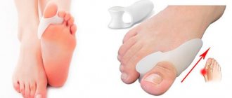
How to get rid of bunions on your feet yourself
Overgrowth of the bunions on the big toes is a consequence of hallux valgus. This disease can cause multiple inconveniences. Not only do bunions cause constant pain, they interfere with wearing your favorite shoes and cause constant discomfort. The reasons that cause such deformations can be very diverse. They can be caused by flat feet, incorrectly chosen shoes and a number of hereditary pathologies.
Hallux valgus is often unknowingly confused with gout. However, these are two completely different diseases, so to correctly determine the disease you need to consult a doctor.
Most of the fair sex suffer from this disease. This can be explained by the fact that women are most often subject to hormonal changes, which can completely change women’s health. In addition, women more often choose uncomfortable shoes, sacrifice comfort for the sake of beauty, thereby provoking the progressive development of the disease.
Existing degrees of the disease
The initial stage may not be felt at all. The finger moves at an angle of about twenty degrees and does not cause any inconvenience. It is precisely because neither pain nor discomfort is felt at this stage that the disease develops, since the required treatment is not carried out.
The second degree of development of the disease is the deviation of the finger by forty degrees. Here, with prolonged loads or wearing shoes for a long time, the bone on the big toe is felt and acute pain increases in the toe.
The third degree already brings with it severe and almost constant pain in the area of the thumb, although the finger is also displaced by forty degrees, as in the second stage.
The fourth stage is characterized by a displacement of the finger by fifty degrees or more. The patient experiences pain non-stop. And here, at this stage, it is already difficult to do something at home; only surgery can correct the deformity.
Ways to solve the problem
If your closest relatives have a problem with hallux valgus on the big toes, then we can safely assume that you may also encounter this disease, so you need to carefully monitor your feet and immediately begin to take action at the slightest deviation of the bones in the area of the big toes.
Also, if you have been diagnosed with flat feet, then you should more carefully monitor the deviation in the degree of the bones on the big toes.
As soon as you realize that you feel at least slight discomfort, and your everyday shoes, which previously did not cause you any trouble, begin to put a little pressure in the area of the bone, it is better to immediately consult a doctor. Doctors will conduct a diagnosis and determine whether you have the disease or its absence.
If the bones deviate by at least twenty degrees, you need urgent treatment so as not to provoke the development of the disease to the next stages. Doctors usually prescribe wearing a hallux valgus splint. This method can slow down the deformation and even stop it completely. Both doctors and specialists from orthopedic salons, where they are usually sold, will help you choose a splint.
If you have discovered the disease at a late stage, the third or fourth stage, then it is unlikely to be possible without surgical intervention. There is no need to be afraid of this. Surgeons perform real miracles. Of course, the postoperative period will need to be endured, it may cause some inconvenience. But the result will exceed all your expectations. You will finally get rid of the pain that interferes with your life; finally, you will be able to walk in the shoes that you like, and not just in soft slippers or shoes made of soft elastic leather, which also cause pain.
Disease prevention
If you know for sure that you are predisposed to hallux valgus, then you should regularly visit an orthopedist in order to identify the disease in the early stages, and be attentive to your feet.
No one except you will notice deviations at the right time. Author: K.M.N., Academician of the Russian Academy of Medical Sciences M.A. Bobyr
A few words about prevention
The best prevention for growths is a healthy lifestyle: a balanced diet, regular consumption of fresh vegetables and fruits, giving up bad habits, sweets and fried foods. Wear protective gloves before handling chemicals. Avoid stress and lack of sleep.
If you are a schoolchild or student, you have a lump on your finger and it hurts - you probably write too much and hold the pen incorrectly. In this case, the growth is an ordinary callus that will go away over time. There is no need to put a band-aid on it or try to get rid of it by other methods. It's better to buy a soft-bodied pen, try to hold it a little differently and not squeeze too much with your fingers.
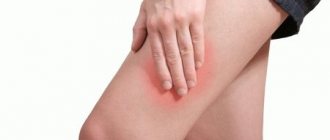
Growths on the knee, ankle and hip joints
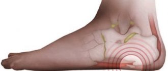
A hard growth on the ankle joint may be a heel spur. This is rough bone tissue that forms at the attachment point of the plantar fascia. With regular injury to tendon tissue, scar deformation and deposition of calcium salts begin. The formation of a heel spur leads to acute pain when walking.
Growths on the knee joint in young people and adolescents can appear as a result of traumatic exposure. These may be hollow bone cysts or calluses. In old age, growths on the joints of the knees almost always indicate the development of gonarthrosis (deforming osteoarthritis of the knee joint). The diagnosis is confirmed using an x-ray.
Almost all growths on the hip joints are a consequence of the development of coxarthrosis. Deforming osteoarthritis leads to thinning and rupture of cartilaginous synovial tissue. The head of the femur begins to injure the acetabulum. Cracks form on the surfaces, which are quickly filled with calcium salts. Characteristic spines and growths appear.
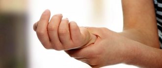
Growths on the knuckles of the fingers
Gradually developing growths on the joints of the fingers can be single (solitary) or multiple. If the growths on the joints of the hands are of a solitary nature, damage to the periosteum or the head of the phalanx of the finger cannot be ruled out. As a result of the crack, a rough bone callus can form, and the patient may not even be aware of the severe injury he has suffered.
Multiple growths on the joints of the fingers most likely indicate a systemic failure in the metabolic and immune system. This may be a sign of the development of gout, systemic lupus erythematosus or rheumatoid polyarthritis. The denser and more painful the growths, the higher the likelihood of the inflammatory etiology of this pathological process.
Sudden growths on the joints of the hand may be a sign of inflammation or intra-articular hemorrhage. The inflammatory reaction can be aseptic and develop under the influence of autogenic factors. Thus, primary signs of the formation of growths often appear during periods of acute colds. This is aching in small and large joints, their redness and slight swelling. After the cold passes, the discomfort in the joints persists. And after a few weeks, you can already feel the deformation of bone tissue under the skin.
Before treating joint growths
It is very important to conduct a full examination before treating growths on the joints. A malignant neoplasm detected in a timely manner can be successfully treated in modern medicine.
The examination is prescribed:
- an x-ray that will allow you to differentiate the structure of the neoplasm;
- puncture (if the neoplasm is soft and fluctuation is felt upon palpation);
- Ultrasound of soft tissues;
- MRI and CT examination.
After an accurate diagnosis is made, treatment is developed. It can be conservative or surgical. It depends on what is found during differential diagnosis.