Osteoarthritis of the foot is a chronic disease of the joints, which entails the destruction of articular cartilage and bone deformation. It is degenerative-dystrophic in nature and is manifested by pain when walking, limitation of movements and a sharp decrease in the supporting function of the leg. It is also characterized by enlargement of the joint due to stable swelling of the soft tissues. The incidence of the disease increases with age. For statistics - primary or secondary arthrosis of the foot, the treatment of which is required immediately, is detected during examination in 85% of women and men. Age group – after 55 years.
Mechanism of disease development
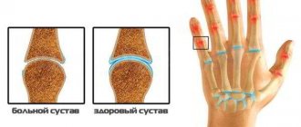
Arthrosis begins with impaired blood circulation in the subcartilaginous layer of the periosteum.
Because of this, the nutrition of the cartilage deteriorates, it loses its elasticity, becomes thinner, and cracks appear on it. The amount of synovial fluid decreases, due to strong friction, the joints become inflamed. Progressive arthrosis is very dangerous. If treatment for arthrosis of the hand or foot is not started in time, the joints begin to take on unnatural, ugly shapes and may lose mobility.
The disease often occurs in women during menopause. Hormonal levels change in the female body and less estrogen is produced. Because of this, the body loses a lot of moisture, which makes cartilage and joints more brittle.
Types of arthrosis development
For treatment of arthrosis of the foot joints to be successful, it is necessary to make a correct diagnosis. Each stage of disease development requires a different set of therapeutic measures. Doctors distinguish the following stages of pathology development:
- First. At this stage, noticeable fatigue of the lower extremities appears, as well as “pulling” pain after long walks or excessive physical exertion. At this stage, the patient may not even suspect that he is developing a disease, because he attributes the symptoms to banal fatigue. But if you do not visit a doctor, the disease will begin to gain momentum;
- Second. All the symptoms of the first stage begin to appear more actively. The pain becomes longer and more severe. Calluses may appear on the heels due to improper foot position, the knuckles become thicker;
- Third. The joints undergo serious changes, so the person begins to limp severely. Joint mobility decreases. X-rays show that the gaps between the joints are significantly reduced or disappear completely.
To make a diagnosis, the doctor must conduct an examination and diagnosis. It includes:
- taking an anamnesis, when the doctor asks the patient about all his complaints;
- X-rays and other examinations that will make it possible to accurately determine the condition of the joints, cartilage and bones, and also identify possible changes in their structure;
- examinations that will allow you to study the condition of muscle tissue;
- tests to find out about the state of the body and the nature of inflammation in it;
- measuring the size of the foot so that you can track the dynamics of treatment for arthrosis of the toe or any other joint.
Kinds
Depending on the location, arthrosis is of two types:
- joints of the hands and fingers.
- small joints of the foot, big toe.
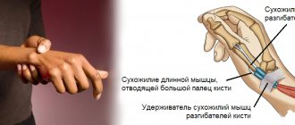
Arthrosis of the joints of the hands and fingers
Arthrosis of the hands is often found among typists, pianists, programmers, and secretaries. The disease affects the interphalangeal joints. With arthrosis of the fingers, the joint tissues dry out and crack. Subcutaneous small nodules often appear in the joints of the hands. Usually they don't disappear anymore.
As the disease progresses, the bone tissue becomes denser and growths appear on it. They cause severe pain when moving your fingers. In advanced cases, a person loses the ability to move the hand and fingers.
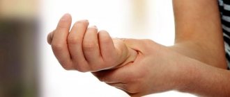
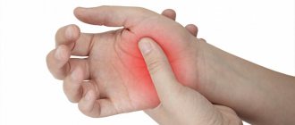
Symptoms of the disease: crunching in the joints when moving the fingers, pain in the fingers after exercise, seals on the joints of the fingers, swelling of the periarticular tissues.
Doctors at the Health Workshop treat arthrosis of the hand using non-surgical methods.
Arthrosis of the foot joints
Ballerinas, dancers, gymnasts, wrestlers, and jumpers are susceptible to arthrosis of the feet. The disease affects the joints of the toes. Bone growths appear on the small joints of the foot. Because of them, it is painful for a person to move his foot. As the disease progresses, the deformation of the joints is very noticeable, the person practically cannot move the foot.
Arthrosis of the big toe is the most common type of arthrosis of the feet. When the disease occurs, a “bone” forms in the area of the thumb joint. Osteoarthritis of the big toe occurs due to wearing tight shoes.
Symptoms of arthrosis of the foot: pain in the foot after physical activity, crunching in the joints when moving, an increase in the size of the joints. When walking, a person limps and rests on the outer edge of the foot.
When treating arthrosis of the foot, doctors at the Health Workshop use laser therapy, magnetic therapy and other methods.
Treatment of arthrosis of the foot: drugs and necessary procedures
Treatment of arthrosis of the foot is a complex of therapeutic and preventive measures that are prescribed by the doctor after examination. The main treatments include the following procedures and medications:
- Physical rehabilitation, physiotherapy, medical. massage, physical therapy, mechanotherapy. Cycling and swimming in the pool are beneficial.
- Drug therapy. Non-steroidal PVPs are prescribed: indomethacin, iboprufen, piroxicam, ortofen and others. Intra-articular injections of steroid drugs are given.
- In particularly serious cases, surgical intervention is prescribed.
Prevention of the disease is important: timely elimination of factors contributing to the development of the disease, reducing the load on the affected joint, getting rid of excess weight.
In our clinic, we accurately diagnose the disease and prescribe competent and effective treatment for arthrosis of the foot, heel and joints.
Do not delay in contacting specialists. Timely therapy will help quickly solve the health problem. arthrosis
Causes
Arthrosis can be primary and secondary.
Primary arthrosis appears due to problems with the cartilage tissue of the joint, namely:
- genetic disorders in the composition of joint cartilage tissue;
- joint hypermobility;
- dysplasia, flat feet, wide feet.
Secondary arthrosis is the result of joint damage and other diseases. Among its reasons:
- injuries, microtraumatization of joints;
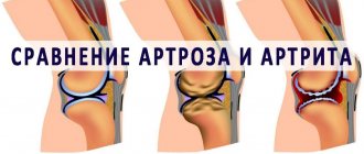
- acute and chronic arthritis, synovitis;
- metabolic disorders, lack of calcium, phosphorus and other minerals;
- gout, psoriasis, rheumatoid arthritis;
- diabetes mellitus and other diseases of the endocrine system.
Carrying out the operation
Arthrodesis of the ankle joint according to the traditional method is performed under general anesthesia in an open manner. Surgical procedures under arthroscope control can be performed under spinal anesthesia. The session requires an average of 2-3 hours of intraoperative time. Let's consider the principle of classical tactics.
- A pneumatic tourniquet is applied to the lower third of the thigh. Next, access is created by making a linear skin incision along the joint with a scalpel. The incision is approximately 10 cm.
- At the next stage, an opening and reliable supination of the joint is performed, which will facilitate the work with the next manipulations.
- The surfaces of the tibia and talus are then prepared. Preparation includes resection of cartilage tissue with a surgical chisel and removal of ossification.
- Then the foot is removed from the vicious position. The tibial element and the talus component are tightly juxtaposed with each other in a position that is convenient from a physiological point of view. The achieved position is secured by a metal structure of the required type.
- The surgical tracts used are closed at the final stage using layer-by-layer suturing of soft tissues, leaving drainage.
In cases of severe deformity, fibular osteotomy may be used. Extensive bone losses are compensated by grafts - fragments of similar biological material taken from the patient from the iliac crest.
If external fixation systems were used, for example, the Ilizarov apparatus, plaster is not used. When installing internal metal implants, a cast is placed on the operated limb. Until ankylosis occurs, the patient is in a plaster cast. The rate of bone fusion in each individual patient may differ due to the physiological characteristics of the body. The joint is completely fused and immobilized 3-6 months after surgery.
Risk factors
Risk factors for the disease include:
- hereditary predisposition;
- heavy physical labor, heavy load on the finger joints;
- overweight, excessive alcohol consumption;
- long-term wearing of uncomfortable tight high-heeled shoes;
- sedentary lifestyle;
- hypothermia of the feet.
If your work often puts stress on the joints of your fingers and toes, or you have relatives who suffer from arthrosis of small joints, you should think about prevention. We will talk about it at the end of the article.
Possible consequences and complications
Serious complications can only be avoided by choosing an experienced specialist and by 100% following all the doctor’s recommendations after surgery. In rare cases, the following complications are observed:
- Bone graft rejection.
- Anemia due to large blood losses.
- Uneven fusion of joints.
- Injuries to nerve endings during surgery.
- Infection, accumulation of pus and intoxication.
- Thrombosis.
If you experience pain, bleeding, cramps, fever or numbness, immediately contact your doctor for diagnosis and elimination of complications.
Stages of arthrosis of small joints
There are three stages of the disease.
- The first stage of the disease is accompanied by periodic pain in the joints of the fingers or toes during active movement or work. The pain subsides after rest. The joints may begin to crack, and swelling may form near them.
- Second stage of arthrosis. The disease progresses, the pain intensifies and becomes constant. Moving your fingers causes pain. Heberden's and Bouchard's nodes form in the joints. Their appearance is accompanied by burning and pain. After a few months, the redness and swelling disappear, and the nodules become hard.
- The third stage of arthrosis of small joints is the most serious. The fingers and toes become deformed and are almost impossible to move. The area around the affected joint becomes red and swollen. Sharp pain in the joints is felt not only when moving, but also at rest. A person walks with difficulty, going up and down stairs becomes a challenge for him.
Complications of ankle arthrodesis
The incidence of complications after a standard operation with a wide opening of the joint, as clinical experience shows, is an order of magnitude higher than after arthroscopic interventions. Here is some comparative data on negative reactions for two types of procedures (without the use of external fixators) detected during the first 3 weeks:
- phlebothrombosis is detected in 22% of cases after open ankle arthrodesis, in 1.8% after minimally invasive intervention;
- wound infection develops in approximately 12% of patients, while the risks after arthroscopy are practically absent (<0.1%)
- necrosis of surrounding tissues, respectively, in 17% and 0.2%;
- wound hematomas and seromas: 22% and 0.9%.
Persistent swelling.
Intraoperative blood loss after standard arthrodesis is 250 ml, after arthroscopic arthrodesis - about 120 ml. Failure of ankylosis after 6 months is determined in 5%-6% of people who underwent the procedure according to the traditional scenario, and in 0.5%-0.9% of patients who underwent arthroscopy with intramedullary fixation. Skipping the topic of comparison, we note that after any type of artificial ankylosis there are increased risks associated with the formation of arthrosis in other joints of the limb and shortening the leg length to 3 cm.
Our doctors
Poltavsky Dmitry Ilyich
Traumatologist-orthopedist
Experience 28 years
Make an appointment
Marina Vitaly Semenovich
Traumatologist-orthopedist, head of the minimally invasive traumatology and orthopedics service
Experience 36 years
Make an appointment
Zubikov Vladimir Sergeevich
Traumatologist-orthopedist, Doctor of Medical Sciences, doctor of the highest category, professor
44 years of experience
Make an appointment
Samilenko Igor Grigorievich
Traumatologist - orthopedist, doctor of the highest category
24 years of experience
Make an appointment