Osteophytes
- bone growths that form on articular surfaces are not an independent disease. More often they indicate the presence of some underlying pathological process in the musculoskeletal system.
At the same time, osteophytes themselves are, perhaps, one of the most striking diagnostic symptoms for an orthopedist or rheumatologist. Bone outgrowths are clearly visible during X-ray examination of diseased joints, and also serve as a cause for aggravation of a whole complex of symptoms - pain, inflammation, joint deformation.
Osteophytes are bone growths that indicate a disorder in the human musculoskeletal system.
Left untreated, osteophytes begin to grow, pushing apart and injuring the soft tissue with every movement in the joint. In advanced cases, they completely block the joint
- but even then the pain does not subside due to compression and irritation of the nerve endings.
Inflammation becomes chronic and plagues patients, including
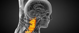
at night , affecting the quality of sleep and mood. The condition of the joints also worsens, ligaments and muscles weaken. After all, osteophytes are essentially foreign bodies that disrupt the natural anatomical position of the ligaments and interfere with the normal passage of nutrients into the tissue. Osteophytes of the spine can lead to compression of the nerve roots, which directly affect the functioning of the heart, lungs, genitourinary system and other internal organs, as well as a person’s ability to move and self-care.
To prevent severe impairment of motor function and chronic inflammation, osteophytes should be treated in advance.
. And in some cases, even think about their prevention. What should you know about this invisible enemy?
Treatment at Top Ikhilov
For osteophytes of the cervical spine, doctors select the optimal treatment option for the patient, which consists of conservative and/or surgical techniques.
Conservative therapy of cervical osteophytes in Top Ichilov:
- Drug treatment
. To eliminate pain, the clinic uses non-steroidal anti-inflammatory drugs, as well as muscle relaxants. If pain due to osteophytes is chronic, then the use of antidepressants is also allowed. - Physiotherapeutic treatment
. Treatment methods such as electrophoresis and ultraphonophoresis allow drugs to be injected into the affected area to relieve pain and inflammation. Physiotherapy for osteophytes of the cervical spine is used to eliminate muscle spasms, pain and improve metabolic processes in tissues. - Exercise therapy
. Therapeutic exercises are an integral component of the complex treatment of osteophytes of the cervical spine. Exercises are also actively used during the rehabilitation period, which helps strengthen the muscles surrounding the spine. - Acupuncture and manual therapy
. To relieve muscle tension and pain, Top Ikhilov also conducts sessions of manual therapy and acupuncture. - Wearing orthopedic structures
. This therapy is carried out to bring the affected vertebrae to normal condition.
Osteophyte surgery at the Top Ichilov Clinic:
Surgical methods for treating cervical osteophytes include:
- Fasectomy
is an operation in which the doctor removes the facet cartilage tissue on which the osteophytes are located. This operation prevents the further appearance of osteophytes in the cervical spine. - Foraminotomy
is an operation that increases the space in the area where the nerve root exits the cervical spine. Cervical foraminotomy significantly relieves the symptoms of nerve compression due to osteophytes. After the operation, the patient's symptoms such as pain, stiffness, tingling and weakness of the upper extremities disappear. - Laminectomy
is an operation to remove a bone plate. Such an operation is rarely performed for osteophytes. Surgery is appropriate for spinal stenosis, since in some cases osteophytes compress it, which ultimately leads to myelopathy and disruption of the bowel and bladder. - Laminotomy
is an operation to expand the gap between the nerve roots. Thanks to this operation, compression of the spinal cord is eliminated. - Corpectomy
– removal of part of the vertebra and adjacent intervertebral discs. Thanks to this operation, compression of the spinal cord is eliminated. To restore the integrity of the spine after a cervical corpectomy, a bone graft is installed.
All operations on the cervical spine in Top Ichilov are performed using minimally invasive techniques. Such operations are low-traumatic and are not accompanied by large blood losses. You can also read reviews on how osteophytes in the cervical spine are treated in Israel on our website. You will read the stories of real patients who managed to get rid of this problem in our clinic.
Causes of osteophytes
Osteophytes form on bones in cases where the joints require compensatory support. If a joint is unable to perform its shock-absorbing function due to benign wear and tear or chronic diseases, the edges of the bones begin to touch and rub against each other. This leads to irritation of the periosteum (periosteum). Since the periosteum plays a huge role in the blood supply (and therefore nutrition) of the surface layers of bone, they begin to experience nutritional deficiencies and become vulnerable to mechanical abrasion. The periosteum also participates in the processes of bone tissue formation (it is thanks to it that the so-called callus grows on injured areas of the bone). The combination of these two factors (a decrease in bone strength and irritation of the areas that are responsible for its growth) results in the formation of osteophytes - first in the form of tubercles or small protrusions. Subsequently, they can take almost any form - a spike, a hook, a visor and others.
Cause the development of osteophytes
can:
- degenerative-dystrophic processes and diseases of bone and cartilage tissue
; - injuries
(not only bones and joint capsule, but also ligaments and soft tissues near the joint); - metabolic and endocrine diseases
(diabetes mellitus, gout, rickets); - congenital and acquired curvatures of posture and feet
(scoliosis, lordosis, kyphosis, flat feet); - inflammatory diseases of bone tissue
(tuberculosis bone infection, brucellosis); - autoimmune diseases
(rheumatoid arthritis, psoriasis); - unnaturally high load on the joints
(excess weight, professional and amateur sports, professions associated with physical labor, shifting the center of gravity during pregnancy); - oncological diseases
(both tumors with primary localization in the bone and metastases of other neoplasms).
The risk of osteophyte formation is increased by genetic predisposition, bad habits and poor environmental conditions. The progression of the problem can be affected by any factor that directly affects bone strength (for example, hormonal imbalances, stress, age-related changes, physical inactivity).
Most often, osteophytes are observed in older people, but the onset of the pathological process often occurs at the age of 40 years. Therefore, starting from this milestone, you should begin to prevent the problem and eliminate its possible causes
.
Inflammation of bone tissue
Acute and chronic inflammatory processes in bone tissue, periosteum and even adjacent articular structures (for example, tendonitis, fasciitis) can provoke the growth of osteophytes at almost any point in the affected joint. The inflammatory process is usually caused by infections (tuberculosis, osteomyelitis, brucellosis, borreliosis) and autoimmune diseases (rheumatoid arthritis, psoriasis). Patients with sources of chronic infection in the body are at risk. Inflammatory diseases of the joints are also common due to complicated infections of the genitourinary organs (gonorrhea, syphilis), intestines (salmonellosis, yersiniosis), and less often - the respiratory system (pneumonia and others).
The inflammatory process is characterized by a chaotic arrangement of osteophytes in those places where the infectious pathogen has most damaged the joints - this is how its picture differs from the degenerative-dystrophic one (in this case, osteophytes are formed on those surfaces that are most susceptible to wear and tear).
Degenerative processes in bone tissue
The most common degenerative diseases of articular cartilage and bone tissue are osteoarthritis, osteochondrosis and spondylosis. In deforming arthrosis, osteophytes can appear as an independent phenomenon without bone degradation. Most often they are observed at the edges of the articular surfaces (so-called marginal osteophytes). Despite the preservation of the bone, the growths should be treated as early as possible: over time, they can become so massive that they almost completely close the lumen of the joint space. This problem is called joint fusion and can only be treated surgically. In some cases, removal of massive osteophytes is impossible, so the joint is completely replaced (if osteophytes have grown on the vertebrae, their arches are removed).
Bone fracture
When an untreated or improperly healed bone fracture occurs, the load on the joint is distributed incorrectly. This leads to “misalignment”, in which one side of the articular surface wears out more and more and begins to become overgrown with osteophytes. Another scenario for the development of pathology is also possible: with severe or splintered bone injuries, a tear of the periosteum is possible. In this case, she begins to ossify
and
become overgrown with osteophytes
,
calcification
(calcification)
of the tendons
. Not only fractures and cracks, but also severe bruises of the periosteum and dislocations can provoke the development of osteophytes.
It does not matter how long ago the injury was received - the process of formation of osteophytes can be delayed in time for many years and even decades
.
That is why it is so important to promptly contact a traumatologist even with a severe bruise
- an unattended crack or even a microcrack in the bone, unsuccessful scarring of internal tears and other pathologies that “go away on their own”
can cause long-term asymptomatic dystrophy
.
The elbow and knee joints are especially susceptible to the formation of such post-traumatic osteophytes, and the hip joints are a little less common.
Prolonged stay in a forced position
Staying in an awkward position may be due to an unergonomic workspace or sleeping area. Incorrect body position from an anatomical point of view leads to increased muscle tone and destruction. This worsens the shock-absorbing properties of the muscle corset. Excessive physical exertion, which occurs when maintaining an uncomfortable position for a long time, causes the muscles to contract strongly, thereby injuring the periosteum and leading to microscopic ruptures of the bone capsule. The normal innervation and trophism of some areas is also disrupted due to compression of nerve endings and blood vessels. The combination of these factors forces the body to adapt and protect the periosteum in places where it is exposed to atypical loads. Since excess loads contribute to accelerated wear of synovial cartilage, the problem is complemented by a degenerative-dystrophic process - and the growth of osteophytes accelerates.
It should be noted that athletes, office workers, painters and masons, and salespeople often face the problem of staying in a forced position for a long time. Osteophytes of this kind occur in the lower back, hand joints, hip joints, calcaneal tubercle, knees, ankles, elbows and spine.
Staying in one position for a long time is a common cause of osteophytes.
Tumor diseases of bone tissue
Osteophytes can occur as a consequence of both benign and malignant tumors. Benign formations more often provoke the formation of the so-called. spongy osteophytes designed to protect synovial cartilage where its growth is impaired. In malignant tumors, osteophytes often take on the appearance of spurs and visors.
When malignant tumors metastasize (especially to the vertebral body), a severe form of osteophytes
- dense lumps occupy a large area and are difficult to treat.
Endocrine diseases
Diseases of the endocrine system (hypo- and hyperthyroidism, acromegaly), hormonal imbalances (for example, ovarian dysfunction) and age-related hormonal changes (after menopause) also have a negative impact on the human skeletal system. Osteophytes often occur as a result of neurogenic factors (stress, depression, psycho-emotional stress) or long-term use of hormonal drugs (especially with self-medication and incorrect dosage selection).
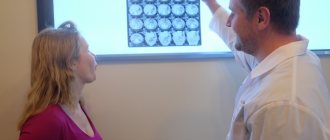
Diagnostics
Diagnosis of the cervical spine in Israel takes 3 days. During this time, doctors manage to establish a diagnosis and prescribe a set of treatment measures.
First day – consultation with a doctor
Diagnosis of any disease in Top Ikhilov begins with a consultation with the attending physician. It is noteworthy that you can get a second opinion from an Israeli specialist without visiting Israel. To do this, you will need to send diagnostic data to our clinic, after which you will be scheduled for a video consultation with our doctor. If after the video consultation you decide to be treated in our clinic, then the initial in-person consultation will be free for you.
During the initial examination, the doctor examines medical documents in detail, asks the patient about the symptoms of the disease and examines the patient. Then the doctor refers the patient to undergo instrumental examinations of the cervical spine.
Second day – instrumental diagnostics
- X-ray studies of the cervical spine.
- CT and MRI.
- Myelography.
- Electromyography.
- Electroneurography.
- Other research methods.
Third day – doctors’ conclusions
On the third day of diagnostic activities, an expert group of doctors Top Ichilov studies the diagnostic data in detail, establishes an accurate diagnosis and determines the treatment tactics for cervical osteophytes.
Possible consequences
During the development of thoracic osteochondrosis, various complications may appear regarding internal organs. Most often, the development of diseases of the heart and blood vessels is noted. This is due to irritation of nerve receptors in the chest and neck area.
Occasionally the appearance of:
- disturbances in the functioning of the duodenum;
- problems with digestion;
- dyskinesia in the gallbladder.
To avoid the development of such health problems, treatment for osteochondrosis of the thoracic spine should be started on time.
Advantages of treatment at Top Ikhilov
- Qualified doctors with extensive experience.
- Advanced medical equipment for the diagnosis and surgical treatment of spinal diseases.
- Minimally invasive and robotic spine surgeries, after which patients recover in a matter of days.
- Loyal pricing policy, the possibility of stage-by-stage payment for each service performed.
- Extensive experience working with foreign patients. Availability of an international department and a staff of professional translators (if necessary).
- Accompanying a foreign patient at all stages of diagnosis and treatment of the disease.
- The opportunity to combine treatment with a holiday in Israel.
- 5
- 4
- 3
- 2
- 1
(8 votes, average: 4 out of 5)
Thoracic osteochondrosis: symptoms, sensation, factors in the formation of the disease
Let's take a closer look at the symptoms and treatment methods for osteochondrosis of the thoracic spine. Most often, the formation of the disease in the thoracic region is caused by insufficient load on the muscles (physical inactivity), which causes excessive stress on the discs between the vertebrae.
The formation of osteochondrosis in the chest can begin under the influence of the following factors:
- bad habits;
- scoliosis and poor posture;
- genetic predisposition;
- sedentary lifestyle;
- frequent significant physical activity;
- history of spinal injuries;
- great stress and psychological stress.
The functions of the discs between the vertebrae in the sternum are equally negatively affected by increased physical activity and lack of movement.
GOUT
Gout. It is considered as a metabolic disease and is characterized by paroxysmal joint pain. Along with acute attacks of pain, gout is characterized by the deposition of salts (sodium urate) in the tissues, resulting in the formation of gouty nodules. In addition to uric acid, they contain a small amount of lime.
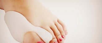
Favorite places for salt deposition are the auricles, cartilaginous covers of articular surfaces, especially the metatarsophalangeal joint of the big toe, wrist, and knee joints. This does not exclude the possibility of salt deposits in other joints. In addition to cartilage, the process affects the synovial membrane, periosteum, and tendons.
The joints thus change noticeably. They become swollen, red and painful to the touch. With a long course of the disease, persistent changes in the joints may occur: the ligaments thicken, effusion appears in the joint, and it becomes painful. When moving, a slight crunching sound is heard. Gouty nodes can enlarge over time and change the shape of the joint. In addition to the joints, gout affects blood vessels, usually the vessels of the kidneys and heart; Sclerotic changes occur in the vessels.
Treatment. Patients with gout need dietary, medication and physiotherapeutic treatment. Laser therapy is performed both on the blood (in the absence of contraindications) and locally on the area of the affected joints.
How to make a diagnosis
The diagnostic search process always begins with a conversation with the patient. During it, disturbing complaints are clarified, they are asked about their intensity, and they are interested in whether there have been any injuries in the past. Also at the appointment, an examination of the affected area is required. The doctor does not ignore changes in the configuration of the joint; he notes the presence of swelling, signs of redness and other negative changes.
Instrumental techniques play a significant role in the diagnosis of bone defects.
- Radiography. The main diagnostic option. Allows you to see characteristic non-anatomical outgrowths that look like spines and hooks. A. X-ray of the ankle.
- CT. A method to understand what structure osteophytes have. It also helps in detecting pathology at the initial stage, when the X-ray picture may not be enough to make a diagnosis.
- MRI. Used in the diagnosis of osteophytes of the ankle joint, excluding controversial cases.
b. MRI of the ankle joint.
General symptoms and characteristic signs
- pain; may be dull, pressing or stabbing in nature;
- impaired mobility of a limb or back, which will develop gradually over several weeks or even months;
- deformation;
- edema.
At the very beginning of the development of pathology, a person does not feel pain, so he is in no hurry to see a doctor. Only as the disease progresses, when a pronounced degenerative process and destruction of cartilage is observed, will the patient experience the first unpleasant sensations.
The patient feels a stabbing or aching pain, which will worsen even more when the osteophytes compress the nerve endings. Pain may occur during movement. Less commonly, it appears when coughing or sneezing.
When osteophytes grow to a sufficiently large size, joint mobility is impaired due to blocking of articular movements by bone formations.
Due to thickening of the joint capsule, contracture is observed. The patient can no longer move normally, especially if osteophytes develop in the joints of the legs.
In advanced disease (at the third stage of osteoarthritis), a person develops joint deformity due to a pronounced increase in osteophytes, which take on the load of the joint itself. Total destruction of cartilage occurs.
- edema;
- skin redness;
- gait disturbance, lameness (if the knees, femurs, feet are affected).