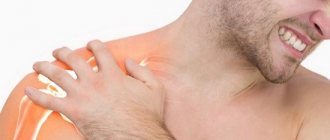
Back pain manifests itself in different ways, but it always causes discomfort to a person and negatively affects the ability to work. Taking painkillers can temporarily dull the discomfort, but will not get rid of the cause of its occurrence, which may be associated with both diseases of the spine and diseases of the internal organs. Any pathological processes can lead to disorders that seriously impair the quality of life. A comprehensive Check-up “Back Pain” examination will help you avoid serious health consequences and start treatment for back diseases on time. In-depth diagnostics make it possible to establish the cause of pain and undergo treatment according to an individual program.
Get specialist advice or make an appointment
+7 (499) 400-47-33
Causes
People aged 40+ are at risk. But some factors influence the appearance of the problem even in young years. The list of causes of the disease includes:
- obesity;
- sedentary lifestyle;
- suffered spinal injuries with displacement;
- scoliosis and flat feet;
- pregnancy;
- constant load on the spine.
Congenital anomalies of the spinal ridge, oncology in this area, and prolonged stress contribute to the development of the disease. Regardless of the factor, our clinic’s specialists will select the optimal treatment plan for spinal osteochondrosis and help you get rid of the disease.
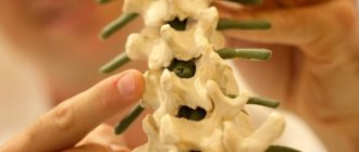
CT scan of the spine shows:
- Texture, structure, density of bone tissue. Any damage, tumors and pathological changes (including spinal osteochondrosis, metastases);
- Spinal canal and intervertebral foramina;
- Vertebral processes;
- Spinal cord.
What diseases will a CT scan of the spine show, and in what cases is it better to do an MRI? Let's look at it in this article.
Stages of development
Initially, changes in the spine do not cause problems, but over time, the functioning of all body systems occurs. The disease has 4 stages:
1. The standing height of the diseased intervertebral disc decreases. Cracks in the annulus fibrosus occur. Pathology does not go beyond one component.
2. The muscles and ligaments attached to the disc come closer together. They lose tone and sag. The defect affects nearby vertebrae. They gain extra mobility. The vertebral segment becomes increasingly less stable. The vertebrae can shift and slip, causing spondylolisthesis.
3. Unnatural protrusion occurs, the development of arthrosis and dislocations in the intervertebral discs.
4. The affected parts try to adapt to new conditions, get rid of excess mobility using the body’s strength and protect the spine. As a result, osteophytes are formed - bone growths. They injure the nerve roots of the spinal cord.
The last stage, in the absence of treatment for spinal osteochondrosis, manifests itself in the form of severe aching pain in the problem area, but then the pathology takes on a hidden form. And the patient stops feeling discomfort. A bone shell is formed on the vertebrae, which “walls up” the entire motor segment.
Neurological manifestations of back pain: problems and solutions
M.V.Putilina
Department of Neurology, Federal University of Internal Medicine, Russian State Medical University, Moscow
Neurological manifestations of back pain (dorsalgia) account for 71-80% of all diseases of the peripheral nervous system. Dorsalgia is characterized by a chronic course and periodic exacerbations of the disease, in which various pain syndromes are leading.
Moreover, according to a number of researchers, in 80% of cases acute pain regresses on its own or as a result of treatment within 6 weeks, but in 20% of cases it takes a chronic course [1, 2]. Hereditary predisposition, microtrauma, and incorrect motor stereotype lead to degeneration of the vertebral motor segment.
The degenerative process may involve various structures of spinal motion segments: intervertebral disc, facet joints, ligaments and muscles. Tissue deformations arising under the influence of static-dynamic loads are the cause of constant irritation of pain receptors.
In cases of concomitant damage to the spinal roots or spinal cord, focal neurological syndromes may appear [3, 4]. In dorsalgia, the determining factor is the appearance of severe pain syndromes associated with irritation of the nerve endings of the sinuvertebral nerves located in the soft tissues of the spine [5].
Currently, the pathogenesis of spinal pathology as the main cause of pain has been well studied, but many problems associated with dorsalgia have not been solved [6, 7].
Clinical manifestations
The initial stage of degenerative-dystrophic diseases of the spine has scanty clinical signs. Patients complain of moderate pain in the corresponding part of the spine, which occurs or intensifies with movement, changes in statics (flexion, extension, rotation), physical activity, and prolonged stay in one position [8].
During this period, it is difficult to make a correct diagnosis and prescribe adequate therapy; very often, doctors’ prescriptions are limited to the use of anti-inflammatory drugs and analgesics [6, 9]. Currently, there is an obvious overdiagnosis of spinal pathology as the main cause of pain. The role of myofascial syndromes in the origin of pain is usually underestimated (from 35 to 85% of the population suffers) [10]. The essence of myofascial pain syndrome is that the muscle suffers primarily, and not after morphological or functional disorders in the spine. Any muscle or muscle groups can be involved in the pathological process. Muscle spasm leads to increased stimulation of the nociceptors of the muscle itself. A spasmed muscle becomes a source of additional nociceptive impulses (the so-called vicious circle “pain - muscle spasm - pain”).
Several years after the first exacerbation, the pain has a clear localization, patients note heaviness, stiffness and stiffness in the affected segment of the spine, and there is pronounced tension in the back muscles. Subsequently, periods of activation of the process are observed more often and become longer [11, 12]. In cases of recurrent course, clinical manifestations during the exacerbation period are characterized by severe pain, sharp symptoms of tension, due to which the patient is unable to care for himself. During the period of regression, neurological manifestations begin to decrease, but the pain continues to be intense, there remains a significant limitation in the range of motion in the corresponding part of the spine, and the symptoms of tension are less pronounced than in the acute stage. As a rule, the patient is not able to fully care for himself and cannot perform work. During the period of incomplete remission, the pain is moderate, sometimes intermittent, the limitation of the range of motion of the corresponding area of the spine can be significant, a forced posture remains, the patient is able to care for himself, but his ability to work is limited. During the period of complete remission, periodic mild pain and a slight limitation in the range of motion of the corresponding area of the spine, the absence of tension symptoms, are noted, while the patients’ ability to work is preserved.
Clinically, the disease manifests itself in the form of a reflex syndrome (occurs in 90% of cases) and compression syndrome (detected in 5-10% of cases) [2, 4]. Reflex syndromes arise due to irritation of pain receptors (nociceptors) of the posterior longitudinal ligament as a result of one or more pathological factors and are accompanied by reflex blocking of the corresponding vertebral motor segment due to muscle tension (in particular striated muscles) with the creation of a muscle “corset”. Compression syndromes are caused by the mechanical effect of a hernial protrusion, bone growths or other pathological structure on the roots, spinal cord or arteries. Compression syndromes, in turn, are divided into radicular (radiculopathy), spinal (myelopathy) and neurovascular (for example, vertebral artery syndrome).
Problems
The lack of sufficiently effective care for patients with spinal diseases, which are usually chronic in nature, with alternating remissions and exacerbations, leads to a loss of confidence in the doctor [13]. In this case, the following problem arises - the lack of doctor-patient interaction and the latter’s confidence in the incurability of his disease. The passivity of the doctor is unacceptable, as it can lead to the psychosocial death of the patient long before his biological death. At the same time, the task of conducting adequate outpatient treatment is of particular importance due to the fact that currently used methods do not always take into account etiopathogenetic factors, the characteristics of sanogenetic reactions in a particular patient, often lead to “failure of compensatory reactions” [8] and worsen the process of rehabilitation events [4]. To solve this problem, first of all, it is necessary to remember that back pain can be both primary, associated with degenerative changes in vertebral structures, and secondary, caused by pathological conditions. Therefore, the main task of the doctor when examining a patient with acute back pain is to separate musculoskeletal pain from pain syndromes associated with somatic or oncological pathology.
Diagnostics
Diagnosing the neurological manifestations of back pain is a difficult task for a doctor, but with the proper use of additional examination methods, it can be easily solved. We cannot abandon traditional methods of X-ray diagnostics and neuroimaging methods (computer and magnetic resonance imaging), laboratory tests (general blood and urine tests, biochemical tests) [6]. In case of difficulties, electroneuromyographic studies are used: lesions of the peripheral neuron corresponding to a given nerve and a decrease in the speed of impulse transmission along the nerve distal to the place of its compression are determined.
Therapy
Treatment of degenerative-dystrophic diseases of the spine is one of the most pressing problems of modern neurology. In patients with this type of pain syndrome, without identifying the pathophysiological mechanisms, it is impossible to choose the optimal treatment strategy. When determining treatment tactics, it is necessary to take into account the localization, nature and severity of clinical manifestations of pain. In recent years, the pharmacological arsenal of treatments for patients with vertebrogenic pathology has significantly improved [1, 14]. However, the problem of back pain is still far from being solved. Drug treatment of neurological manifestations of dorsalgia is a complex task that requires deep knowledge of the pathogenesis and clinical manifestations of the disease. Treatment of patients should be comprehensive, using medications and non-drug therapy methods.
Principles
The main principles of drug therapy are early initiation, pain relief, and a combination of pathogenetic and symptomatic therapy. Therapeutic measures differ in the acute and interictal periods of the disease. First of all, measures are taken to relieve or reduce pain [1, 3].
In case of acute pain, it is necessary to recommend the patient to bed rest for 1-3 days. Drug therapy should be started immediately in the form of nonsteroidal anti-inflammatory drugs (NSAIDs), analgesics, and muscle relaxants, since the first and main task is rapid and adequate pain relief [9, 15]. When treating acute back pain, significant regression of pain should be expected within 1-2 weeks.
The long-standing policy of limiting physical activity, up to strict bed rest, has now been somewhat revised: for moderate pain, partial limitation is recommended, and for severe pain, the period of bed rest is reduced to 1-3 days. In this case, the patient must be taught the “correct” motor behavior: how to sit, how to stand up, how to walk, not to carry heavy objects, etc. If therapy is ineffective, other drugs in optimal doses can be tried within 1-2 weeks. Persistent pain for more than 1 month indicates a chronic process or an incorrect diagnosis of back pain.
The presence of compression syndrome is an indication for the prescription of anti-ischemic drugs: antioxidants, antihypoxants, vasoactin drugs. The issue of using antidepressants is decided individually for each patient.
NSAIDs
NSAIDs remain the first choice for pain relief. The main mechanism of action of NSAIDs is inhibition of cyclooxygenase (COX)-1, 2, a key enzyme in the arachidonic acid metabolic cascade, leading to the synthesis of prostaglandins (PGs), prostacyclins and thromboxanes [9, 14]. Due to the fact that COX metabolism plays a major role in the induction of pain at the site of inflammation and the transmission of nociceptive impulses to the spinal cord, NSAIDs are widely used in neurological practice. All anti-inflammatory drugs have anti-inflammatory, analgesic and antipyretic effects, are able to inhibit the migration of neutrophils to the site of inflammation and platelet aggregation, and also actively bind to serum proteins.
Features of the action
Differences in the action of NSAIDs are quantitative, but they determine the severity of the therapeutic effect, tolerability and side effects in patients. The high gastrotoxicity of NSAIDs, which correlates with the severity of their sanogenetic effect, is associated with non-selective inhibition of both COX isoforms. Currently, there are two groups of NSAIDs depending on their effect on COX. Non-selective NSAIDs block both constitutional COX-1, which is associated with the gastrointestinal side effects of these drugs, and inducible COX-2, the formation of which activates anti-inflammatory cytokines. Selective drugs act primarily on COX-2. Non-selective NSAIDs include: lornoxicam (Xefocam), ibuprofen, indomethacin.
Selective COX inhibitors include: nimesulide (Nise®), meloxicam, celecoxib.
Complications
At the same time, the use of NSAIDs is associated with a wide range of side effects, the risk of which seriously reduces their therapeutic value, primarily the problem of the negative impact of these drugs on the gastrointestinal tract (GIT), the development of NSAID gastropathy. In patients regularly taking NSAIDs, the risk of developing these complications is 4 or more times higher than in the population, and amounts to 0.5-1 cases per 100 patients. Moreover, according to long-term statistical data, every 10th patient who develops gastrointestinal bleeding while taking NSAIDs dies [12, 16].
In 20-30% of patients taking NSAIDs in the absence of significant damage to the gastrointestinal mucosa, various dyspeptic symptoms appear - gastralgia, nausea, a feeling of “burning” or “heaviness” in the epigastrium, etc. In addition, specific side effects of NSAIDs include increased the risk of developing cardiovascular accidents - myocardial infarction and ischemic stroke. However, the benefits of using NSAIDs as an effective and affordable treatment for dorsalgia significantly outweigh the harm associated with the risk of developing dangerous complications. First of all, this is due to the fact that specific complications can be successfully prevented. Most side effects occur in people with so-called risk factors. For NSAID gastropathy, this is age over 65 years, a history of peptic ulcers (the greatest danger is observed in patients who have previously suffered gastrointestinal bleeding), as well as concomitant use of drugs that affect the blood coagulation system [17]. Risk factors for cardiovascular complications are comorbid diseases of the heart and blood vessels - coronary heart disease, arterial hypertension not compensated by treatment. Taking these factors into account and using adequate preventive measures (prescribing proton pump inhibitors if there is a risk of developing NSAID gastropathy) can significantly reduce the risk of developing NSAID-associated complications.
Choosing a doctor
In recent years, on the Russian pharmacological market, a huge number of original drugs have been supplemented by an order of magnitude larger number of generics. It is not easy for a practicing doctor to make a choice among this variety, given the aggressive advertising, as well as the abundance of heterogeneous and sometimes biased information. When choosing an NSAID, your doctor should consider the following:
- Price/quality ratio of drugs.
- The spectrum of action of drugs in various forms of release.
- Background diseases of the patient.
- Patient finances.
Nise®
One of the most successful drugs available on our pharmacological market is the drug Nise® (nimesulide) produced by Dr. Reddy's Laboratories Ltd. (India). The drug is used for rapid relief of moderate or severe acute pain, has anti-inflammatory, analgesic and antipyretic effects [15]. Reversibly inhibits the formation of PGE2 both at the site of inflammation and in the ascending pathways of the nociceptive system, including the pathways of pain impulses in the spinal cord. Reduces the concentration of short-lived PGN2, from which PGE2 is formed under the action of prostaglandin isomerase.
A decrease in the concentration of PGE2 leads to a decrease in the degree of activation of EP-type prostanoid receptors, which is expressed in analgesic and anti-inflammatory effects. It has a slight effect on COX-1 and practically does not interfere with the formation of PGE2 from arachidonic acid under physiological conditions, thereby reducing the number of side effects of the drug. Nise® suppresses platelet aggregation by inhibiting the synthesis of endoperoxides and thromboxane A2, and inhibits the synthesis of platelet aggregation factor. It also reduces the release of histamine and reduces the severity of bronchospasm caused by exposure to histamine and acetaldehyde [15, 16, 18-21]. The drug inhibits the release of tumor necrosis factor a, which causes the formation of cytokines. It is able to slow down the synthesis of interleukin-6 and urokinase, thereby preventing the destruction of cartilage tissue. Blocks the synthesis of metalloproteases (elastase, collagenase), preventing the destruction of proteoglycans and collagen of cartilage tissue. It has antioxidant properties and inhibits the formation of toxic oxygen breakdown products by reducing the activity of myeloperoxidase. Interacts with glucocorticoid receptors, activating them through phosphorylation, which also enhances the anti-inflammatory effect of the drug.
Properties and effect
Due to its high bioavailability, already 30 minutes after oral administration, the concentration of the drug in the blood reaches ~50% of the peak, and a clear analgesic effect is noted. After 1-3 hours, the peak concentration of the drug occurs and, accordingly, the maximum analgesic effect develops [19-21]. When applied topically, it causes a weakening or disappearance of pain at the site of application of the gel, including pain in the joints at rest and during movement, reduces morning stiffness and swelling of the joints and helps to increase range of motion. The drug is effective both for short-term relief of acute dorsalgia and for long-term treatment of chronic pain syndrome over many months.
A study was conducted in Finland in which 102 patients with acute back pain received nimesulide 100 mg 2 times a day or ibuprofen 600 mg 3 times a day for 10 days. Nimesulide was superior to the control drug in terms of pain relief and effect on spinal function. Moreover, among patients receiving nimesulide, side effects from the gastrointestinal tract occurred in only 7%, and among those taking ibuprofen - in 13% [18]. Scientists have concluded that nimesulide is superior in its tolerability to traditional NSAIDs, since it relatively rarely causes dyspepsia and other gastrointestinal complications. The most important advantage of the drug Nise® is its affordable price and good tolerability, proven by a series of post-registration studies [17]. Thus, when using nimesulides, the frequency of such common side effects as ulcers of the upper gastrointestinal tract and drug-induced liver damage, and the cardiotoxic effect that some highly selective NSAIDs are “burdened” are minimized. This allows us to recommend Nise® for use in general medical practice. Moreover, this drug has several dosage forms. Nise® is available in tablet and form for topical use, which allows you to individualize and optimize the targeted therapeutic effect of the drug. Since the drug on a gel matrix penetrates tissues quickly and in greater concentration, it is possible to combine local and systemic forms of the drug to achieve a better (greater) therapeutic effect.
In conclusion, we note that the problems of neurological manifestations of back pain are still far from a final solution, but their further study will make it possible to develop new strategies for the diagnosis and treatment of dorsalgia.
Literature
- Alekseev V.V. Treatment of lumbar ischialgic syndrome. RMJ. 2003; 11 (10): 602-4.
- Voznesenskaya T.G. Pain in the back and limbs. Pain syndromes in neurological practice. Ed. A.M.Veina. M.: Medpress, 1999; With. 217-83.
- Popelyansky Y.Yu., Shtulman D.R. Diseases of the nervous system. Ed. N.N.Yakhno, D.R.Shtulman. M.: Medicine, 2001; With. 293-316.
- Shtulman D.R., Levin O.S. Neurology. Handbook of a practicing physician. M.: Medpress-inform, 2002; With. 70-90.
- Kuznetsov V.F. Vertebroneurology. Clinic, diagnosis, treatment of spinal diseases. M., 2004.
- Levin O.S. Diagnosis and treatment of neurological manifestations of spinal osteochondrosis. Cons. Med. 2005; I (6): 547-55.
- Borenstein D. Epidemiology, etiology, diagnostic evaluation and treatment of low back pain. Intl. honey. magazine 2000; 35: 36-42.
- Bogduk N, Mc Guirk B. Medical management of acute and chronic low back pain. Amsterdam: Elsevler, 2002.
- Nasonov E.L. Non-steroidal anti-inflammatory drugs (prospects for use in medicine). M., 2000.
- Belova A.N. Myofascial pain. Neurol. magazine 2000; 5 (5): 4-7.
- Gatchel RJ, Gardea MA. Lower back pain: psychosocial issues. Their importance in predicting disability, response to treatment and search for compensation. Neurologic clinics 1999; 17: 149-66.
- Porter R.W. Management of Back Pain. Second edition. Chuchill Livigstoner Longman group UK Limited. 1993.
- Podchufarova E.V. Chronic back pain: pathogenesis, diagnosis, treatment. RMJ. 2003; 11 (25): 1295-401.
- Nasonov E.L., Tsvetkova E.S. Selective cyclooxygenase-2 inhibitors: new prospects for the treatment of human diseases. Ter. arch. 1998; 5:8-14.
- Balabanova R.M., Belov B.S., Chichasova N.V. and others. The effectiveness of nimesulide in rheumatoid arthritis. Pharmateka. 2004; 7:5-8.
- Bennett A. Nimesulide is a well established cyclooxygenase-2 inhibitor with many other pharmacological properties relevant to inflammatory diseases. In: Therapeutic Roles of Selective COX-2 Inhibitors. Ed. JRVein, RMBotting. William Harvey Press; p. 524-40.
- Karateev A.E., Yakhno N.N., Lazebnik L.B. etc. Use of non-steroidal anti-inflammatory drugs. Clinical recommendations. M.: IMA-PRESS, 2009.
- Pohjolainen T, Jekunen A, Autio L, Vuorela H. Treatment of acute low back pain with the COX-2-selective anti-inflammatory drug nimesulide: results of a randomized, double-blind comparative trial versus ibuprofen. Spine 2000; 25 (12): 1579-85.
- Pelletier JP, Mineau F, Fernandes JC et al. Two NSAIDs, nimesulide and naproxen, can reduce the synthesis of urokinase and IL-6 while increasing PAI-1, in human OA synovial fibroblasts. Clin Exp Rheumatol 1997; 15: 393-8.
- Rainsford K. Current status of the therapeutic uses and actions of the preferential cyclo-oxygenase-2 NSAID, nimesulide. Inflammopharmacology 2006; 14 (3, 4): 120-37.
- Wober W, Rahlfs V, Buchl N et al. Comparative efficacy and safety of the non-steroidal anti-inflammatory drugs nimesulide and diclofenac in patients with acute subdeltoid bursitis and bicipital tendinitis. Int J Clin Pract 1998; 52 (3): 169-75.
Source consilium-medicum Neurology No. 1/2011
Cost of treatment for spinal osteochondrosis
| Services list | Price, rub |
| Appointment (examination, consultation) with an orthopedist - traumatologist, chiropractor, osteopath, kinesiotherapist, test session | 1 600 |
| Initial appointment (examination, consultation) with a neurologist, Doctor of Medical Sciences | 2 200 |
| Kinesi taping 1 zone | 1 100 |
| Magnetic Vacuum Acupuncture | 1 700 |
| Intra-articular Injection (diprospan) | 4 000 |
| Intra-Articular PRP Injection | 6 500 |
| PRP therapy for tendonitis, ligamentitis, contractures | 6 500 |
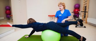
| Kinesitherapy | |
| Individual lesson with an instructor | 2 700 |
| Individual lesson with a doctor | 3 900 |
| Physiotherapy | |
| HIVAMAT session up to 15 min, 1 unit. body | 2 200 |
| HIVAMAT session up to 15 min, 2 units. body | 3 200 |
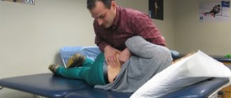
| OSTEOPATHY AND MANUAL THERAPY | |
| Manual therapy 1 department (15 min.) | 2 700 |
| Soft manual techniques (45-60 min.) | 7 500 |
| Osteopathic techniques (30-75 min.) | 7 500 |
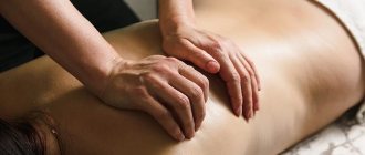
Cost of services in the direction of “Medical massage”
| Services list | Duration | Price, rub |
| Back + Legs Massage | 55 min. | 3700 |
| Therapeutic back massage | 25-40 min. | 3000 |
| Neck massage | 15 minutes. | 2200 |
| Massage of the thoracic spine | 15 minutes. | 2200 |
| Massage of the lumbosacral spine | up to 30 min. | 2200 |
| Massage of the collar area + one upper limb (arm) | 30 min. | 3000 |
| Neck massage + two upper limbs | 40 min. | 3900 |
| Neck massage + chest massage | 40 min. | 3900 |
| Massage of one upper limb | 15 minutes. | 2200 |
| Massage of two upper limbs | 30 min. | 3000 |
| Chest massage | 15 minutes. | 2200 |
| Massage of the lumbosacral spine + one lower limb (leg) | 30 min. | 3000 |
| Massage of the lumbosacral spine + two lower limbs | 45 min. | 3900 |
| Massage of one lower limb | 20 minutes. | 2200 |
| Massage of two lower limbs | 30 min. | 3000 |
| Cupping massage (dynamic setting) | 25 min. | 2200 |
| Therapeutic back massage + cupping massage | 40 min. | 3900 |
| General therapeutic massage | 120 min. | 6900 |
| Modeling massage (for weight loss) (Thighs, Buttocks, Abdomen) | 45 min. | 5800 |
| Anti-cellulite full body massage | 90 min. | 7200 |
| Head massage | 20 minutes. | 2200 |
| Neck and collar area massage | 20 minutes. | 2200 |
Contact us
Call now
8 (495) 803-27-45
Make an appointment through our service
Make an appointment
Symptoms of osteochondrosis
With degenerative-dystrophic tissue damage, abrasion of the vertebrae themselves, articular surfaces and intervertebral discs occurs. The disease manifests itself as back pain, but it can spread to other parts of the body. There are discomfort in the legs, numbness in the fingers and hands, pain in the genital area, head, and heart.
More often in women, the disease manifests itself as pain in the lumbar region. But often a problem is diagnosed in the neck, chest, or several areas at the same time. Activity decreases and fatigue increases, both mental and physical. The sensitivity of the upper and lower extremities is impaired.
With pathology of the cervical spine, the most common symptoms are headache, a sharp drop in vision and eye fatigue. Damage to the lumbosacral region threatens dysfunction of the reproductive system.
Classification of spinal diseases
All diseases of the spinal column are classified into two categories: the type of changes and location.
Depending on the nature of the changes, these could be:
- degenerative diseases of the spine (osteochondrosis, spondylosis and others);
- abnormal tissue development;
- spinal injuries;
- the occurrence of neoplasms;
- inflammation;
- changes affecting intervertebral discs (protrusion, hernia).
Diseases are classified according to location:
- cervical region;
- thoracic region;
- lumbar region.
In each of these departments, a variety of changes can occur, leading to the development of diseases.
Consequences
Lack of proper attention to the problem in the early stages threatens serious disorders in all body systems. The patient experiences severe pain, a feeling of a heart attack, dizziness, nausea, and chills. In advanced cases, spinal stroke, brain damage, and the development of vascular and heart pathologies may occur.
It is important to consult a doctor at the first symptoms of osteochondrosis for treatment without medications. We invite you to our Innovative Medical Center. Uncontrolled use of painkillers will only worsen the situation.
What can be seen with MSCT of the spine?
MSCT of the spine
MSCT examination of the spine allows you to detect:
- changes in tissue density and structure. This is how neoplasms (tumors) and intervertebral hernias are detected;
- violations of the integrity or displacement of the vertebrae;
- bone growths.
MSCT is significantly superior to classical radiography in terms of the quality of the resulting image and is the best method for visualizing bone structures. MSCT is also used to identify pathologies of the spinal cord, but MRI is considered the most informative examination of soft tissues. When examining the spine, these methods (MSCT and MRI) can complement each other, allowing the doctor to assess the condition of the vertebrae, intervertebral discs, spinal cord and soft tissues surrounding the spine.
Diagnostics
To identify the problem and prescribe the most effective method of treating osteochondrosis, an anamnesis is collected. All patient complaints are taken into account. A visual inspection is carried out. It includes:
- body position assessment;
- examination of the skin;
- palpation and tapping of areas where the patient complains of pain;
- determination of skin sensitivity.
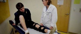
Next, an examination of the most affected part of the spine is prescribed. The patient is referred for an X-ray examination, computed tomography (CT) or magnetic resonance imaging (MRI). With the help of procedures, the degree of damage to the back area, the condition of the nerve processes, and blood vessels are revealed.
The Innovative Medical Center also uses ultrasound to diagnose problems. Ultrasound is informative, safe and considered a less expensive way to detect pathology.
MRI of the spine: how the procedure works
Before the examination, you must remove all metal accessories, watches, and place bank or other magnetic cards. The patient is placed on a special table that will be located inside the scanner. During the procedure, you need to maintain a stationary position for better image quality.
Equipment for MRI
The doctor retires to the next office, but maintains contact with the patient via two-way communication and sees him on the monitor. The scanner maintains a comfortable temperature, good air circulation and lighting. You can use headphones with pleasant music.
After the procedure, you can relax and drink coffee while waiting for a description of the images. This will take 15-20 minutes.