High stress, excess weight and unbalanced nutrition are just some of the factors that provoke the development of joint dysplasia in dogs. This is a common disease accompanied by gradual atrophy of the muscles and limbs of pets. To learn how joint dysplasia develops and is treated, see below.
Read in this article
- What is this?
- Main causes of joint dysplasia
- Risk groups: what you need to know?
- What symptoms occur with dysplasia?
- Development of dysplasia: stages
- Diagnosis of the disease in dogs
- Treatment of joint dysplasia
- Surgery
- Prevention of dysplasia
What is this?
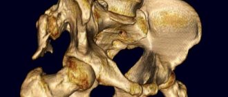
Joint dysplasia is the abnormal formation and development of joints, leading to impaired mobility and degenerative changes. In the initial stages, the disease entails deformation of the pet’s joint and then bone tissue.
An incorrectly formed or damaged joint, when rubbed, “erases” the cartilage tissue, causing severe pain. Gradually, the process affects the health and strength of the bones, preventing the dog from fully moving and leading an active lifestyle.
Note! According to statistics, dysplasia usually affects the hip joints. This is due to the fact that they bear the greatest load when running and jumping.
Main causes of joint dysplasia
Joint dysplasia in dogs is a common disease.
It requires timely diagnosis and effective treatment in a veterinary clinic. The first thing to do is to identify the cause of the disease. Among the most common:
- Heredity
Pets suffering from dysplasia have offspring that already have congenital damage to the hip and sometimes elbow joints. The genetic cause is detected in 70-75% of cases of disease, therefore it is the most common.
- Intense physical activity
In combination with an ill-designed regimen, they lead to overload: skeletal tissue does not have time to develop as quickly as the muscle corset is formed. All this provokes deformation of the surface of the joints and their damage.
- Physical inactivity
In some cases, the cause of the development of dysplasia is low mobility and constant keeping of the pet in an enclosure. Hypodynamic syndrome, combined with excess weight, places additional stress on the supporting apparatus.
- Poor nutrition
Lack of vitamin D, calcium, magnesium, as well as a number of essential amino acids - all this leads to disruption of mineral metabolism not only in bone, but also in connective tissue. Result: gradual damage and deformation of the joints.
Note! Another dangerous factor that provokes the development of dysplasia and is included in this group is an excess of phosphorus in the diet.
- Injuries, dislocations and sprains
A fracture or other injury can trigger the development of the disease. This cause is quite rare and usually results in the formation of a rare type of stifle dysplasia in dogs of any weight.
Features of nutrition of a sick animal
Most pets with dysplasia suffer from obesity, so they must be put on a low-calorie diet with a weekly fasting day. When reviewing your current diet, you must:
- Give up economy-class dry food or improve the quality of natural products.
- Exclude prohibited foods from the menu (sweets, smoked foods, pickles, canned food, flour).
- Make sure you have adequate protein intake.
- Reduce your usual portions of food to smaller amounts that allow you to lose excess weight.
With natural feeding, it is recommended to introduce dietary supplements into the diet, and with dry feeding, choose veterinary food with chondroprotectors.
Risk groups: what you need to know?
Heredity is a common, but not the only cause of joint dysplasia. The disease can be transmitted not only genetically, but arises and develops gradually. The following dog breeds are particularly susceptible to dysplasia:
- Saint Bernards,
- retrievers,
- newfoundlands,
- east european shepherd dogs,
- Mastino-Neapolitan,
- rottweilers,
- labradors,
- German Shepherds.
Note! All of the above breeds are characterized by large body weight, increased growth and, as a rule, intense physical activity.
Dysplasia occurs and develops at any age, but most often owners of puppies from 6 to 12 months turn to veterinarians. However, it will be possible to determine the degree of development of the disease in giant breed dogs only at 18 months.
If you are the owner of a large breed puppy who is growing quickly and is overweight compared to his peers, is very active and likes to play on slippery floors (ice), then he is very likely to develop hip dysplasia.
The only sure way out if you suspect joint dysplasia in a dog is to contact a specialized veterinary clinic in Moscow.
Consultation with a pediatric orthopedist
Treatment of congenital anomalies of the limbs is surgical. Parents should take this issue extremely responsibly, understanding the importance of early seeking medical help. Often, the outcome of treatment is influenced by the age of the child at which the operation was performed and the qualifications of the surgeon. Regardless of the type of congenital defect, medical consultation should be obtained as early as possible, in the first months of the child’s life. At the CONSTANTA Clinic in Yaroslavl you can make an appointment with an experienced hand surgeon, pediatric traumatologist-orthopedist Torno T.E.
During the consultation, the doctor:
- carefully examine the child (pathological changes are noticeable already in the first days of the baby’s life);
- determine the severity of the deformation;
- select the tactics of medical treatment;
- will calculate the exact cost of medical care.
What symptoms occur with dysplasia?
The first thing you should pay attention to is your pet's activity. If earlier your puppy willingly participated in fun, games and walks, now he quickly gets tired and increasingly tries to rest. Many owners note that their dogs, even in the initial stages of the disease, are afraid to go down stairs or, conversely, to go up them. Some puppies experience lameness, but this goes away with rest.
10 symptoms, the appearance of which indicates the development of joint dysplasia in dogs and requires immediate contact with a veterinarian:
- reducing the pet’s physical activity and avoiding long walks in the fresh air with the owner,
- a sick animal increasingly lies on its side, so it is almost impossible to see it while lying on its stomach,
- even after a full sleep and a long rest, it is difficult for the pet to get up, he may wheeze,
- when forced to run, jump and even walk, you can notice the extension of the hind limbs (“rabbit running”),
- stiffness occurs in any, even the simplest movements (for example, it is difficult for a pet to even give a paw),
- the affected hip, elbow or knee joints swell and swell, they are painful when palpated,
- the dog often lies down, stretches out its paws and turns them in different directions so as not to feel severe pain,
- in puppies there are seals in the affected joints, it is possible that asymmetry is developing that is not typical for the breed,
- the muscles of the back of the body in the last stages of dysplasia atrophy, since there is no load on them,
- the animal becomes less sociable, even simple stroking and attempts to pet the pet can cause aggression.
Have you noticed a few symptoms in your dog? Don't hesitate, contact your veterinarian for help!
Development of dysplasia: stages of the disease
One of the features of joint dysplasia is its gradual development. You can detect the disease by the strange behavior of your pet in the early stages. This will allow you to quickly contact the clinic, timely diagnose the pathology and immediately begin its treatment.
Main stages and their features:
- I degree (mild). The acetabulum flattens, but the bone still “sits” tightly, without causing severe discomfort or pain in the dog. However, there is already a decrease in the pet’s activity. Typically this stage appears at 4-6 months;
- II degree (medium). The flattening is noticeable, depressions and irregularities form on the head of the bone. The joint becomes weaker, but is still considered strong. The pet experiences pain during heavy exertion and long walks;
- III degree (severe). The acetabulum becomes flat, the head of the bone gradually flattens, and the joint begins to collapse. The dog cannot jump or run, throws back its hind legs when resting and is constantly in pain;
- IV degree (very severe). The joint is gradually destroyed, deformation of the hind limbs and muscle atrophy are observed. The paws swell greatly, the lumps are easy to feel, and the animal practically does not stand up.
An abrupt transition from one stage of dysplasia to another is impossible. Deformation of joint, muscle and bone tissue occurs gradually. In the absence of intense provoking factors, the transition from one stage to another can take from several months to several years.
Prognosis for recovery and future life
Prognosis for recovery directly depends on the degree of hip dysplasia diagnosed in the dog:
A.
A physiological norm that excludes the presence of violations.
IN
. The diagnosis is suspicious. The animal requires regular examinations, proper nutrition and adequate exercise.
WITH.
Mild degree with minor impairments. Without compliance with prevention and conservative treatment, periodic dislocations are possible, so the pet’s condition must be monitored.
D
. Pathology of moderate severity, characterized by rapid development. The method of treatment is selected individually.
E.
An advanced form of the disease, accompanied by constant excruciating pain and loss of motor function. Treatment is strictly surgical, since taking medications gives only a temporary effect.
Most often, owners seek help at the third stage (C), when the dog develops characteristic pain at the site of inflammation. The prognosis up to the fourth stage (D) is positive. If all recommendations are followed, a sick pet can live more than 10 years.
Diagnosis of the disease in dogs
Only a veterinarian can diagnose hip or other joint dysplasia in your pet. He performs a general clinical and orthopedic examination, as well as taking x-rays.
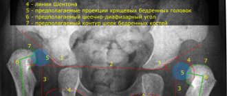
It is performed under general anesthesia so that the dog can be placed in a certain position. The specialist analyzes the received X-ray images, measures angles and calculates indices to make the correct diagnosis.
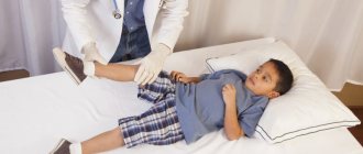
If necessary, special tests are prescribed to determine joint dysplasia. The Ortolani test (creating pressure on the knee joint) or the Bardens test (holding the fingers on the ischial tuberosity) is used.
Together with the diagnosis, the specialist determines the type of lesion of the hip joint: acetabular (in this case, the neck-shaft angle is 135°) or neck-shaft (in this case, the angle is more than 150°).
Treatment of joint dysplasia
At the initial stages of joint dysplasia, conservative treatment is prescribed. It is aimed at reducing the load placed on the musculoskeletal system and muscular frame of the animal, especially in young pets.
Be sure to monitor the dog’s activity by type (running, jumping, etc.), as well as duration. The best exercise is slow walking, using a leash is recommended. The duration of the first walk is 5 minutes, then gradually increases.
Therapeutic treatment includes:
- taking chondoprotectors (drugs aimed at restoring cartilage tissue),
- prescription of painkillers,
- restriction in physical activity,
- adding feed additives,
- weight loss (if you are overweight).
Regular monitoring of the pet's condition during conservative treatment is necessary. On the recommendation of a doctor, vitamin and mineral support is provided for an animal whose body is depleted and weakened as a result of joint dysplasia.
Surgery
If conservative therapy does not produce sustainable results and the progression of the disease in the dog continues, surgical treatment is prescribed. The form of surgical intervention is determined by a specialist, taking into account the weight of the pet, the stage of development of dysplasia and the type of joint deformity.
3 types of surgical treatment that can be prescribed:
- Resection arthroplasty
Indications: animal weighs no more than 30 kg, has symptoms of osteoarthritis, flattened femoral head. During this intervention, the flattened femoral head is removed. After the operation, injury to the acetabulum stops, and therefore pain and discomfort disappear.
- Triple pelvic osteotomy
The intervention is aimed at changing the angle of inclination of the acetabulum, which is why the development of the disease stops. However, this method has limitations: it is prohibited in the presence of complications in the form of osteoarthritis.
- Intertrochanteric osteotomy
It is used for the surgical treatment of individual cases of hip dysplasia, if the cause of its development is deformation of the hip. It has a minimum of contraindications and is characterized by a shortened rehabilitation period.
Note to out-of-towners!
You can get medical advice via Skype or email. by mail, having previously sent the necessary photo and video materials to this address (photos of the child’s hands in different projections, a small fragment of video of finger movements).
Information for out-of-town patients
What do we offer our patients?
From the first call from his parents, every little patient finds himself in a Clinic with modern equipment, competent specialists, responsive staff, sincerely interested in the success of treatment.
Accommodation conditions adapted for children
The Clinic's hospital is equipped with everything necessary for the convenience of children. The beds in the wards have functional equipment. Our employees find a special approach to each child, actively communicate with their parents, answering all questions. Specialists closely monitor the baby’s well-being and give detailed recommendations to parents on recovery after surgery. A souvenir of your stay at the Clinic will be a gift from the doctor - a toy.
Possibility of treatment for free and at your own expense
At the CONSTANTA Clinic you can get the necessary medical care:
- For free. The charitable assistance of Rusfond allows us to provide surgical treatment of congenital malformations without payment from the patient.
- At my own expense. The cost of services obtained at your own expense may vary depending on the complexity and scope of the surgical intervention.
Free personal manager services
The personal manager’s assistance includes information support and solving organizational issues that arise during the preparation and process of treating your child. Such support from a Clinic specialist significantly saves time and is provided by us free of charge.
Clinic Constant. Information for parents of young patients
More information for parents of young patients can be obtained by phone.
Anesthesia for operations in children Treatment of children with the support of the Charitable Foundation
Prevention of dysplasia
The main preventive measure is regular monitoring. Make sure that the weight of the animal does not exceed the established norm, and that the load does not affect its health. Provide your pet with a balanced diet rich in calcium and magnesium. Make sure your puppy does not play on slippery floors or ice. This will avoid injuries, sprains and dislocations that can provoke the development of the disease.
Joint dysplasia in dogs is a common disease. In the final stages and without professional treatment, it can lead to complete deformation and the animal losing its ability to move independently. Remember that the key to successful therapy is contacting experienced veterinarians!


