Juvenile idiopathic arthritis in questions and answers. Diagnostics
1. Examination by a rheumatologist. It happens that parents are offended: one child is prescribed only three tests to make a diagnosis, while another is sent for six months for research. The former believe that they were underexamined, and the latter believe that they were forced to spend a lot of money in vain. In fact, the scope of diagnosis depends on the symptoms, their duration, and the number and location of the joints involved in the disease. If a child comes to me whose symptoms of the disease last two days or only one joint is affected, it is difficult for me to immediately say whether it is JIA or not, and the range of diagnostics will be wide. On the contrary, if a child has several joints affected or the symptoms of arthritis last for several months, I need minimal examinations, more to select therapy than to make a diagnosis.
2. Laboratory tests: rheumatoid factor (RF), antinuclear factor (ANF), blood biochemistry - are of an auxiliary nature. It’s like a scale: we add pluses to the diagnosis scale – the more pluses, the easier it is to diagnose JIA. Up to half of children with JIA have completely normal blood tests and 95% have negative RF. Doctors who are not rheumatologists are sure: arthritis is inflammation, which means there should be changes in the tests. Such a child comes to the pediatrician, he is prescribed a clinical blood test, and they can also check rheumatoid factor and C-reactive protein. The result is everything is normal. What the average doctor says: “You don’t have arthritis, see an orthopedist, chiropractor, etc.” In this regard, I reassure parents: JIA with normal tests has a better prognosis than with bad tests.
3. The main marker of JIA is an increase in antinuclear factor (ANF 1:160 and above), as a sign of “breakdown, failure” in the immune system. It is important that the blood test for ANF is carried out using the immunofluorescence method (with determination of the titer and type of fluorescence). Not all laboratories are capable of performing ANF; it is often replaced with ANA (antinuclear antibodies), which is performed by enzyme immunoassay using a different, cheaper technology. But these are not interchangeable tests.
4. MRI. A clear indication for MRI is damage to one joint (true monoarthritis): we exclude trauma, surgery, orthopedic diseases, osteomyelitis, and tuberculosis. But a high-quality, comprehensive examination in some patients can reveal 2-3 more inflamed joints that the parents did not notice, and then the need for MRI sharply decreases.
5. Ultrasound is a method that can detect the presence of inflammation in the joint (fluid, thickened synovial membranes), but will not answer which type of arthritis it is. We use this method as a screening (indicative) method, as well as to assess the severity of inflammation. The result greatly depends on the qualifications of the specialist who performs the ultrasound of the joints. Ideally, this should be a rheumatologist with a certificate in ultrasound diagnostics, but such specialists can be counted on one hand. It is also important to know children’s ultrasound features of joints in normal and pathological conditions.
6. Bone marrow puncture is performed to exclude blood diseases, primarily leukemia, which may resemble systemic arthritis. Computed tomography - to search for tumors in the chest, retroperitoneum: lymphoma, neuroblastoma, to exclude tuberculosis or other lung lesions, bone lesions.
7. Diagnostic puncture of the joint - necessary to clarify the nature of arthritis, for example, the presence of pus is typical for bacterial inflammation, and blood is typical for hemarthrosis, pigmented villonodular synovitis. In these cases, the fluid is examined - culture, microscopy, tuberculosis testing. The indication for diagnostic puncture is monoarthritis.
Publications in the media
Juvenile idiopathic arthritis (JIA, juvenile rheumatoid arthritis, chronic juvenile arthritis) is a heterogeneous group of diseases united by a tendency towards a chronic progressive course. The term was proposed by the WHO Standing Committee on Pediatric Rheumatology (1994) to replace the previously used terms juvenile chronic and juvenile rheumatoid arthritis.
Statistical data. Incidence: 2–19 per 10,000 children per year. Boys and girls get sick equally often. Etiology unknown. Pathogenesis - see Rheumatoid Arthritis.
Genetic aspects. A high prevalence of Ags HLA-DRВ1*0801 and *1401 was established in patients with polyarthritis, HLA-DRВ1*0101 and 0801 in patients with oligoarthritis. The association of Ag HLA-B27 with the development of arthritis with enthesopathy, as well as HLA-DRB1*0401 with RF-positive polyarthritis, has also been proven.
CLASSIFICATION (Durban, 1997)
Systemic variant - arthritis with/or previous fever for at least 2 weeks in combination with two or more signs: • fleeting, not fixed erythematous rash • generalized enlargement of lymph nodes • hepato- or splenomegaly • serositis. Description • Age of onset of the disease • Characteristics of arthritis during the first 6 months of the disease •• oligoarthritis •• polyarthritis •• presence of arthritis only after 6 months of systemic disease • Characteristics of arthritis after 6 months of the disease •• oligoarthritis •• polyarthritis •• absence of arthritis after 6 months of systemic disease • Features of systemic disease after 6 months • Presence of RF • CRP level.
Persistent/spreading oligoarthritis is arthritis that affects 1–4 joints during the first 6 months of the disease. There are 2 subcategories: • persistent oligoarthritis (affecting no more than 4 joints during the entire period of the disease) • spreading arthritis (affecting more than 5 joints after 6 months of illness). Exclusion factors • Familial psoriasis confirmed by a dermatologist in at least first- or second-degree relatives • Family history confirming the presence of HLA B27-associated diseases in at least first- or second-degree relatives • Positive RF • HLA B27-positive boys with onset illnesses after 8 years • Presence of systemic arthritis . Description • Age of onset of arthritis and psoriasis • Characteristics of arthritis in the first 6 months and at the last clinic visit •• large joints only •• small joints only •• predominance of extremity joints (upper, lower) or its absence •• specific involvement of joints ( hip, cervical spine) •• symmetry of arthritis • Presence of uveitis (acute or chronic) • Presence of ANAT • Ag HLA class I or predisposing alleles.
RF-negative polyarthritis is arthritis affecting 5 or more joints during the first 6 months, in the absence of RF. Description • Age of onset of arthritis • Symmetry of atritis • Presence of ANAT • Presence of uveitis (acute or chronic).
RF-positive polyarthritis is arthritis affecting 5 or more joints within the first 6 months, associated with positive RF based on 2 studies performed over 2 months. Description • Age of onset of arthritis • Symmetry of arthritis • Presence of ANAT • Immunogenetic characteristics.
Psoriatic arthritis - arthritis and psoriasis or arthritis and the presence of 2 of the following signs • dactylitis • nail damage (thimblestone symptom, onycholysis) • familial psoriasis, confirmed by a dermatologist in first-degree relatives. Exclusion factors • Positive RF • Systemic course of arthritis Description • Age of onset of arthritis or psoriasis • Characteristics of arthritis within 6 months from the onset of the disease and during the last visit to the doctor •• only large joints •• only small joints •• predominance of joints of the extremities (upper , lower) or its absence •• involvement of the spine •• involvement of the sacroilial joints •• involvement of the humeroacromial joint •• involvement of the hip joints •• involvement of the sternoclavicular joints •• symmetry of arthritis • Course of the disease •• oligoarthritis •• polyarthritis • Presence of ANAT • Anterior uveitis (specific) •• chronic anterior uveitis •• uveitis, characterized by pain, redness, photosensitivity • HLA typing data.
Arthritis-associated enthesitis - arthritis and enthesitis or arthritis and enthesitis with two of the following: • tenderness of the sacroiliac joints and/or inflammatory back pain • the presence of HLA B27 • a family history indicating the presence of physician-confirmed HLA B27-associated diseases in individuals of the former or second degree relative • anterior uveitis, usually associated with pain, redness or photophobia • onset of arthritis in boys after 8 years. Exclusion factors • Familial psoriasis, confirmed by a dermatologist in at least first- or second-degree relatives • Systemic arthritis. Description • Age of onset of enthesitis and arthritis • Characteristics of arthritis within 6 months from the onset of the disease and at the time of the last visit to the doctor •• large joints only •• small joints only •• predominance of joints of the extremities (upper, lower) or its absence •• involvement spine •• involvement of the sacroilial joints •• involvement of the humeroacromial joint •• involvement of the hip joints • Symmetrical arthritis • Course of the disease •• oligoarthritis •• polyarthritis • Presence of inflammatory bowel disease.
Other arthritis that does not fit into any category/fits into more than one category - arthritis in children of unknown cause, existing for 6 weeks or more and/or • does not meet the criteria of any category • meets the criteria of more than one of the presented categories.
The clinical picture is described in the classification characteristics of each of the forms.
Laboratory data • Normochromic normocytic anemia • Leukocytosis • An increase in ESR and an increase in the concentration of CRP correlate with activity • The concentration of IgM correlates with RF titers, IgA - with the formation of erosions and activity • RF is positive only in 15–20% of patients • ANAT is detected more often in girls with oligoarthritis and uveitis.
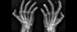
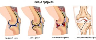
Instrumental data • X-ray examination •• In the early stages there are no changes •• Late stages: osteoporosis, periosteal growths, premature fusion of the epiphyses, erosion, narrowing of joint spaces, ankylosis.
Diagnostic tactics. The diagnosis of JIA, as proposed by WHO, is established in the presence of arthritis of unknown etiology, present for 6 weeks in a child under 16 years of age, with the exclusion of other diseases (congenital joint pathology, etc.).
TREATMENT
General tactics depend on the form of the disease • In the systemic version: NSAIDs, if ineffective - prednisolone 2 mg/kg/day or pulse therapy with methylprednisolone 10-30 mg/kg/day for 1-3 days (especially with myocardial damage). If there is no effect, methotrexate 0.3–0.5 mg/kg/week. In case of development of macrophage activation syndrome - cyclosporine and pulse therapy with methylprednisolone. The use of gold salts and penicillamine is contraindicated • In case of polyarticular form, first use NSAIDs, and when the diagnosis is confirmed, sulfasalazine 30-40 mg/kg (especially with enthesitis) or methotrexate 0.3 mg/kg/week. If ineffective, methotrexate 1 mg/kg IV, or combination therapy (methotrexate, sulfasalazine and/or hydroxychloroquine). In special severe cases, it is possible to prescribe cyclosporine • For oligoarthritis - NSAIDs, if ineffective - intra-articular GCs, if there is no effect within 2-3 months - sulfasalazine 30-40 mg/kg/day, or hydroxychloroquine 5 mg/kg/day, or methotrexate 0.3 mg/kg/week with a gradual increase in dose to 0.5 mg/kg/week.
Mode. Patients should form a movement pattern that counteracts the development of deformities (for example, to prevent ulnar deviation, one should open the tap, dial a telephone number and perform other manipulations with the left hand rather than the right one).
Drug therapy • NSAIDs are used in all cases of JIA •• Ibuprofen for children from 6 months to 12 years 40–50 mg/kg/day (in 3–4 doses), over 12 years of age, doses are similar to adults (1200–1800 mg/day) • • Naproxen is not prescribed to children under 2 years of age, over 2 years of age - 2.5 mg/kg/day • GC is prescribed in the absence of effect from NSAIDs, 1-2 mg/kg orally • Basic drugs • Immunosuppressive drugs •• Methotrexate for spreading oligoarthritis 15–20 mg/m2/week, for seropositive polyarthritis 10 mg/m2/week • Sulfasalazine is not prescribed to children under 2 years of age, over 2 years of age - 40–60 mg/kg/day in 3–6 doses; maintenance dose - 20-30 mg/kg/day in 3 divided doses • Local therapy - see Rheumatoid Arthritis. GC is administered into the joint in doses 2–3 times lower than in adult patients • Intensive therapy for the systemic version: pulse therapy of GC 15–20 mg/kg/day for 3 days.
Non-drug therapy. Plasmapheresis - with a systemic option (the effectiveness continues to be discussed).
Surgery. Synovectomy is rarely used due to the wide range of active drug effects on synovitis. Prosthetics of hip and knee joints, surgical treatment of deformities of the hands and feet are used.
Complications • Amyloidosis • Macrophage activation syndrome sometimes develops as a complication of the systemic form; characterized by fever, weakness, drowsiness, hepatosplenomegaly and often leads to death.
Rehabilitation. Exercise therapy plays an important role. Spa treatment is recommended during periods of minimal activity or remission. To correct deformities, orthoses are used - individual orthopedic devices made of thermoplastic, worn at night. Children often need consultation with a psychologist.
Course and prognosis • In most cases of systemic arthritis of moderate severity, the disease resolves spontaneously • With persistent oligoarthritis, the prognosis is favorable, remission occurs after 4–5 years • With persistent oligoarthritis, the prognosis is relatively favorable • With seropositive polyarthritis, the disease is accompanied by the development of deformities.
Abbreviations • JIA—juvenile idiopathic arthritis.
ICD-10 • M08 Juvenile arthritis
Treatment of juvenile rheumatoid arthritis
Non-drug treatment:
- Physiotherapy.
- Balanced diet: fruits, vegetables, foods rich in polyunsaturated fatty acids.
Drug treatment:
- Non-steroidal anti-inflammatory drugs.
- Glucocorticosteroids.
- Disease-modifying antirheumatic drugs.
- Biological preparations (R – Mab).
- Targeted synthetic drugs.
To better achieve a therapeutic effect, it is necessary to prevent infectious diseases, avoid hypothermia and stress. Home education is recommended for children with systemic onset of JRA and during periods of exacerbation of the disease.
Causes of juvenile rheumatoid arthritis
The nature of the JRA is not known for certain. There are many provoking factors that can cause a restructuring of the immune system, among which are:
- Viruses and bacteria that have antigens similar to connective tissue, which leads to a cross-immune reaction. In other words, the immune system produces antibodies that destroy pathogens, and at the same time, destroy the connective tissue of cartilage, blood vessels, and organ stroma.
- Hypothermia, stress and injury can overactivate the immune system and cause organ damage.
- Hereditary predisposition plays an important role. A risk factor is the presence of any allergic or autoimmune diseases in relatives.
Thus, juvenile rheumatoid arthritis is a multifactorial disease. The existing defect of the immune system is decisive, however, without predisposing factors, hereditary ones may not be realized.