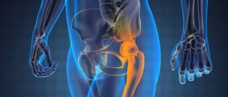
About the femur
The femur (Latin: Femur) is the largest, longest bone in the human skeleton.
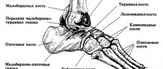
The bone has physiological curves - a saber shape. Both ends of the femur are involved in the formation of large joints - the hip and knee. The upper (proximal end) of the femur is represented by the head of the femur, which rotates in the acetabulum of the pelvic bone to form the hip joint. The lower (distal) end of the bone is represented by two condyles, which are covered with hyaline cartilage and participate in the formation of the knee joint.
Various muscles and capsular-tendon formations are attached almost throughout the entire length of the femur.
Treatment and diagnostic tactics.
In patients with polytrauma, radiographs of the chest, spine, skull, and plain radiography of the pelvis are required to exclude bone-traumatic injuries.
Upon admission, all necessary surgical measures are performed according to damage control tactics (Damage Control surgical tactics).
As a rule, a fracture of the femoral head occurs as part of multiple and combined trauma, and therefore is often not diagnosed.
To determine the nature of damage to the femoral head and acetabulum, in addition to a survey radiograph of the pelvis, radiographs of the inlet and outlet of the pelvis . Both x-rays are performed without repositioning the patient, which is very important in case of unstable hemodynamics; only the x-ray tube is rotated 60 degrees relative to the patient.
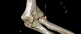
The “gold standard” for suspected fractures of the femoral head is computed tomography ( CT ) of the hip joint with 3D reconstruction, which allows you to clearly visualize all structures of the hip joint - the head, neck of the femur, edges, floor of the acetabulum and other structures. In unstable hemodynamics, performing a CT scan is controversial. The figure shows CT data - a section through the hip joint in the frontal plane, where a comminuted fracture of the femoral head, type 2 according to Pipkin, is clearly identified.
The figure shows on the left a survey X-ray of the pelvis, which shows anterior dislocation of the femoral head, a fracture of the femoral head (type 2 according to Pipkin) - indicated by a white arrow. The avulsion fracture of the femoral head remained fixed to the acetabulum by the round ligament. The right picture shows a CT scan with 3D reconstruction of the same patient. The diagnosis was confirmed by computed tomography.
If there is a dislocation of the femoral head, the dislocation is reduced, but the Pipkin fracture is often not identified. Therefore, if the capacity of the hospital allows for a CT scan and the patient is stable, in case of dislocation of the femoral head, a computed tomography scan of the hip joint should be performed.
A timely diagnosis of a fracture of the femoral head gives a chance to preserve the viability of the femoral head with a timely operation, since the risk of avascular necrosis of the femoral head is directly proportional to the time spent on diagnosis.
Femur fracture
Hip fractures can occur at different levels . As people age, the hip fracture occurs at the upper (proximal) end. This occurs due to deterioration of the nutrition (trophism) of bone tissue at this level.
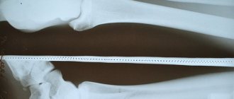
Hip fractures are: pertrochanteric, femoral neck fracture, femoral head fracture. Typically, these types of fractures occur in older people. Fractures of the diaphysis of the femur (body) and the distal end (condyles) of the femur occur due to high kinetic energy directed at this level. This occurs as a result of a strong impact during an accident.
Types of fractures: transverse, oblique, comminuted, multi-comminuted, fragmentary.
Types of bone fractures | |||
| oblique | splintered | spiral | with offset |
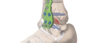
Surgery for pelvic fracture
Surgical intervention is inevitable for displaced pelvic fractures, when the surgeon needs to fasten individual fragments of the damaged bone. For such cases, pelvic surgery provides devices such as knitting needles, screws, metal plates, pins and other metal structures for fixing (connecting) bones.
Surgical manipulation of the pelvic bones using such implants is called osteosynthesis and is carried out using general anesthesia. During the operation, the surgeon will have to carefully examine the internal organs that are located in the pelvic area and eliminate damage resulting from injuries using internal fixation (immersion osteosynthesis). During this manipulation, implants can be installed directly on the bone, inside it, or in combination, while remaining inside the body forever.
Osteosynthesis is also divided into closed (extrafocal) and open, based on the method of its implementation (exposing the manipulation area). In modern pelvic surgery, minimally invasive osteosynthesis is increasingly used, in which the installation of implants is preceded by mini incisions or punctures of the skin.
The use of osteosynthesis for injuries of the acetabulum is not always justified and is determined by the nature of the fracture and its location (wall or floor). In particular, the effect of such surgical intervention in the case of a comminuted fracture of the bottom is much lower than in cases of a large-comminuted fracture of the posterior wall.
In some cases, external fixation is used (percutaneously), in which Ilizarov apparatuses, rod apparatuses and their other analogues are used. They are distinguished by their superficial location in relation to the bone.
Symptoms of a femur fracture
The symptoms of a femur fracture depend on the level of bone damage . When the upper (proximal) end of the bone is fractured, there is deformation of the hip joint area, swelling, the leg is usually adducted inwards and rotated outwards. Sharp pain with minor movements in the hip joint, no movement.
In case of a fracture of the diaphysis and proximal end of the femur, there is deformation of the femoral area, swelling, and pathological mobility of the fragments. One of the serious complications of femoral fractures is massive hemorrhage into the soft tissues of the thigh. With fractures of any segment, the support ability of the limb is impaired.
Rehabilitation after a pelvic fracture
A pelvic fracture is a severe injury to the musculoskeletal system and requires a professional approach from specialists not only in diagnosis and treatment, but also in carrying out rehabilitation measures until the victim’s complete recovery.
Depending on the severity of the fracture, complete recovery from pelvic injuries may take from one and a half to six months. Individual complexes developed specifically for each case and including a number of the following activities will help with this:
- Daily physical therapy to maintain muscle tone, including special exercises to prevent the development of ankylosis, contractures and other complications in the joints.
- Taking special medications that help strengthen bones and nourish them with collagen for complete restoration of the musculoskeletal system.
- The use of ointments, creams and gels that restore joint function and relieve pain and swelling of tissues.
- Therapeutic massage, physiotherapy.
- Walking in the fresh air with a gradual increase in their duration.
- Proper diet. Consumption of foods high in calcium: sea and river fish, dairy products, herbs, vegetables, nuts, persimmons, green beans, poppy seeds, sesame seeds, rose hips.
Treatment of a femur fracture
Currently, the leading method of treating a femur fracture is surgery. The essence of this method is that, as quickly as possible, surgery on the femur allows the patient to begin recovery after a serious injury.
According to statistics, the most common fractures of the proximal femur in older people are pertrochanteric, subtrochanteric and femoral neck fractures. Treatment of fractures in such patients comes down to verticalizing and raising the person to his feet as quickly as possible. The operation of choice for hip fractures in the modern world is hip replacement. A timely operation—total hip replacement—will help avoid severe, rapidly developing complications.
For fractures of the trochanteric region, metal osteosynthesis is most often used. Using special metal structures, bone fragments are fixed, which ensures their consolidation (fusion). For fractures of the diaphysis (body), the choice of surgical method depends on the type of fracture. The use of extraosseous and intraosseous (intramedullary) fixation methods is determined individually.
The main task of the traumatologist is to achieve the most accurate alignment of bone fragments and restoration of limb function using the surgical method. Our center uses the most modern surgical techniques for treating femur fractures .
First aid for a pelvic fracture
Correctly provided first aid at the slightest suspicion of a fracture of the pelvic bones will help to avoid unpleasant consequences, and the speed and professionalism of those nearby will prevent death if the victim has severe pelvic injuries.
In the event that doctors are immediately next to the victim, the first step is to relieve the shock symptom and reduce the pain syndrome. To do this, pain relief is carried out, in particular, the introduction of special medications directly into the fracture site (pain blockades). If the injury is accompanied by bleeding, it is necessary to treat open wounds, apply pressure bandages to bleeding vessels, and in case of significant blood loss, restore blood circulation. Only after this the patient should be carefully transferred to a stretcher and transported to the nearest medical facility.
However, doctors are not always immediately available, which is why up to 30% of victims with isolated trauma are admitted to the hospital in a state of traumatic shock. Statistics show that death in such cases occurs in 6% of cases. This is one third lower than in those hospitalized with multiple pelvic fractures, where shock is observed in almost all victims, and death occurs in 20% of cases, mainly as a result of heavy internal bleeding.
Before doctors arrive, it is especially important to properly prepare the victim for transportation. Below is an approximate algorithm of actions of rescuers located next to the victim:
- Call the doctors, assess breathing, pulse and examine the victim for injuries.
- Provide rest to the victim and, if necessary, immobilize the cervical spine.
- Relieve pain shock with affordable medications, if they are at hand. For those who are conscious and without obvious signs of severe injuries to the pelvic organs, two Analgin tablets and one Diphenhydramine tablet (or Aspirin with butadione), as well as strong sweet coffee, are recommended as an anesthetic.
- Drink plenty of warm fluids to relieve traumatic shock. It is recommended to give the victim warm water (up to 3-4 liters), adding 1 tbsp. a spoonful of table salt and 1 teaspoon of baking soda for every liter.
- Give the patient tincture of valerian (up to 20 drops) and 20 drops of cordiamine (Valocordin or Corvalol) to prevent heart failure and calm down in order to avoid complications during transportation.
- Place the injured person with his back on a hard surface, for example, a shield covered with a mattress or a door removed from its hinges, in the “frog” position: with legs half-bent at the knee joints, raised by about 30 cm, a pillow or an improvised bolster placed under them, knees apart.
- Wrap a scarf, sheet or other available material around the pelvis to prevent further displacement of the broken pelvic bones.
- Cover the limbs, if they are not injured, with heating pads (bottles) with hot water and wrap the victim warmly, regardless of the air temperature.
- Constantly monitor the pulse until the doctors arrive and, if it is difficult to determine, raise the foot end of the improvised stretcher by 30–45 cm.
In cases where it is impossible to call an ambulance, the victim must be transported on his own. In such cases, it is necessary to gently fix his knee joints and feet and strap the injured person to an improvised stretcher.