Movements in the joints[edit | edit code]
Movements in joints
To analyze exercises, it is very important to know the names of movements and understand in which joints they are performed.
Types of joints[edit | edit code]
Planes and directions of the human body
Some bone joints are immobile or allow movement only in a very limited range. For example, the bones of the skull are connected very firmly and do not move relative to each other.
Where the spine connects to the pelvic bone, there is a semi-movable sacroiliac joint that allows for minimal movement. But there is a third category of bone connection - joints. Depending on their structure, size and structure, they allow the bones to perform free movements of a very different nature.
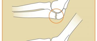
Synovial joints are found in the body more often than others. They are characterized by the presence of an articular capsule that surrounds the junction of the bones on all sides. The inner membrane of the capsule, under the influence of movements performed in the joint, releases synovial fluid, which acts as a lubricant. Typical synovial joints include the shoulder, knee, hip, ankle, hands, feet, and spine. Of all the joints, the knee is the largest, the hip is the strongest, and the shoulder is the most unstable.
Actions of the joints[edit | edit code]
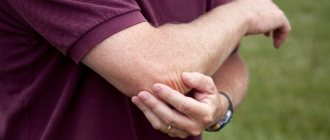
When we perform an action, such as lifting a weight or running, nerve impulses stimulate a specific combination of muscles, and due to their contraction, movement occurs in the synovial joint. For example, when we bend an arm with a dumbbell at the elbow joint, the weight rises because the biceps, attached at one end to the humerus and the other to the bones of the forearm (radius and ulna), contracts and pulls the forearm towards the shoulder.
Direction of movement[edit | edit code]
Most movements have common names regardless of the joints in which they occur. But there are also specific movements that are characteristic only of a particular joint. They take place in a certain anatomical plane. For example, flexion of the arms and legs at the shoulder, hip and knee joints occurs in the same plane. This makes the classification of movements and their analysis simpler and more logical. The table on page 12 shows first the movements common to many joints, and then the specific ones performed only in certain joints.
As a rule, the name of the movement also contains the name of the joint in which it is performed, for example, flexion of the arm at the shoulder joint, extension of the leg at the knee joint, rotation of the spine, lowering of the scapula, etc. Strictly speaking, it would be incorrect to link the movement only with the part bodies. For example, if we say “extension of the leg,” then it is not entirely clear in which joint it is extended—the knee, hip, or ankle.
Usually the movements are paired. If there is movement in one direction, then there must be a reverse movement - if only to return to the starting position. Typical pairs of movements are flexion and extension, abduction and adduction, internal and external rotation, lowering and raising. Reading the analysis of exercises, you will come across these names more than once. The names of the movements are given based on the standard anatomical posture. In this case, “bending the arm at the elbow joint,” for example, will be the same movement regardless of whether the person is standing, lying on his back, or sitting.
| GENERAL MOVEMENTS | Plane | Description | Example |
| Lead | Frontal | Movement directed from the midline of the body | Abduction of the leg at the hip joint |
| Bringing | Frontal | Movement towards the midline of the body | Adduction of the leg at the hip joint |
| Flexion | Sagittal | Reducing the angle between two structures | Pulling the forearm to the shoulder, curling the arms with dumbbells for the biceps |
| Extension | Sagittal | Increasing the angle between two structures | Straightening the arm, returning to the starting position in the same exercise |
| Inward rotation | Horizontal | Rotation of the bone around a vertical axis towards the midline of the body | Bringing your hands together on the upper block |
| Outward rotation | Horizontal | Rotation of the bone around a vertical axis in the direction from the median; body lines | Heels in and toes out |
| Full rotation | All planes | Full rotation of the limb at the shoulder or hip joint | Circular rotation with arms |
| SPECIFIC MOVEMENTS | |||
| 1. Ankle joint | |||
| Plantar flexion | Sagittal | Sock pulling | Standing calf raise |
| Dorsiflexion | Sagittal | Bringing the toes to the shin | Standing calf raise (reverse movement) |
| 2. Wrist joint | |||
| Pronation | Horizontal | Rotate the forearm palm down | Unscrewing the nut |
| Supination | Horizontal | Rotate the forearm palm up | Screwing the nut |
| 3. Shoulders | |||
| Lowering | Frontal | Downward movement of the shoulder blades | Stabilization of the shoulder girdle, for example, when performing a “corner” on the forearms |
| Lifting | Frontal | Upward movement of the shoulder blades, for example. with a shrug | Seated dumbbell press (upward movement) |
| Breeding | Horizontal | Movement away from the spine | Seated chest row (starting position) |
| Mixing | Horizontal | Movement towards the spine | Seated chest row (final position) |
| Inward rotation | Frontal | The upper edge of the shoulder blades deviates outward, and the lower edge inward | Wide grip pulldown |
| Outward rotation | Frontal | The upper edge of the shoulder blades deviates inward, and the lower edge outward | Same exercise, return to starting position |
| 4. Shoulder joint | |||
| Horizontal abduction/extension | Horizontal | Movement of the arm raised to the side backwards | Raising arms while lying on a bench |
| Horizontal adduction/flexion | Horizontal | Movement of the raised arm to the side forward | Same exercise, return to starting position |
| 5. Spine | |||
| Lateral flexion | Frontal | Deviation of the body from the vertical axis to the side | Side bends while sitting on a gymnastic ball |
Authorization
Biomechanics and safety Bobylev V., Zheldakov D.In order to technically perform exercises competently, get the maximum effect from training and, ultimately, avoid injury, it is necessary to understand the biomechanical foundations of movements.
Being in three-dimensional space means the presence of three planes.
- When we move ourselves or a part of our body back and forth, we move in the sagittal plane. The same plane divides our body into two halves - right and left.
- The frontal plane divides our body into the front and back.
- In this plane we move left and right.
- The horizontal plane divides our body into upper and lower parts. The most striking example of movement in the horizontal plane would be turning the head to the right or left.
Bend your leg at the hip while standing and then lying down. The movement is the same, but in what plane is it performed? In order to avoid confusion with the description of movements, scientists have come up with a simple solution - to consider all movements relative to the human body. Therefore, now it doesn’t matter to us whether a person is standing, lying, running or falling on his side, the planes do not change.
“Anatomical position” was chosen as the starting point for describing movements. Why her? All processes in the body (breathing, blood circulation, metabolism) occur most efficiently in it. A person takes an “anatomical position” when he stands with his back straight, his shoulder blades brought towards his spine and his palms turned forward.
All movements are performed by moving one or more bones relative to each other at the points of their connection - joints. The specific shape of the joint allows us to carry out movements in one or more planes.
To perform movements most accurately, you need to know that movement in the joints is performed around a certain axis.
- Flexion - extension is carried out around the frontal axis (mid-lateral), in the sagittal plane.
- Abduction – adduction is performed around the sagittal axis (antero-posterior), in the frontal plane.
- Horizontal abduction - adduction and twisting (rotation) of the spine are performed around a vertical axis, in a horizontal plane.
The axis of rotation of the movement is always perpendicular to the plane of movement. And even if you lie on the floor, or find yourself in a position that cannot be described at all, adduction will not become rotation or abduction. And all because movements are considered relative to the human body, and not to the earth or any object.
Moving bones relative to each other is possible because our muscles have the ability to contract. Depending on how the muscle contracts, we produce movements of a different nature. Let’s use the example of a simple exercise “Bending the arms at the elbow joints using additional weights” to see how this happens. The positive phase of the movement, in which the arm flexes at the elbow joint, is accompanied by contraction, with a simultaneous decrease in the length of the biceps brachii muscle. This type of contraction is called concentric.
- The negative phase of the movement, in which the arm is extended at the elbow joint, is accompanied by contraction of the biceps brachii muscle with an increase in its length. This type of contraction is called eccentric. And there is no contradiction here. The muscle does the work (contraction) allowing the weight to slowly lower rather than fall, although its length increases.
- By accelerating the weight and throwing it, we force the biceps brachii muscle to work in ballistic mode. When, from a pre-stretched state, the muscle, sharply contracting at the beginning of the movement, causes the weight to move by inertia. This type of contraction is the most dangerous due to the inability to control the entire trajectory of movement.
If the body or limb moves in space, then this is a dynamic mode. All types of abbreviations listed apply to this mode.
- By performing the exercise we are already familiar with and stopping the weight halfway, you will force the biceps brachii muscle to work in a static mode, which involves performing work (contraction) without changing the length of the muscle.
In the same mode, all the muscles that hold our body in an upright position (stabilizers) will work. A technically competent exercise based on an understanding of the work of all joints and muscle groups will increase the effectiveness of the exercise and reduce the risk of injury! Always strive for the beauty of the movements you perform! Only beautiful movements are justified from the point of view of biomechanics and anatomy!
* "Straight back" is a term quite often used in sports and fitness. Let's try to decipher this term. A straight back assumes the presence of normal curves of the spine ( moderately expressed S - shaped spine in the sagittal plane) and a neutral position of the pelvis without excessive forward or backward tilt. To avoid spinal injuries, adhere to the following rules:
- To maintain a neutral position of the pelvis and, accordingly, an anatomically correct curve in the lumbar region (lordosis), constantly maintain tone in the abdominal muscles and, if the exercise allows, in the gluteal muscles.
- To effectively control the position of the spine in the thoracic region (kyphosis), it is necessary to adduct the shoulder blades and slightly raise the lower ribs.
In order to ensure the safety of the cervical spine, do not allow excessive tilting and, especially, throwing your head back.
More information about the biomechanical parameters of the simulator
The ability to adjust the position of the blocks (rollers) in accordance with the width of your shoulder or hip joints makes the exercises performed as close as possible to natural movements. The correctly selected distance between the blocks optimally matches the forces of gravity and muscle resistance in any phase of movement. This design feature of the simulator (protected by two patents) sharply reduces the risk of injury to working joints and muscles.
In addition, the presence of blocks adds additional instability, causing stabilizers, such as the abdominal and back muscles, to become more active. Thus, the energy intensity of the exercises performed increases significantly.
The concept of biomechanics of movements
Physical training
The science of the laws of movement of humans and animals is called biomechanics. For the correct and justified use of physical therapy in the treatment of patients, it is necessary to understand the patterns of human movements.
To register the state of the human body, it is customary to distinguish between three main (imaginary) planes of the body:
- sagittal, or anteroposterior, divides the body or any part of it into left and right halves (sections), and the sagittal plane passing through the middle of the body is called the median plane;
- the horizontal plane crosses the body transversely, dividing it into the head (cranial) and caudal (caudal) sections. A horizontal plane drawn on any limb divides it into proximal (closer to the body) and distal (further from the body) sections;
- the frontal plane (parallel to the surface of the forehead) divides the body and its parts into anterior (ventral) and posterior (dorsal) sections.
All three planes are perpendicular to each other.
The following movements are distinguished in the joints: flexion, extension, abduction (outward), adduction (inward), rotation or rotation (turning inward and outward). Turning inward is also called pronation, turning outward is called supination. In some joints (for example, in the shoulder, hip, wrist), circular movements are also possible.
When two bone links are connected through a joint (biokinetic pair), the possibilities of movement are determined by the structure of the joint, the action of the muscles, and the limiting action of the capsule and ligaments of the joint.
Joint mobility depends on age, gender, individual characteristics, and the functional state of the nervous system. Women and young people have more mobility.
N. A. Gukasova (1997) systematized data from one of the main areas of biomechanics—dynamic human anatomy—for exercise therapy specialists. Dynamic anatomy studies the functions of muscles and the effect of body gravity on its position both at rest and during movement (static and dynamic).
For exercise therapy specialists, the correct choice of starting positions in therapeutic gymnastics procedures, the selection of special and general developmental exercises, and their dosage are important. This requires knowledge of the biomechanics of the laws of leverage, “degrees of freedom,” and the state of muscle tone—synergists and antagonists.
Human movements are carried out according to the laws of levers. Levers are individual bone links of the human body, for example, the bones of the shoulder, forearms, femurs, bones of the legs, feet, head, and spine. Each bone link is usually acted upon by two forces: muscle force and the force of gravity of the given bone link. Depending on the location of the application of forces in relation to the fulcrum of the lever or the axis of rotation, “balance levers”, “speed levers” and “force levers” are distinguished.
A lever of the first kind is a “balance lever”, in which the forces are located on both sides of the fulcrum (axis of rotation) and are directed in one direction. The lever arm is considered to be a perpendicular, lowered from the fulcrum of the lever (axis of rotation) to the direction of muscle force or gravity; The lever arm corresponds to the distance from the fulcrum of the lever to the point of application of forces (Fig. 1).
Levers of the first kind: a - with equal shoulders; b - with unequal shoulders. O - axis of rotation, or fulcrum; MA - muscle strength; TV is the force of gravity of a given bone link; OA - lever arm of muscle force; OB - gravity lever arm
According to the law of the first kind of lever, movements of the head and spine occur. With an asymmetric change in muscle strength and the force of gravity of a bone link, an imbalance in the lever occurs, and this is clinically manifested by a violation of posture in the sagittal or frontal plane.
For the correct choice of physical exercises, it is necessary to know the reasons that caused the imbalance function. Thus, with unilateral radiculitis, it is necessary to reduce muscle tone on the affected side of the muscles that move the head and spine, using relaxation exercises, corrective starting positions, and massage. In case of postural disorders in children or weakened patients, on the contrary, exercises and massage techniques should be used that help strengthen weakened muscle groups and increase their tone. In patients after partial or complete amputation of a limb, the severity of the bone link decreases, so exercises that help build muscle mass on the side of the amputation should be used, and the patient should be taught to relax the muscles of the opposite limb and use a prosthesis to restore the equality of torques of forces.
When the head or torso is bent, tilted, or turned, the lever is thrown out of balance. The rotational moment of one force becomes greater or less than the rotational moment of another force. The body returns to the state of initial equilibrium when the initial equality of the moments of rotation of forces is restored.
Movements of the limbs occur primarily according to the law of the lever in type II . A type II lever is a lever in which the forces applied to it are located on one side of the fulcrum or axis of rotation and are directed in different directions. This lever has two varieties depending on which force (gravity or muscle) will be located closer to the fulcrum (axis of rotation). If the force of gravity is closer to the point of support and the arm of its lever is less than the arm of the lever of muscle force, then such a lever of the second kind is called a “lever of force” (Fig. 2, a). If the muscle force is located closer to the fulcrum and the arm of its lever is smaller than the arm of the gravity lever, then such a type II lever is called a “speed lever” (Fig. 2, b). The movement of the foot during the rise on the toes is an example of movement according to the law of “leverage of force”. In this movement, the fulcrum is the heads of the metatarsal bones, the force of gravity of the body passes through the hip joints, femur bones, tibia, talus bones and presses down, and the muscles of the back of the lower leg counteract the force of gravity and strive to keep the body in a state of balance when it is standing on its toes . In this case, there is an equality of the rotational moments of gravity and muscle force. If the muscles located along the back of the lower leg are weak, the patient will not be able to maintain balance while standing on his toes, since the torque of the muscle force will be less than the torque of gravity. In this case, the balance will be disrupted, and the person will strive to stand on his full foot. Therefore, exercises are necessary to strengthen the muscles located on the back of the lower leg, i.e. muscles - flexors of the foot (triceps surae, plantaris, tibialis posterior, flexor digitorum longus, peroneus longus and brevis). In addition, the patient should be advised to reduce body weight (lose weight) if his weight exceeds normal values.
Levers of the second type: a - force lever; b - speed lever. O - axis of rotation, or fulcrum; MA - muscle strength; TV is the force of gravity of a given bone link; OA - lever arm of muscle force; OB - gravity lever arm
An example of a movement according to the law of the “speed lever” is bending the arm at the elbow joint.
Knowledge of the laws of levers of the first and second kind helps to correctly select the most optimal starting positions (IP) for performing physical exercises. Thus, to strengthen the rectus abdominis muscles, it is necessary to gradually increase the torque of gravity of the torso by changing the length of the lever arm, or the mass of the torso, or both. Changing the lever arm can be achieved by changing the position of the torso. Flexion and extension of the torso are first performed in the starting position, sitting on a chair, on the floor. This ensures a gentle effect on the rectus abdominis muscle. As you adapt, you should gradually move on to more stressful exercises: lying on your back, lying on your back across a gymnastic bench, touching your head and feet to the floor. This change in IP makes it possible to gradually increase the leverage of the gravity lever of the body. You can increase the impact on the rectus abdominis muscle by increasing body weight by performing exercises with weights (dumbbells, medicine balls) and resistance (rubber bandages). Lengthening the gravity lever arm while increasing gravity itself is another option for improving the strength of this muscle.
Another example. A patient has consequences after a fracture in the upper third of the humerus. There is a need to strengthen the abductor muscles of the shoulder (middle deltoid muscle, supraspinatus muscle) to restore mobility in the shoulder joint. In the first half of the course of treatment, easier conditions should be created for these weakened muscles by reducing the moment of rotation of the gravitational force of the arm and the arm of the gravity lever. The lever arm of the arm's gravity force can be reduced by performing abduction at the shoulder joint with the arm bent at the elbow joint. In this case, the lever arm of the gravity force of the hand will decrease by almost 2 times.
To reduce the weight of the limb, the patient’s arm should be abducted with the help of the methodologist’s hand or by hanging it on Straps, using blocks, or performing exercises in water. In the second half of the course of treatment, it is necessary to strengthen and increase the strength of the abductor muscles of the shoulder. For this exercise, you should perform it with a full lever (abduction of the straight arm to the side), with an extended lever of the gravity of the limb (exercises with clubs, gymnastic sticks), with weights, i.e. increasing the gravity of the limb (dumbbells, medicine balls), with resistance (rubber bandages, expanders).
To restore mobility in a particular joint, you need to know the possible directions of its movements. A free, unfixed body has six degrees of freedom:
three forward movements in the directions:
- anteroposterior (forward, backward),
- transverse (right, left),
- vertical (up, down);
three rotational movements around the above axes.
In the musculoskeletal system, all bone links are connected to each other.
Triaxial joints have three degrees of freedom; these are the most mobile joints. These include the articulation of the neck with the spine, the articulation of the vertebrae with each other, the shoulder, brachioradial, sternoclavicular, and hip joints. These joints are spherical or nut-shaped in shape. They allow movements around three mutually perpendicular axes (antero-posterior, transverse, vertical) and within three mutually perpendicular planes (frontal, sagittal and horizontal). The planes of motion are located perpendicular to the axes of rotation.
In the triaxial joints of the limbs, abduction, adduction, flexion, extension, rotation inward, outward are possible; for the spine - tilts to the right, left, flexion, extension, turns to the right, left.
Biaxial joints (wrist, joint of the first finger, metacarpophalangeal joints, knee) have less mobility and two degrees of freedom. In shape they are ovoid, ellipsoidal, saddle-shaped. The following movements are possible in them:
- transverse axis, sagittal plane (flexion, extension);
- axis anteroposterior, plane frontal (abduction, adduction).
In the carpometacarpal (saddle) joint of the first finger of the hand, the following movements are possible: opposition (opposition) and reposition (reverse movement).
Circular movements are possible in biaxial and triaxial joints.
Uniaxial joints (humeral-ulnar, radioulnar, interphalangeal, ankle, chopar) have one degree of freedom. They are block-shaped or cylindrical in shape. The following movements are possible in them:
- axis transverse, plane sagittal;
- if the axis of the joint goes obliquely, for example, in the radioulnar, chopard joints, outward rotation (supination) and inward rotation (pronation) are possible.
Circular movements in uniaxial joints are impossible.
All large muscles that carry out movements in the joints are united from an anatomical point of view. At the same time, functionally they are not the same. Different sections of the same large muscle perform different movements. Thus, flexion of the arm at the shoulder joint is carried out by the anterior bundles of the deltoid muscle, abduction of the arm to the side to the horizontal by the middle bundles, extension by the posterior bundles. The upper part of the pectoralis major muscle raises the humerus, its lower part lowers it. This knowledge for patients with impaired movement functions helps to purposefully select exercises to train certain portions of the muscles.
Muscle tone is the involuntary tension of muscles at rest. Muscle tone provides the ability to take different body positions in space. A muscle can be in four states: rest, contraction, relaxation, stretching. Any movement is carried out as a result of the friendly work of muscles. Synergists refer to muscles involved in unidirectional movement. Antagonists are muscles that produce movement in the opposite direction. Synergists, for example, are the muscles - the ulnar and radial flexors, which perform a unidirectional movement - flexion of the hand, and the muscles - the ulnar and radial extensors - their antagonists, since they carry out the oppositely directed movement - extension of the hand.
The tone of the muscles changes during all movements: when bending, the tone of the flexor muscles increases and the tone of the extensor muscles decreases by the same amount.
In exercise therapy there are three main types of muscle work:
- inferior dynamic (resistance),
- overcoming dynamic,
- static.
The first and second types of work are carried out in an isotonic mode, the third - in an isometric mode.
Measuring joint movements
Rotation angles are measured using measuring instruments. The simplest of them is called a goniometer, or goniometer; it consists of a protractor with a 180° scale connected to two jaws. One of the branches is movable (Fig. 3). When measuring, the axis of the protractor is aligned with the axis of the joint, and the branches are placed along the axis of the articulating proximal and distal segments.
Measuring knee movements using an inclinometer
For continuity and comparability of measurement results and to eliminate errors, the same measurement techniques are required (Fig. 4, 5, Table 1). The angle of maximum extension-flexion of a joint in one plane is called the range of motion.
The position of the goniometer when measuring mobility in the joints: a-d - shoulder; d, f - elbow; g, h - hip
The position of the goniometer when measuring mobility in the joints: a, b - wrist; c, d - knee; d, f - ankle
Table 1. Measuring range of motion in selected joints
| Position of the angle meter rotation axis (see Fig. 4, 5, point 0) | Position of the goniometer branches | ||
| Movement in a joint | 1 branch (see Fig. 4, 5, line 0-A) | Branch II (see Fig. 4, 5, line O-B) | |
| Flexion, extension, abduction in the shoulder joint (see Fig. 4, a-d) | Head of humerus | Acromion - the highest point of the ilium | Acromion - humeral condyle |
| Flexion and extension at the elbow joint (see Fig. 4, e-f) | Humeral condyle | Humeral condyle - acromion | Humeral condyle - styloid process of the radius |
| Flexion and extension in the wrist joint (see Fig. 5, a) | Styloid process of the ulna | Along the outer edge of the ulna | Along the outer edge of the fifth metacarpal bone |
| Abduction and adduction in the wrist joint (Fig. 5, b) | Between the distal ends of the bones of the forearm | Midway between the ulna and radius bones | In the middle between the third and fourth fingers |
| Flexion and extension at the knee joint (see Fig. 5, c, d) | Lateral condyle of the femur | Lateral condyle of the femur - greater trochanter | Lateral condyle of the femur - lateral malleolus |
| Flexion and extension at the ankle joint (see Fig. 5, e, f) | Medial malleolus | Medial malleolus - medial condyle of the femur | Medial malleolus - middle of the 1st metatarsophalangeal joint |
When measuring movements in the shoulder joint, 0° is taken as the initial value with the arm lowered and the jaws of the protractor closed. When measuring movements in the elbow, wrist, hip and knee joints, 180° is taken as the initial value. Measurements in the ankle joint are usually taken from the initial value of 90°. The average normal mobility in the joints of the limbs is presented in table. 2.
Table 2. Average mobility in some joints of the limbs (in rnanvcax from the starting position)
| Joint | Flexion - extension | Adduction - abduction | Internal - external rotation |
| Brachial | 180 — 60 | 0 — 180 | 90-90 |
| Elbow | 145 — 0 | — | — |
| Radioulnar | — | — | 90 — 90 |
| Radiocarpal | 90 — 80 | 20 — 45 | |
| Hip | 125 — 15 | 10 — 45 | 45 — 45 |
| Knee | 130 — 0 | — | — |
| Ankle | 45 — 20 | — | — |
Movements of the body in the sagittal, frontal and horizontal planes - tilts, turns, rotations - are carried out thanks to movable joints between the vertebrae. The mobility between them is small, but in total it turns out to be significant. The cervical and lumbar spine are the most mobile, the thoracic spine less so. The following movements of the body are possible: flexion and extension (bending forward and bending back), bending to the sides (right and left), rotation around a vertical axis (turning right and left) and circular movements.
The starting position for measuring movements in the joints of the cervical spine is sitting on a chair with the torso and head straightened; The measurement is carried out according to the position of the head. Movements in the thoracic and lumbar regions are measured in a standing position with legs slightly apart and arms hanging freely along the line of the spinous processes. When measuring rotation in the lumbar region, it is necessary to fix the pelvis, having first seated the patient “astride” on the seat of a chair. The movements of the spine are determined both in degrees (which is more difficult) and by the maximum movements of various sections.
In the cervical spine, flexion normally occurs until the chin touches the sternum, extension until the back of the head is horizontal, bends to the sides until the auricle touches the shoulder, and with maximum rotation, the chin touches the acromion. A trained adult, when bending forward, can touch the floor with his fingertips without bending the knee joints; when bending to the side, the fingertips, sliding along the outer surface of the thigh, can touch the corresponding knee joint. The normal range of motion in the cervical spine is considered to be: extension 70°, flexion 60°, lateral rotation 45°. The lateral tilts in the thoracic and lumbar regions together amount to 50°. The total amplitude of flexion and extension in the lumbar spine reaches 80°. Total movements of the entire spinal column are possible within the following limits: up to 160° - flexion, up to 45° - extension, total amplitude of movement in the frontal plane - up to 165°, rotation in each direction - up to 120°.
Initial body positions for performing exercises
The patient's posture can make it easier, more difficult or aggravating to perform physical exercises, and depending on the purpose of the training, it can help increase its effectiveness. The starting position for persons with reduced functionality should simplify and facilitate the execution of movements. In physical therapy, various IPs are used, but more often - lying, sitting and standing positions. The supine position is used more often for patients on bed rest for certain diseases of the spine; lying and sitting - for weak patients and with diseases of the lower extremities, standing - for stronger, more active patients, with diseases of the upper extremities.
The starting position is lying down - the simplest pose, or lightweight IP. In this position there is no struggle against gravitational forces, maximum relaxation of the skeletal muscles is possible, stability of balance is ensured by a large area of support and a low position of the overall center of gravity of the body. The upper and lower limbs are free to perform movements. The lying position, depending on the patient’s condition and the nature of the upcoming exercises, can be on the back or stomach, and for special indications - on the side.
The initial sitting position is a body posture with a significant area of support. In this position, the balance of the body is relatively easily (automatically) maintained, and significant static muscular efforts of the lower extremities are eliminated. This pose allows for significant movements of the free segments of the limbs and includes muscle groups of the neck and torso in the exercises.
In the initial standing position, a high position of the general center of gravity of the body and a small support area are noted. Various parts of the nervous system are involved in regulating this (vertical) body position. Vertical posture depends on the relative position of various parts of the body, constant impact, gravity and other external forces. It is maintained due to the contraction of certain muscles, which automatically, at the subconscious level, correct deviations of the body from the equilibrium position. The quadriceps muscles of the thighs, extensors of the hip joints, muscles of the legs and feet, abdomen, torso, neck, etc. are involved in maintaining the vertical position of the body. The feet rest on the heel bones and the heads of the metatarsal bones. Varieties of standing postures depend on the size of the support area, the position of the general center of gravity and the line of gravity, and have varying degrees of balance stability (for example, the position of the legs: one in front of the other, the feet on the same line, on the toes, in the “at attention” position, etc.) . The most stable and easily controlled position is a standing position with legs wide apart.
Stands, hangs, walking, running, squats, jumping, jumping from the point of view of biomechanics
Supports refer to body positions with unstable balance. The most typical is the prone position. The body is straightened and occupies an inclined position, the head is held straight, the cervical spine is in a state of slight extension. The upper limbs are straightened, located almost at a straight angle to the body and are in contact with the supporting surface. The lower limbs are also straightened, but are at an acute angle to the supporting surface. All parts of the body form a closed kinematic chain. The degree of balance stability is relatively high, since the support area is of significant size, and the height of the overall center of gravity is small (30-35 cm). Therefore, in this position it is possible to perform various movements with the movement of body parts without disturbing balance.
Body positions with upper support include various hanging positions. These provisions are stable. The simplest of them are “clean” hangs with straight arms. The human body occupies a straightened vertical position. The arms are raised up, straightened and fixed to the apparatus. The force of gravity tends to stretch the body. The hang counteracts the force of muscle pull. The operation of the movement apparatus in this position is complex, since it is performed under conditions unusual for the body. Hanging exercises are considered strength exercises. If the leg support is used in the whiskey position (mixed hang), then the body weight is evenly distributed into muscle groups, and the breathing function is not impaired. Mixed hangs are widely used in exercise therapy.
Walking is a normal human motor activity. It is a constant alternating activity of the legs. When one leg, resting on the ground, serves to support and subsequently push away the body (the support phase of one leg), the other, raised and hanging in the air, moves forward (the swing or swing phase of the other leg). Each leg sequentially goes through both phases - support and transfer. Two steps make up a cycle.
Running is a cyclic step movement, a complex reflex motor act that requires the participation of all the skeletal muscles of the body, significant tension in the nervous system and sufficient physical training of a person. It can be dosed according to speed, duration, step width.
Squats are exercises performed primarily by working the muscles of the lower extremities. The feet can rest on the support area with the entire plantar surface or only on the heads of the metatarsal bones and toes. Exercises can be facilitated by supporting the hands on the front surfaces of the thighs, supporting them with some object. The amount of load is dosed by the depth of the squats, tempo and number of repetitions. Exercises can be complicated and burdened by the load on free hands.
Jumps are characterized by free flight of the body in the air as a result of repulsion from the supporting surface. The main work is performed by the muscles of the lower extremities, auxiliary - by the muscles of the trunk and upper extremities. The exercise is ensured by the simultaneous contraction of large muscle groups of the lower extremities, a large range of motion in the large joints of the legs and shoulder joints.
Jumping jacks are simple jumps in place. The main load during jumping falls on the flexors of the foot. When jumping, the ankle joint uses the maximum range of motion. The muscles of the hip and knee joints play a supporting role. Movements in these joints occur with a small amplitude.
| Back | Table of contents | Further |