Human anatomy is undoubtedly a major core subject for study in medical schools. Despite the fact that normal human anatomy is a discipline that stood at the origins of the development of medicine, a large number of scientific works still appear that make adjustments to modern anatomical atlases.
It would seem that human anatomy cannot change so quickly with the course of evolution, but our understanding of it is constantly improving as new research methods appear - evidence of this is provided by ever new versions of the anatomy atlas.
Atlas of anatomy Sinelnikova R.D. in 4 volumes, this is perhaps the most authoritative and time-tested source of knowledge on this topic. It is constantly republished, delighting us with its visual illustrations and text accessible to everyone. Many students tried to download Sinelnikov’s atlas for study, but the links either did not work, or there was a virus in the folder... We solved this problem by making a website dedicated to this source.
The main goal of studying human anatomy is to create a fundamental knowledge base for students for further study of other medical disciplines. It is difficult to imagine mastering a curriculum in physiology, pathological physiology, pathological and topographic anatomy, operative surgery, and a whole range of clinical disciplines without a thorough study of normal human anatomy.
It is very important for a student to have a visual image of the material studied; for this it is necessary to study human anatomy in pictures. The main feature of this science. of course, is the structuring of its sections and subsections, as well as a clear systematization of the entire nomenclature.
Thus, we can distinguish the following directions that correspond to each system:
- osteology (section on the bones of the human skeleton). Studies the skeleton, both as a whole mechanism and as individual bones. There is also a study of age-related changes in bones.
- syndesmology (joints, ligaments). An extremely important section for future orthopedists and traumatologists.
- myology (muscular system). He studies not only the structure, but also the development and physiology.
- splanchnology (internal organs). Includes the anatomy of the endocrine, digestive, respiratory, excretory and genitourinary systems.
- angiology (vessels and their derivatives). Information is presented on the structure of blood and lymphatic vessels.
- neurology (central and peripheral nervous system). An extremely important section for successful diagnosis of diseases and perhaps the most difficult.
- aesthesiology (the science of the sense organs). Everything about vision and hearing. And also about taste, olfactory and tactile sensitivity. Closely related to neurology.
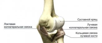
Radioulnar (distal) joint (art. radioulnaris distalis).
Classification. It is a cylindrical, uniaxial joint.
Structure.
The joint
is formed by the articulation of the articular circumference of the head of the ulna and the ulnar notch of the radius.
The distal radioulnar joint is separated from the wrist joint by a triangular-shaped disc. The capsule and ligamentous apparatus are common to the wrist joint. Functions
.
The proximal and distal radioulnar joints together form a combined cylindrical (rotator) joint: the radius rotates around the ulna. Movements in these joints are carried out simultaneously around the vertical axis (pronation - supination).
Radiocarpal joint (art. radiocarpea) – fig. 7.
Classification. The structure of the joint is complex. The shape of the articular surfaces is elliptical with two axes of movement.
Structure.
The joint
is formed by the carpal articular surface of the radius, the articular disc and the articular surfaces of the carpal bones: scaphoid, lunate, triquetrum.
The capsule is attached along the edge of the articular surfaces, capturing the radius, ulna and bones of the proximal row of the wrist. Strengthened by ligaments: carpal collateral radial and ulnar, radiocarpal palmar and dorsal (ligg. collaterale carpi radiale et ulnare, radiocarpeum palmare et dorsale).
Functions
.
Movements in the joint around the frontal (flexion and extension of the hand) and sagittal (abduction and adduction of the hand) axes.
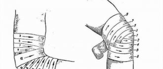
Rice. 7. Joints and ligaments of the hand. Frontal cut: 1 - radius bone, 2 - radiocarpal joint, 3 - radial collateral ligament of the wrist, 4 - midcarpal joint, 5 - intercarpal joint, 6 - carpometacarpal joint, 7 - intermetacarpal joint, 8 - interosseous intercarpal ligaments , 9 – collateral ulnar ligament, 10 – distal radioulnar joint, 11 – articular disc, 12 – ulna.
Joints of the hand - fig. 8.
Midcarpal joint (art. mediocarpea). The joint is formed by the articular surfaces of the first and second rows of carpal bones. Complex, closer to elliptical in shape. The ligamentous apparatus and, in most cases, the capsule are common with the wrist joint.
Intercarpal joints (artt. intercarpeae) are located between the individual bones of the wrist, flat, inactive.
Carpometacarpal joints (artt. carpometacarpeae) are formed by the articular surfaces of the second row of carpal bones and the articular surfaces of the bases of the metacarpal bones. The carpometacarpal joints of the II-V fingers are closer to elliptical in shape and have little mobility.
Rice. 8. Connections of the bones of the hand, palmar surface: 1 – ulna, 2 – distal radioulnar joint, 3 – ulnar collateral ligament of the wrist, 4 – pisiform bone, 5 – hook of the hamate, 6 – carpometacarpal palmar ligament, 7 – metacarpal palmar ligaments, 8 - deep transverse metacarpal ligaments, 9 - metacarpophalangeal joint, 10 - interphalangeal joints, 11 - deep digital flexor tendon, 12 - superficial digital flexor tendon, 13 - carpometacarpal joint of the thumb, 14 - capitate bone , 15 – radial collateral ligament of the wrist, 16 – palmar radiocarpal ligament, 17 – radiocarpal joint, 18 – radial bone, 19 – interosseous membrane of the forearm.
Carpometacarpal joint of the 1st finger (art. carpometacarpea pollicis)
is a simple, saddle-shaped joint.
Formed by the articular surfaces of the trapezium bone and the base of the first metacarpal bone. The capsule is attached along the edge of the articular surfaces. Functions
.
Movements in this joint are carried out around two axes: sagittal and frontal.
Around the sagittal axis - adduction and abduction of the thumb relative to the index finger, and around the frontal axis - flexion with simultaneous opposition to the other fingers and extension of the thumb. Intermetacarpal joints (artt. intercarpeae) are formed by the adjacent lateral surfaces of the bases of the II-V metacarpal bones. These joints are flat; in them there is a slight displacement of the bones relative to each other during flexion and extension of the hand.
Metacarpophalangeal joints (artt. metacarpophalangeae) are formed by the articular surfaces of the heads of the metacarpal bones and the bases of the proximal phalanges. The articular surfaces of the heads are rounded, and the articular sockets are ellipsoidal. The capsule is attached along the edge of the articular surfaces. Strengthened by ligaments: lateral, ligg. collateralia (collateral), palmar,
ligg.
palmaria (contain fibrous cartilage), deep transverse metacarpal,
lig.
metacarpea transversa profunda. Functions
.
In the joints, movements around the frontal axis are possible - flexion and extension of the fingers, around the sagittal axis - abduction and adduction of the fingers.
Interphalangeal joints (artt. interphalangeae). Classification.
Simple,
typical trochlear, uniaxial joints . Structure. Formed by the articular surfaces of the articulating phalanges (the head and base of adjacent bones participate in the formation of the joint).
The capsule is attached along the edge of the articular surfaces. The joints are strengthened by a complex of collateral and palmar (ligg. collateralia et palmaria) ligaments.
Functions
.
In the joints, movement around the frontal axis is possible - flexion and extension of the phalanges of the fingers.
Method for restoring ligaments of the distal radioulnar joint
The invention relates to medicine, namely to traumatology and orthopedics, and is intended for the restoration of ligaments of the distal radioulnar joint. Access to the joint is provided through two incisions along the radial and ulnar edges of the lower third of the forearm. A tunnel is formed along the dorsal and volar surfaces above the bones of the forearm. The autograft is passed through the tunnel around the bones of the forearm. Then, with the forearm bones in a neutral position, the head of the ulna is set into place. The forearm bones are fixed transosseously with two Kirschner wires proximal to the head of the ulna. Along the radial edge, above the radial bone, the ends of the tendon autograft are sutured in the wound side by side, followed by immobilization of the forearm. The method ensures restoration of the correct relationship of the forearm bones, reliable fixation, and reduction of trauma. 4 ill.
The invention relates to medicine, namely to traumatology and orthopedics, and is used in the restoration of ligaments of the distal radioulnar joint.
For persistent (fixed) chronic dislocations of the head of the ulna with severe limitation of movements, resection of the distal end of the ulna 2-2.5 cm long is appropriate. However, this operation, which was considered almost universal, has a number of disadvantages due to the loss of support for the ulnar half of the wrist joint - instability with possible ulnar movement of the hand, weak grip [1].
There are known methods of surgical treatment aimed at restoring the stability of the radioulnar joint with the creation of new ligaments from tendons, fascia, etc. One of these methods - the H. Milcha method [2] - was taken by us as a prototype. The operation is performed using four incisions: ulnar, radial, volar and dorsal. Using a drill with a diameter of 0.5 cm, two tunnels are created in the radius. From the fascia lata of the thigh, sewn together in the form of a tube, a ligament is restored, which is passed through the bone canals made. The lower end of the ulna is pressed against the plane of the radius joint, the free ends of the tube are pulled together from the fascia and sutured together.
The method is traumatic and technically very difficult. In addition, over time, the tube from the fascia lata of the femur atrophies (thins), stretches and cannot withstand the functional load, which leads to instability of the joint.
The essence of the invention is expressed in a set of essential features sufficient to achieve the desired technical result, which consists in improving treatment outcomes by reducing the invasiveness of the operation and increasing the physiological capabilities of the operated joint.
Remembering that with isolated displacement of the head of the ulna, damage to the palmar and dorsal radioulnar ligaments and the ulnar collateral ligament of the wrist occurs, we perform reconstructive surgery with the creation of new ligaments from the tendon.
The essence of the invention is that the method for restoring the ligaments of the distal radioulnar joint includes access to the joint, the formation of channels under the flexor and extensor tendons, passing an autograft through them, bending around the bones of the forearm, restoring the normal relationship of the radius and ulna, subsequent fixation and immobilization of the limb in plaster bandage. Access to the joint is provided through two incisions along the radial and ulnar edges of the lower third of the forearm. A tunnel is formed along the dorsal and volar surfaces above the bones of the forearm, after which, in the neutral position of the forearm bones, the head of the ulna is reduced into place. In this case, the bones of the forearm are fixed transosseously with two Kirschner wires proximal to the head of the ulna. Along the radial edge, above the radius, the ends of the tendon autograft are sutured in the wound side by side, followed by immobilization of the forearm.
The proposed method is made of two cuts instead of the four proposed in the prototype. It is also less traumatic because the radius bone is not injured during the operation. Compared to the prototype, the method is technically easy to implement.
The tendon autograft, used to pass through the canals around the bones of the forearm, is mechanically stronger, does not atrophy or stretch, which allows it to withstand greater physical loads.
Low-traumatic fixation with two wires for a period of 6 weeks allows you to maintain the correct relationship of the forearm bones in the distal radioulnar joint.
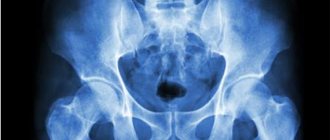
The proposed method is illustrated by the following radiographs, where figure 1 is a view of the joint before surgery, figure 2 is a view of the joint in two projections after surgery, figure 3 and figure 4 show both radioulnar joints of the patient a year after surgery.
The method is carried out as follows.
A tunnel is formed using a forceps from incisions along the radial and ulnar edges of the lower third of the forearm, proximal to the head of the ulna, along the dorsal and volar surfaces above the bones of the forearm. A tendon autograft is passed through this channel around the bones of the forearm. After this, in the neutral position of the forearm bones, between supination and pronation, the head of the ulna is pressed (reduced) into place, the bones of the forearm are fixed transosseously with two Kirschner wires 5-6 cm proximal to the head of the ulna. In the wound along the radial edge of the forearm, above the radius, the ends of the tendon autograft are sutured together side by side. Immobilization of the forearm and fixation with pins for a period of 6 weeks, followed by physiomechanical therapy on an outpatient basis.
Clinical example
Patient Mur-va O.Yu., born in 1983, medical history No. 2146, was admitted to the hand surgery department on June 01, 2001 with a diagnosis of chronic rupture of the ligaments of the distal radioulnar joint (see Fig. 1). Two years before treatment, she fell on her right arm and was treated with a plaster splint.
On 06/07/01 an operation was performed - plastic surgery of the ligaments of the distal radioulnar joint of the right forearm. From two incisions along the ulnar and radial edges of the lower third of the forearm, a tendon graft from the palmaris longus muscle and the superficial flexor of the fourth finger was passed through a tunnel formed around both bones (directly above them). The head of the ulna was reduced and fixed with two knitting needles for 6 weeks (see Fig. 2). In the wound, along the radial edge of the forearm, above the radius, the ends of the tendon autografts are sewn together side by side. Stitches on wounds. Immobilization with a plaster splint for 6 weeks. After removing the splint and removing the wires, the patient underwent physiomechanical therapy on an outpatient basis. The patient was examined a year later: she had no complaints and was satisfied with the result of the operation. A comparative picture of both joints is presented in Figures 3 and 4.
Information sources
1. A.I. Ashkenazi. Surgery of the wrist joint. Moscow, Medicine, 1990, pp. 215-216.
2. B. Boychev et al. Operative orthopedics and traumatology. Sofia, 1961, pp. 311-312.
A method for restoring the ligaments of the distal radioulnar joint, including access to the joint, the formation of canals under the flexor and extensor tendons, passing through them an autograft around the bones of the forearm, restoration of the normal relationship of the radius and ulna bones with subsequent fixation, immobilization of the limb with a plaster cast, characterized in that access to the joint is made from two incisions along the radial and ulnar edges of the lower third of the forearm, a tunnel is formed along the dorsal and volar surfaces above the bones of the forearm, after which, in the neutral position of the forearm bones, the head of the ulna is set in its place, the bones of the forearm are fixed transosseously with two Kirschner wires proximal to the head of the ulna bones, along the radial edge, above the radius, the ends of the tendon autograft are sutured side-by-side in the wound, followed by immobilization of the forearm.