At present, unfortunately, there is no tendency to reduce the incidence of tuberculosis among the population. Ukraine continues to be among the “top ratings” in terms of tuberculosis prevalence statistics.
Quite often we come across leaflets, posters or brochures explaining what pulmonary tuberculosis is. I want to talk a little about other manifestations of this disease in humans. For example, in large joints: hip, knee, shoulder , damage to which can go undiagnosed for a long time and be masked as external manifestations of arthrosis.
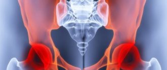
Tuberculous coxitis (tuberculosis of the hip joint)
According to statistics, the most common joint affected by tuberculosis is the hip joint.
Symptoms
Tuberculous coxitis is characterized by a long-term chronic course and periodic exacerbations. An exacerbation usually occurs when the intraosseous focus of tuberculosis breaks into the joint cavity. Patients may complain of low-grade fever, weakness, fatigue, and lameness. The pain can be in both the hip and knee joints, which naturally complicates diagnosis. During an exacerbation of the disease, swelling is observed in the joint area and enlarged lymph nodes can be felt.
Diagnostics
Most often, at the initial stages, classical X-ray diagnostics are used; it is better to use digital radiography. Using the image, bone atrophy, local, and later widespread osteoporosis can be diagnosed.
In cases where making a diagnosis may be difficult or simply requires a more detailed diagnosis and a 3-dimensional view of the lesion, spiral computed tomography (SCT) can be used.
Causes
The reasons for the development of this pathology can be considered:
- Disturbance of metabolic processes in the body.
- Development of an autoimmune reaction. At the same time, the body’s protective cells begin to destroy themselves.
- Past infectious disease in acute form.
- Large loads on the joints, frequent fatigue.
With the development of coxitis, the synovial bursa of the joint becomes inflamed. Further, the disease can progress in different ways. Coxitis in adults and young patients must be treated. This disease is considered very dangerous, since its progression can have very dangerous consequences - sepsis, chronic pathologies of connective fibers, and even leukemia.
Tuberculosis drives (tuberculosis of the knee joint)
The knee joint is immediately behind the hip joint in terms of incidence according to observations.
Symptoms
There is also a temperature of about 37 degrees, fatigue, lameness, pain that increases during loads on the leg, thickening of the skin fold, swelling of the joint, and limitation of its function. A fistulous tract may even form behind the joint, in the popliteal fossa.
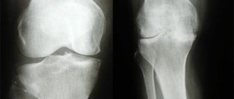
Diagnostics
When using classical x-rays, it is also possible to observe atrophy of the bones that form the knee joint, and uneven osteoporosis can be determined. A detailed study can reveal a lesion in the bone that grows towards the joint, followed by destruction of the articular surface.
The use of SCT immediately answers many questions of interest: the exact number of lesions, their size, structure and content, the presence of small lesions not visible on x-rays, a 3D view of the location of lesions in the bones around the joint.
Coxitis in ankylosing spondylitis: correlations with disease activity and quality of life
Introduction. Ankylosing spondylitis (AS) is a systemic chronic inflammatory disease of the axial skeleton with frequent involvement in the pathological process of entheses and peripheral joints, as well as other organs and systems [4]. In a number of patients, AS proceeds very aggressively with the rapid development of ankylosis in the sacroiliac joints (SIJ) and spine [1, 2]. One of the common factors of poor prognosis in AS is coxitis. Coxitis is an inflammatory process in the hip joint in which a narrowing of the joint space occurs with or without the formation of osteophytes [3].
According to B. van der Cruyssen et al. [9, 10], who analyzed materials from registers of patients with AS, the frequency of coxitis ranges from 24 to 36% depending on the duration of AS, and the need for endoprosthetics is 8%. At the same time, according to a multicenter cross-sectional study conducted in Russia [5], clinical signs of coxitis were observed in 56% of cases, and in 7% of cases joint replacement was required.
Coxitis often develops in patients with AS in childhood and adolescence and in most cases is bilateral; in adults, coxitis often develops in the first 10 years of the disease [5,6].
According to the latest domestic modern research in this area: in the hospital group of patients with AS, the frequency of coxitis reaches 51% [8].
Patients with AS with coxitis have higher disease activity and more pronounced functional impairment than patients without damage to the hip joint. Coxitis causes a significant decrease in working capacity. Ultrasound examination (ultrasound) allows in some cases to carry out differential diagnosis with similar clinical manifestations between synovitis and enthesitis localized in this area [3].
The relevance of assessing quality of life is shown in a number of diseases, however, this issue has not been sufficiently studied in patients with ankylosing spondylitis. The scarce data presented in the foreign literature, with ambiguous results and different methodological approaches, do not allow us to form a holistic picture of the quality of life of patients with ankylosing spondylitis in the Russian population. In addition, the available literature does not provide works on an in-depth analysis of possible factors influencing the quality of life of this category of patients. Therefore, it was decided to compare the quality of life of patients with ankylosing spondylitis with the presence of coxitis and patients without involvement of the hip joints in the inflammatory process.
Purpose of the study. To compare indicators of disease activity and quality of life in patients with AS with and without the presence of coxitis, to determine the possibilities of using ultrasound of the hip joints as a screening method for early diagnosis of complications for all patients with Bechterew's disease.
Materials and methods. The study was conducted on the basis of the rheumatology department of the State Healthcare Institution of the State Clinical Emergency Hospital No. 25 in Volgograd. The cross-sectional study included 56 patients with confirmed AS (meeting the modified New York 1984 criteria) [11].
Among the patients participating in the study, 46 were male, the average age was 32.4 years, the average duration of the disease was 14.7 years, the average age of onset was 22.8 years, and 46% of patients had disabilities.
The study was conducted within the framework of the scientific topic of the candidate's dissertation “Complex analysis of factors of progression and resistance to therapy in ankylosing spondylitis” and was approved by the ethics committee of Volgograd State Medical University.
All patients underwent a general clinical and laboratory examination using the following indices: BASMI (Bath Ankylosing Spondylitis Metrology Index), BASFI (Bath Ankylosing Spondylitis Functional Index), BASDAI (Bath Ankylosing Spondylitis Disease Activity Index). Quality of life was assessed using the HAQ (Health Assessment Questionnaire). To assess the condition of the hip joints, an ultrasound examination was performed.
To evaluate statistical data, the STATISTICA 10 program (StatSoft, USA) was used.
Results and discussion. Of the 56 patients, ultrasound signs of coxitis were found in 27 (48.22%), in 8 (14.28%) patients coxitis was combined with enthesitis in the greater trochanter, in 21 (37.5%) patients changes in the hip area were not detected. joints.
The overwhelming number of patients had positive HLA-B27, in six patients this antigen was negative (in 1 the presence of coxitis was noted), 2 patients did not undergo testing. All patients were divided into groups:
Group 1 – patients with coxitis;
Group 2 – with coxitis in combination with enthesitis of the greater trochanter;
Group 3 – patients without signs of involvement of the hip joint in the inflammatory process.
Table 1. Characteristics of the medical history of patients with ankylosing spondylitis, according to the patient registration card (M, CI)
Notes: hereinafter N – quantity, M – arithmetic mean, CI – confidence interval.
All examined patients were hospitalized and had fairly high disease activity. Our results are consistent with previous studies on the overall frequency of occurrence of coxites (48.22% in this study, 56% [2] and 74% [3] in similar studies).
Table 2. Indicators of activity, quality of life and ultrasound characteristics studied (M, CI)
Notes: TCO—thickness of the cervical-capsular region, THC—thickness of hyaline cartilage.
A decrease in the thickness of hyaline cartilage (HCT) (N>2.0 mm) and an increase in the thickness of the cervical capsular region (CCT) (N up to 7.0 mm) were reliable markers in ultrasound examination for the diagnosis of coxitis.
According to table. 1, the first group of patients (coxitis) had an earlier onset (18.4 years), a later age at diagnosis (34.9 years), a much longer duration of the disease (17.95 years) and, as a result, higher rates of both disease activity (BASDAI=6.5), and functional and metrological indices (BASFI=5.5 and BASMI=5.0, respectively), as well as low quality of life values (HAQ=1.68). The second group of patients with a combination of coxitis and enthesitis of the greater trochanter has higher rates of the studied parameters (BASDAI = 6.5, BASFI = 6.5, BASMI = 6.5, HAQ = 1.82), probably due to the small number of the general group of subjects and the severity of clinical manifestations at the time of the study. The third group of patients showed the lowest studied indicators on scales and quality of life questionnaire (BASDAI=4; BASFI=3.5; BASMI=4; HAQ=1.12), which is most likely explained by the later onset and earlier detection of the disease in comparison with patients from three groups with coxitis, in which an inverse relationship is observed: earlier onset and later diagnosis.
Instrumental ultrasound indicators corresponded to the measured indices and quality of life. In patients with coxitis: THC = 1.46 mm, TShO = 9.8 mm, Coxitis + Enthesitis: THC = 1.56 mm, TShO = 10.0 mm and in patients without coxitis: THC = 2.3 mm, TShO =5.0 mm. Accordingly, according to table. 2, the more pronounced the ultrasound changes were, the worse the activity indicators, metrological, functional index and quality of life.
During the study, a separate cohort of patients emerged. Among the examined patients: out of 27 (48.22%) with identified coxitis, 9 (16.08%) had signs of coxitis according to ultrasound, but had no clinical manifestations (did not experience pain or limitation of range of motion in the joint).
Table 3. Comparative characteristics of patients diagnosed with coxitis with and without clinical manifestations
According to the comparative characteristics described above, patients without clinical manifestations of coxitis have an earlier diagnosis (28.75 years), a shorter duration of the disease (12.5 years) and a later onset of the disease (26.1 years) than patients with severe clinical manifestations (35.2 years, 16.35 years, 21.15 years, respectively).
Conclusion. Patients with AS with coxitis, especially in combination with enthesitis of the greater trochanter, have significantly higher rates of disease activity, severe dysfunction and low quality of life than the comparison group without coxitis.
Our data fully reflects modern ideas about the need for early and timely diagnosis of AS, for the earliest possible start of rational pharmacotherapy, which will subsequently ensure the highest possible quality of life for these patients.
Gratitude. The author expresses his gratitude to Natalya Valerievna Nikitina for her assistance in conducting an ultrasound of the hip joints.
Surgery
In order for the treatment of childhood coxitis to be as effective as possible, it is necessary to begin the patient’s recovery with complete immobilization (immobilization) of the hip joint. To do this, you need to use special plaster casts. The doctor also prescribes medications for the child. If such treatment does not give the desired result, and the pathology continues to progress, the doctor prescribes surgery. This procedure will eliminate the source of inflammation and restore the functionality of the joint. Today there are several types of surgical operations for coxitis, namely:
- Necrectomy – removal of dead tissue;
- Arthroplasty. Most often used for adult patients.
- Intra-articular resection. This operation is less traumatic, and after it is performed the patient has every chance of completely restoring the motor activity of the joint.
- Extra-articular resection. During this operation, the joint capsule is not opened, which makes the procedure less effective. It is not recommended to use this technique for the tuberculous form of coxitis, as it will not bring the desired result.
- Corrective osteotomy. The joint is given the desired physiological position.
After surgery or effective treatment with medications, the patient must undergo an additional course of rehabilitation. At this stage, you need to do therapeutic exercises, massage, and physiotherapy. Remember that the sooner you see a doctor and begin treatment, the greater the likelihood of a full recovery. Do not self-medicate, as this can only worsen the patient's condition.