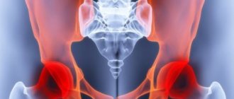
Movable areas of the body experience increased stress and “wear out” over time, which in some cases is revealed by a preventative MRI study. The hip joint has a complex anatomical structure, and the branching of the nerve canals in its area leads to the spread of pain to the leg, back or groin area. This feature makes diagnosis difficult, and patients often do not associate pain with problems in the bone joint. At the first signs of discomfort, you should consult a doctor to avoid further spread of inflammation and critical consequences.
Description and symptoms of pathologies of the articular zone of the musculoskeletal system
Prolonged manifestations of pain are the reason for an MRI of the hip joint. The bone joint itself is not penetrated by nerve fibers, so some diseases can be asymptomatic for a long time. Soreness appears at the stage of damage or compression of the nerve canals passing in the “hinge” region of the body. At the initial stage of the appearance of disorders, unpleasant sensations arise in a limited area of the connection of the femur with the pelvis. If the disease is ignored, the source of inflammation grows and spreads to neighboring tissues: muscle and tendon fibers, cartilage formations, nerve and blood tracts.
A person may experience the following sensations:
- prolonged radiating pain that does not go away within several days, the cause of which can be identified on an MRI of the hip joint;
- night pain leading to insomnia;
- the desire to change positions as often as possible in order to relieve the pelvic joint with constant discomfort in it.
These signs serve as a reason for a thorough diagnosis, and emergency clinical care is required if the patient complains of the following conditions:
- Irradiation to the groin. Through the conductive canal of the nerve, pulsating sensations spread to the lower abdomen and groin area; with inflammation of adjacent fibers, the sciatic nerve is affected.
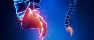
- Shootings in the lower part of the back. They can be both sharp and dull, pulsating, leading to limited movement of the body and pelvis.
- Spread of pain to the leg, including the knee area. It may manifest itself as muscle weakness or itchy sensations in the skin.
- A “hinge” clutch that prevents free movement of the limb. It is a sign of arthritic and arthrosis lesions.
- Partial or complete lack of mobility associated with the destruction of pelvic tissues or trauma experienced.
- Lameness associated with low intensity pain. It requires correction, since a change in motor habit leads to deformation of the entire musculoskeletal system.
Most patients describe the phenomenon of crunching sounds when changing body position or movement. A kind of crunching sound is produced by individual ligaments. If the sound is not accompanied by unpleasant sensations, there is no need to worry. Additional pain requires contacting a medical specialist.
Diagnostic tests
To determine the cause of pain in the lateral surface of the thigh and hip joint, when sitting and lying on the side, the following studies are carried out:
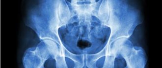
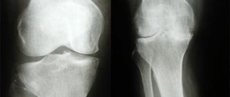
- Hardware methods. Includes computed tomography and magnetic resonance imaging, x-ray examination of the joints and spine. The x-ray is taken in three projections.
- Instrumental methods - arthroscopy, with simultaneous biopsy of the affected tissue.
- Laboratory tests reveal signs of inflammation in the joints and metabolic disorders. The cause of joint destruction is identified.
To make a diagnosis, the accompanying symptoms of pathology of the joints and spine are taken into account. At the initial visit, the doctor detects pathological signs:
- Hip pain, acute, chronic, wandering, permanent.
- Hip discomfort depends on the time of day.
- Irradiation of pain occurs in the groin area, lumbar back, and along the lower limb.
- The pain when lying on the side changes in character when turning over or sitting. When turning, bending, walking, running, the pain subsides or intensifies.
- Painful sensations affect the surface of one thigh, or both at once.
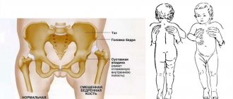
Women indicate the presence of complicated childbirth, intense training. A child's pain may be caused by a vaccination.
Hip pain
Causes of pain in the hip joint
High loads placed on the natural “hinges” of the human body lead to the development of various diseases. Both old and recent traumas contribute to subsequent disorders. One of the common anomalies is a fracture or destruction of the femoral neck, adjacent pubic joint or sacral bone, which can be recognized not only on x-rays, but also on MRI. The hip joint suffers from severe bruises, muscle and ligament sprains.
In some cases, mechanical abrasion of cartilage fibers occurs. This pathology affects people who are forced to engage in heavy physical labor at a professional level, or who engage in sports with a high level of physical stress. Problems may also arise in persons with congenital anomalies in the development of this part of the body. Inflammatory processes also lead to tissue and nerve damage.
The pelvis and its adjacent joints suffer from systemic and degenerative diseases. Bone fibers are susceptible to necrosis if the blood supply to the department is disrupted. Endocrine disruptions lead to the development of joint pathologies, manifested in the following diseases:
- Arthritis, arthrosis and coxarthrosis - abrasion and inflammation of the cartilage layers and outer surfaces of the bones.
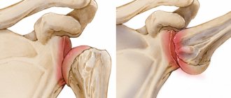
- Bursitis is inflammation as a result of mechanical damage or internal infection of the joint capsule, accompanied by severe swelling and sharp pain.
- Tendinitis is an inflammatory irritation of the ligamentous apparatus, covering both the articular areas of the pelvis and the rest of the leg, down to the toes.
- Anomalies of bone growth due to congenital predisposition and the influence of other pathologies.
- Irradiation from nearby organs of the genitourinary system or gastrointestinal tract.
- Tumor formations that put pressure on nerve endings.
If any of these diseases are suspected, a targeted examination is prescribed to differentiate pathologies and identify the root cause of the painful condition.
Diagnosis of diseases using hardware and MRI of the hip joint
The first step in identifying disorders in the joints of the musculoskeletal system is to contact an orthopedic traumatologist. If necessary, the doctor will resort to the participation of other highly specialized specialists: surgeons, oncologists, rheumatologists or neurologists. At the initial stage, it is necessary to do standard laboratory tests that will determine whether an inflammatory process is occurring in the body of the person being examined.
MRI of the hip joint is rarely performed as a primary hardware study. The area is first checked using radiographic imaging and ultrasound scanning. These diagnostic methods help to detect obvious anomalies occurring in the bones and soft tissue adjacent formations. Most often, this is the result of injuries, fractures, muscle or tendon ruptures.
If the cause of pain is more complex processes that are not detected by traditional diagnostic methods, the pelvis is scanned using tomographic methods - CT and MRI. Visualization using a computer scanner allows you to specifically study the condition of the skeleton, the inner surface of the “hinge” zones, the degree of integrity of the blood and nerve channels, as well as recognize the presence of tumors of various natures. An examination using a nuclear resonance unit allows you to assess the condition of muscles, cartilage and other less dense fibers. The layering of bones will not interfere with MRI imaging of the hip joint.
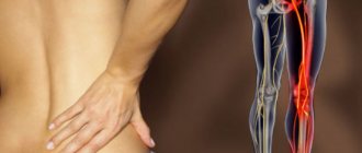
Inner thigh hurts when walking
What to do if the inner side of the thigh hurts when walking, which doctor should you contact to diagnose the diagnosis and prescribe treatment? The first thing to do in such a situation is to temporarily eliminate any physical activity. There is no need to continue to perform your professional duties through pain. Be sure to make an appointment with an orthopedist as soon as possible.
This specialist will conduct an initial examination and manual examination, and perform a series of diagnostic functional tests. This will allow him to make a preliminary diagnosis and provide the necessary medical care. He will then develop a plan for conducting the survey. It may include:
- X-ray of the hip and knee joint (to exclude the possibility of developing osteoarthritis, deposition of calcium salts, instability of the position of the heads and condyles of bones, etc.);
- an X-ray image of the lumbosacral spine to exclude the possibility of a decrease in the height of the intervertebral spaces, which corresponds to the second stage of development of osteochondrosis (also in the image, an experienced doctor will be able to see deforming osteoarthritis of the intervertebral joints, instability of the position of the vertebral bodies and intervertebral discs, etc.);
- MRI of the lumbosacral spine, hip and knee joint (necessary to assess the condition of soft tissues, such as cartilage, ligament, tendon, muscle, connective);
- Doppler ultrasound of the blood vessels of the lower extremities (allows you to diagnose atherosclerosis, varicose veins, obliterating endarteritis, thrombosis, vasculitis, diabetic angiopathy and other pathologies);
- electromyography and electroneurography.
Based on the results of all studies, the doctor will be able to make an accurate diagnosis and prescribe appropriate treatment.
Treatment and pain relief
The primary symptoms of each described disease are identical, but this does not mean that the treatment will also be the same. After identifying the root cause of the anomaly, specific therapy is prescribed, aimed at eliminating the source of the disease.
The following recommendations are general in restoring functionality after MRI of the hip joint:
- reducing physical activity on the affected limb, increasing the duration of rest;
- the use of drug therapy aimed at relieving inflammation, pain, spasms and regeneration of cartilage tissue, general strengthening of the body;
- influencing the pelvis using physiotherapeutic methods (during the remission period), visiting a massage room and conducting a course of therapeutic exercises;
- deciding on surgical intervention if purulent or bloody inclusions, fluids, or malignant formations are found in the joint cavities.
During primary treatment, it is important to get rid of debilitating pain, which will help improve your quality of life and overall well-being. Standard analgesics can be used, but provide only temporary relief. To enhance the effect, it is recommended to limit physical activity, massage the sore area, and apply a cool compress if the cause of the pain is an injury. On the recommendation of a physician, you can turn to non-steroidal anti-inflammatory drugs.
Evaluation of treatment effectiveness using MRI of the hip joint
After undergoing restorative manipulations, it is important to carry out repeated diagnostics to verify the correctness of the prescribed treatment course. The best way to compare the rate of tissue regeneration is MRI. If there are images from a previous study, the functional diagnostician compares the stage of the disease, identifies the absence or presence of relapses (tumors or infections affecting the pelvis), and migration of metastases.
After surgery, the composition of the fluid in the joint cavities and the degree of fiber restoration are examined. In some cases (with cancer of the bone), it is necessary to remove the hinge part of the hip and replace it with a prosthesis. The implant material is a metal alloy, so MRI scanning of the hip joint is contraindicated. An alternative is the same informative examination as a computer scan.
Hardware examination can be carried out in specialized diagnostic centers containing tomography rooms. You can select the nearest medical facility on the website of the Moscow Unified Recording Center. An expanded list of clinics makes it easier to compare by ratings, location addresses, and prices for services. Mark the best offers and sign up for diagnostics through the service. This will open access to additional discounts on the selected type of tomography.
Diagnosis of pathology
Painful sensations can occur in the presence of various diseases. It is not recommended to try to get rid of unpleasant symptoms on your own. It is very important to establish the correct diagnosis, and then undergo the prescribed treatment for hip joints in Moscow in a specialized clinic.
To determine the provoking factor in the development of unpleasant symptoms, doctors use various diagnostic methods. First, a visual inspection and palpation of the painful area is performed. The doctor also collects medical history data.
Based on patient complaints, diagnostic procedures are prescribed:
- Laboratory testing of blood and urine.
- Biochemical blood test.
- Radiography.
- Tests for rheumatoid factor.
- Proteinogram.
- CT scan.
- Magnetic resonance imaging.
- Determination of vascular patency.
- Ultrasonography.