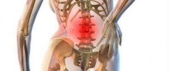
Cervical osteochondrosis is a disease that affects the vertebrae and intervertebral discs. Cervical osteochondrosis refers to deforming dorsopathies. Involutive changes in the discs are observed as early as 20 years of age. At the same time, they become more sensitive to stress, less elastic, and lose lubricating fluid.
Most often, the pathology occurs in the elderly, but currently there is a significant increase in incidence among children and young people. Neurologists at the Yusupov Hospital identify cervical osteochondrosis using the latest diagnostic tests. After clarifying the diagnosis, complex therapy is carried out with the most effective medications, physiotherapeutic procedures and innovative methods of physical rehabilitation.
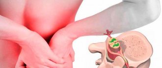
The name of the disease consists of two Greek terms “osteon” (bone) and “chondros” (cartilage). Cervical osteochondrosis begins with changes in the central part of the disc. The intervertebral disc loses moisture and decreases in size, this leads to the convergence of the vertebral bodies and pinching of the nerve roots and blood vessels. The vertebrae receive nutrients from surrounding tissues, which causes harm to the body. Compression of nerves and blood vessels leads to a protective muscle spasm, which, as the disease progresses, becomes a cause of pain.
Which doctor treats this disease?
At the Yusupov Hospital, the treatment of osteochondrosis is the field of activity of neurologists. However, if symptoms of neck osteochondrosis appear, you may contact a general practitioner. A neurologist will select medications for cervical osteochondrosis that have the least burden on the body, which is important during drug therapy.
To determine the presence of a pathological process in cartilage tissue and cervicobrachial osteochondrosis, the patient is sent for a comprehensive examination to the diagnostic center of the Yusupov Hospital. Tactics on how to treat cervical osteochondrosis are being developed in accordance with research results.
Interdisciplinary collaboration also makes it possible to treat the patient's comorbidities. In addition, when contacting the Yusupov Hospital, the patient receives full information support: a treatment plan, an extract on the cost of services, information about consultations with specialists and diagnostic measures.
Make an appointment
First aid
Of course, you should first do a spinal x-ray or other test to determine an accurate diagnosis. First, in case of severe pain, you can take a painkiller: “Tempalgin”, “Dolaren”, “Analgin”. If this group of drugs does not help relieve pain, you can take Ibufen, Diclofenac or Nise, but keep in mind that such drugs have a negative effect on the gastrointestinal tract. Therefore, this can only be an emergency, and then immediately consult a doctor.
You can also apply pain-relieving ointments. It is allowed to take diuretics if swelling appears in the area of inflammation. You can also use a Shants collar, but you should not wear it for a long time.
Causes
Cervical osteochondrosis develops under the influence of various provoking factors. No specific cause of cervical osteochondrosis has been identified. Often the disease is associated with metabolic disorders and aging of the vertebrae.
Researchers suggest that cervical osteochondrosis develops for the following reasons:
- Excessive load on the spine. A high load on the spine is observed when wearing incorrect shoes, flat feet, obesity, and prolonged sitting;
- Metabolic disorders. Deficiency of vitamins, minerals, and calcium metabolism disorders can cause degenerative processes in the vertebrae;
- Congenital and acquired anomalies of the development of the spine and ligamentous apparatus (thickening of the ligaments, lumbarization, sacralization);
- Pathologies of the gastrointestinal tract leading to insufficient absorption of nutrients;
- Infections, intoxication;
- Injuries, bruises, spinal fractures, as a result of which the blood supply and innervation of the spinal column are disrupted, which causes their degenerative disorders;
- Stress;
- Wearing shoes with heels;
- Pregnancy, especially multiple pregnancy;
- Autoimmune connective tissue lesions, pathological structure of collagen types 1 and 2;
- Occupational hazards (lifting heavy loads, prolonged vibration, working in a sitting position with constant head tilt);
- Atherosclerotic and other changes in the vertebral arteries;
- Curvature of the spine (kyphosis, scoliosis, kyphoscoliosis).
An important risk factor for the development of cervical osteochondrosis is family history. This fact proves the presence of osteochondrosis in children when the spine is not yet overloaded.
Causes of cervical osteochondrosis
Today it is impossible to name the exact causes of degenerative processes occurring in intervertebral discs. There is no confirmation that cervical osteochondrosis is an aging phenomenon.
Numerous studies conducted by scientists from different countries have found that osteochondrosis of the cervical spine has predominantly provoking factors.
Among the predisposing causes of the development of osteochondrosis of the neck are:
- low level of activity, inactivity and a predominantly sedentary lifestyle;
- types of work that involve a static load on the cervical spine;
- excess body weight, insufficient level of physical development;
- disruption of connective tissue development processes;
- old spinal injuries;
- spinal deformity, use of insufficiently comfortable pillows and mattresses for rest;
- genetic predisposition.
The vast majority of reasons are in one way or another related to the natural processes of aging of body systems, as well as the likely development of pathologies of bone and cartilage tissue.
Degrees
Thanks to the special structure of the spine, it is able to perform its functions. The main structural unit is considered to be the spinal motion segment (SMS). It consists of two adjacent vertebrae, an intervertebral disc and a muscular-ligamentous apparatus. Osteochondrosis leads to dystrophic-degenerative processes, first in the intervertebral disc, then in the vertebra. When one vertebra is damaged, its functions are provided by adjacent ones. This leads to increased load and loss of mobility of the affected segment.
Doctors distinguish several stages in the development of cervical osteochondrosis:
- First degree of cervical osteochondrosis. Since the intervertebral disc is deprived of its own blood supply and receives nutrients from surrounding tissues, it is susceptible to degenerative changes. Osteochondrosis at the 1st stage of development is characterized by destruction of the nucleus pulposus and cracks in the fibrous ring. Clinically, this is manifested by acute or persistent local pain in the neck (cervicalgia) and stiffness;
- Osteochondrosis of the second degree of the cervical spine. At this stage, the destruction of the fibrous ring continues, pathological mobility and instability of the vertebrae appear. Patients complain of pain in the neck, aggravated by physical activity, tilting the head or in a certain position;
- The third stage of the disease is characterized by complete destruction of the fibrous ring. The nucleus pulposus is not fixed. Intervertebral hernias may occur, which cause severe pain. At this stage, due to poor fixation of the SMS, spinal curvature may form;
- At the fourth stage of the disease, the intervertebral disc is replaced by connective tissue, and other adjacent segments are affected. Spondyloarthrosis and arachnoiditis develop. The joints become completely immobile - ankylosis develops. Bone tissue grows around the affected area - osteon is formed. With the fourth degree of cervical osteochondrosis, clear symptoms are observed: severe pain that radiates to the arm, sternum, to the area between the shoulder blades, sensitivity disorders.
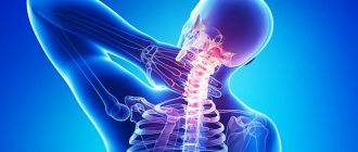
How does cervical chondrosis manifest: symptoms
The vertebrae of the neck are located very close to each other. Any damage or displacement to the side leads to pinching of nerve fibers and blood vessels. All this has a direct impact on how cervical chondrosis manifests itself . The main signs of the disease are:
- sharp pain in the neck, radiating to the chest, back of the head, shoulder blades, shoulders, temples;
- increased pain and the appearance of a crunch when turning the head;
- muscle weakness, limitation of motor activity of the upper extremities;
- decreased neck mobility;
- dizziness;
- hearing impairment, decreased visual acuity;
- general weakness, decreased performance.
Without treatment, cervical chondrosis continues to progress . The processes of dystrophy and degeneration of the vertebrae and intervertebral discs in the upper segment of the spine are irreversible.
Symptoms and signs
Signs of cervical osteochondrosis in the initial stages may be nonspecific: dizziness, headaches, weakness, crunching when moving the head. As the disease progresses, the following symptoms develop:
- Severe pain in the neck and shoulders;
- Numbness of the hand;
- Dizziness;
- Increased blood pressure;
- Impaired coordination of movements;
- Increased sweating.
There are several syndromes that appear with the development of a pathological condition of the muscles of the back and cervical spine:
- Cervical migraine syndrome.
- Vertebral artery syndrome.
- Hypertension syndrome.
- Cardiac syndrome.
- Radicular syndrome.
They occur when nerve endings are injured, arteries and veins are compressed during the development of the disease. The most dangerous complication is considered to be vertebral artery syndrome. There is a disruption of blood flow through the artery supplying the brain and spinal cord. The patient's hearing decreases, vision decreases, and constant dizziness develops. The patient may lose consciousness while moving due to a sudden disruption of blood flow.
As a result of compression of the nerves responsible for the innervation of the muscles of the chest and diaphragm, pain appears in the heart area, not associated with heart disease, but at the same time tachycardia, arrhythmia and hypotension may develop. Compression of the veins leads to the development of hypertensive liquor syndrome. Intracranial pressure increases, nausea, vomiting, and severe headache appear due to impaired blood flow from the brain.
As a result of compression of the neck, radicular syndrome develops - severe pain appears in the neck, shoulders, shoulder blades, and back of the head. With this syndrome, the arms and neck area become numb. With cervical migraine syndrome, the patient experiences severe pain in the back of the head, which is often accompanied by nausea and vomiting.
Reflex syndromes occur when the spinal roots are not yet affected. Patients complain of pain in the neck, head (especially the back of the head), and arms on one or both sides. Reflex pain, unlike radicular pain, is not combined with sensory disturbances. Cervicalgia can be dull and aching. Acute sharp “shoots” of pain are called cervicago. There is muscle spasm and pain, pain in the paravertebral points. Signs of cervical osteochondrosis intensify in an uncomfortable position, when tilting the head, coughing, or physical activity. Signs of epicondylosis, glenohumeral periarthrosis and shoulder-hand syndrome appear due to nerve impulses from the annulus fibrosus of the affected segment, which causes compensatory muscle spasm.
Radicular syndromes are accompanied by impaired motor activity and sensitivity. In this case, nerves and blood vessels are infringed, venous and lymphatic outflow in the pathological focus is disrupted as a result of a decrease in the intervertebral canal. The pain with radicular syndrome is acute and intense. A common cause of pinched spinal nerves is the formation of a hernia. In the area of the pathological focus, muscle tone decreases. With radiculoischemia, in addition to nerves, blood vessels are compressed.
If the phrenic nerve is involved in the pathological process, cardiac syndrome occurs. It manifests itself as a burning, acute pain in the left half of the chest with radiation to the arm and interscapular region. The name of the syndrome is due to the fact that the nature of the pain is similar to an attack of angina. The main difference between pain during angina pectoris is that it is relieved after taking nitroglycerin, can occur at rest and is combined with interruptions in heart rhythm (tachycardia, arrhythmia).
Signs of cervical osteochondrosis depend on the location of the pathological process. When the upper cervical vertebrae are affected, the blood supply to the brain is disrupted due to compression of the cerebral arteries. This leads to headaches (especially in the occipital region), dizziness, fainting, and high blood pressure. Dizziness with cervical osteochondrosis is caused by a decrease in blood flow to the inner ear. Patients also experience nausea and vestibular and ocular symptoms.
With combined damage to the vertebrae, they speak of cervicothoracic osteochondrosis. The disease is manifested by the following symptoms:
- Dizziness;
- Pain in the neck and arm;
- Tingling, crawling sensation on the upper limb;
- Intercostal neuralgia.
Make an appointment
Why arthrosis of the cervical vertebrae needs to be treated
If the treatment of osteoarthritis of the cervical vertebrae is delayed, the disease can lead to the following complications:
- compression of nerve endings and, as a result, severe pain;
- pinching of the spinal canals;
- stroke.
Rare, but accurate: arthrosis of the cervical vertebrae can lead to stroke
Diagnostics
Cervical osteochondrosis is a chronic disease that can lead to the formation of hernias and compression of the spinal cord. Therefore, it is important to establish an accurate diagnosis in a timely manner and begin therapy. To identify cervical osteochondrosis, the following types of instrumental diagnostics are used:
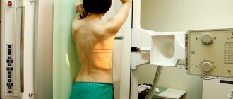
- Spondylography or radiography of the spine. This research method is painless, highly informative and does not require special training. An X-ray of the spine allows you to evaluate its anatomical and functional features. In the picture, attention is paid to the structure of the vertebrae, their relationship to each other, the distance between them, the lumen of the spinal canal;
- Computed tomography - provides information mainly about the condition of bone tissue, allows you to identify narrowing of the spinal canal and disc herniation;
- Magnetic resonance imaging - allows you to determine changes in soft tissues. The MRI image clearly shows changes in the intervertebral discs and spinal cord.
At the Yusupov Hospital, the patient undergoes a comprehensive examination. Doctors take into account the individual characteristics of his body and concomitant diseases. An important advantage of the neurology clinic is the availability of modern, high-quality equipment and specialized specialists: neurologists, neurosurgeons, oncologists.
How it develops
The cervical region includes 7 vertebrae, the most mobile of the entire spinal column. If certain pathological processes occur in the body, the cervical vertebrae, protected by an elastic cartilage “plate” (disc), become covered with osteophytes (bone growths). The cartilage, in turn, becomes thinner and destroyed, and the adjacent soft tissue atrophies.
The intervertebral disc can bulge and put pressure on nearby tissue or nerves. The vertebrae change their shape, which leads to spinal deformities. If they are pronounced, the patient develops deforming cervical arthrosis.
Sometimes the cause of neck arthrosis is... flat feet!
Drug treatment
Treatment of osteochondrosis of the cervical spine consists of drug and non-drug therapy. Even after complete recovery, to exclude relapses of the disease, neurologists at the Yusupov Hospital carry out preventive measures. In the acute period, for the treatment of cervical osteochondrosis, doctors prescribe medications to patients from the following pharmacological groups:
- Non-narcotic analgesics (analgin, baralgin, trigan). They are taken orally or administered intramuscularly to quickly achieve an effect;
- Nonsteroidal anti-inflammatory drugs (diclofenac, ibuprofen, ketanol);
- B vitamins in large doses.
In order to reduce fluid retention in the area of the spinal root and surrounding tissues, diuretics (furosemide, Triampur, Lasix) are used. Antihistamines (diphenhydramine, suprastin, pipolfen) potentiate the effect of analgesics. Muscle spasms are eliminated by muscle relaxants (sirdalud, miorix, mydocalm, flexen). For prolonged severe pain, neurologists perform a nerve block.
To improve metabolic processes in the intervertebral disc, chondroprotectors (alflutop, evalar) are used. These drugs increase the content of glycosaminoglycans, increase the firmness, elasticity and shock absorption of the intervertebral discs.
Anti-dizziness pills
Patients often experience dizziness with cervical osteochondrosis. To reduce them, doctors prescribe non-steroidal anti-inflammatory drugs. NSAIDs belonging to different groups differ in their mechanism of action and effect, so only a qualified specialist can determine the appropriate drug.
It is important to remember that medications for cervical osteochondrosis cannot be taken without a doctor’s prescription. Nonsteroidal anti-inflammatory drugs have side effects, so before prescribing them, the neurologist determines the presence of contraindications in the patient and the required dosage. Drugs for dizziness in cervical osteochondrosis can improve the patient’s quality of life.
Injections for osteochondrosis
Injections for osteochondrosis of the cervical spine help relieve pain during an exacerbation. With this method of drug administration, the effect occurs quickly. Neurologists use various injections.
Nurses administer drug solutions subcutaneously, intramuscularly or intravenously. During the period of exacerbation of the disease, drugs that are administered by injection for cervical osteochondrosis have an exclusively symptomatic effect. To treat the disease, contact the neurology clinic of the Yusupov Hospital.
Make an appointment
Headache treatment
Headache is a symptom that occurs with various disorders. However, cervical osteochondrosis is characterized by attacks of intense headaches. Head movements increase symptoms, so to eliminate them, doctors prescribe analgesic tablets and non-steroidal anti-inflammatory drugs.
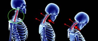
Degrees of development of cervical osteochondrosis and their characteristic symptoms
The process of development of the disease proceeds quite slowly, but at the same time it has 4 clearly visible stages:
- 1st degree cervical osteochondrosis – discomfort and minor pain during prolonged walking or static position. The beginning of the formation of compactions in the intervertebral discs;
- Osteochondrosis of the cervical spine, degree 2 – intense pain. In order to eliminate pain, manual therapy and appropriate medications are used;
- 3rd degree cervical osteochondrosis – the presence of severe lesions of the connective tissue of the affected area;
- Osteochondrosis of the cervical spine, grade 4, is the final stage of the development of the disease, characterized by damage to almost all parts of the spine.
The last stage of development of the disease requires surgical intervention, which entails a long recovery.
Non-drug therapies
Complex non-drug therapy for cervical osteochondrosis of the spine includes:
- Protective mode - if the roots are pinched, patients lie on a hard surface,
- Massage;
- Physical therapy;
- Spinal traction;
- Physiotherapeutic procedures.
Massage for cervical osteochondrosis is used to reduce pain and swelling, improve peripheral blood supply, and eliminate muscle spasms. A contraindication to performing this procedure is the presence of acute pain. Massage the neck and back in the direction of lymph outflow. Particular attention is paid to the interscapular and paravertebral zones.
Therapeutic exercises for osteochondrosis of the cervical spine are aimed at eliminating muscle spasms and strengthening the muscular frame. Since instability of the vertebrae often occurs in the cervical spine, the exercise therapy instructor conducts individual classes, during which he teaches the patient how to safely perform exercises. Some authors recommend conducting physical therapy classes in a Shants collar.
To improve the mobility of the cervical vertebrae, rehabilitation experts recommend performing the following exercises:
- Flexion and extension of the neck. Bend your head forward toward your sternum without pulling your shoulders forward and then back. Hold the incline for 3 seconds, repeat each exercise 8-10 times;
- Neck turns. Turn your neck first to the left until it stops, then to the right, without changing the position of your shoulders and the level of your chin;
- Lower your head all the way down. Then tilt your head back without changing the level of your shoulders. Hold the position for 5 seconds.
The following exercises have been developed to strengthen the neck muscles:
- Place your hand on the back of your head. Tilt your head back, resting on your hand;
- Place your hand in the temporal region. While tilting your head, resist with your hand;
- Place your hand on your forehead, resisting it, tilt your head forward;
- With your right hand, tilt your head to the side, your left hand should be behind your back. Repeat the exercise on the other hand.
Autogravity therapy is the exact name for the spinal traction procedure. It is carried out using special devices. The goal of therapy is to reduce muscle spasm and restore the correct position of the vertebrae. To avoid complications, spinal traction is performed by a doctor.
To improve blood supply to the pathological focus, relieve swelling and eliminate pain, the following physiotherapeutic procedures are used in the Yusupov Hospital:
- Diadynamic currents. During this procedure, low-frequency currents are applied using a special device, which stimulate the muscles, relieve spasm and pain. They have a positive effect by improving tissue trophism;
- Ultraviolet irradiation. Under the influence of UV radiation, vitamin D metabolism improves, calcium content increases, bone tissue becomes stronger;
- Exposure to ultrasound - used to accelerate blood flow, antispasmodic and reparative effects. Ultrasound is capable of penetrating deep into tissues; sometimes it is used for better absorption of medicinal substances;
- Amplipulse therapy - allows you to relieve pain by blocking nerve impulses from the source of pain.
In the acute period of the disease, which lasts 4-7 days, painkillers, antispasmodics, and irritants are used to reduce pain. The patient is provided with rest. Immobilization of the cervical spine is carried out using a Shants collar. Exercise therapy and massage are contraindicated. Ultraviolet radiation is used.
The duration of the subacute period is 29 days. After complete recovery, the patient should rest for several days. Then you can begin a course of rehabilitation therapy. In the chronic course of the disease, the patient is prescribed muscle relaxants, chondroprotectors, B vitamins, and for pain - analgesics, NSAIDs. Physical therapy classes and massages are provided. The patient is given physiotherapeutic procedures (amplipulse, alternating current exposure), and spinal traction is performed.
At the Yusupov Clinic, doctors have extensive experience in successfully treating cervical osteochondrosis. Patients are given the opportunity to undergo a full course of rehabilitation: physiotherapeutic procedures, massage, spinal traction. The neurology clinic employs specialists of the highest medical category, professors, who use proprietary methods for the treatment of osteochondrosis.
How to treat osteochondrosis of the cervical spine?
Treatment of cervical osteochondrosis is determined by a specialist and depends on the degree of development of the pathology, the form of its course and the characteristics of the clinical manifestation of the disease.
Today, the most effective methods of treating the disease are:
- treatment with conservative methods, including medicinal/non-medicinal;
- surgical intervention;
- complex combination of techniques.
Physiotherapy
Involves the impact of physical factors on the affected area. With an integrated approach and proper implementation of all procedures, improvements become clearly noticeable already in the second or third month of treatment.
Among the most popular areas of physiotherapy prescribed for the treatment of cervical osteochondrosis are:
- electrotherapy;
- shock wave therapy;
- magnetic therapy;
- ball therapy;
- laser therapy;
- vibration massage.
Neck massage for osteochondrosis
Massage should be performed carefully, without the use of force. Violation of massage technique can cause negative consequences.
The starting position for the massage is the “lying on your stomach” or “sitting with a straight back” position.
All existing massage techniques are based on techniques such as:
- stroking - influencing the surface layers of the skin. It is performed with the palms of the hands and fingertips downwards from the back of the head, to the level of the upper third of the back;
- squeezing - influencing the deep layers of skin in the upper third of the back. Performed with two fingers (thumb and index), carried across the neck;
- rubbing – the main goal is to warm up the skin and increase blood flow in the desired area;
- kneading – affects deep-lying tissues; it must be used with caution, as improper use can aggravate the situation.
Therapeutic exercises for cervical osteochondrosis
There are certain exercises for the treatment of cervical osteochondrosis. The most effective of them include:
Self-extension
Starting position: sitting/standing with a straight back.
Execution order: while maintaining the starting position, try to lower your shoulders as low as possible, while stretching your head up.
Intensity of execution: at least 10 times (2-5 seconds each), at least 3 times a day.
Self-massage
Equipment: terry towel.
Starting position: sitting/standing, wrap a towel around your neck, and grab its ends with your hands.
Procedure: pull the ends of the towel one by one, gently kneading the neck muscles.
Important! During the exercise, you must ensure that the towel does not slip and rub your neck.
Gymnastics (flexion/extension, turns, bends)
Starting position: sitting/standing with a straight back.
Execution order: from the starting position, perform smooth flexion/extension, rotation or tilt of the head, first in one direction, then in the other.
Intensity of execution: 5-7 movements in one direction.
Nutrition
Proper nutrition for osteochondrosis is an important condition for achieving remission. The progression of cervicothoracic osteochondrosis stops with diet and therapeutic measures. Neurologists at the Yusupov Hospital know how to treat osteochondrosis of the cervical spine, so they create a complex of treatment measures, including procedures, exercise therapy, proper nutrition and lifestyle changes.
Many patients turn to neurologists with the question of how to treat osteochondrosis of the cervical spine and whether there are any dietary restrictions. Specialists at the Yusupov Hospital create individual nutrition programs that take into account the patient’s preferences. The diet for osteochondrosis is based on balanced, low-fat foods that are rich in nutrients. The patient's daily diet includes foods high in calcium.
What is osteochondrosis of the cervical spine?
Cervical osteochondrosis is a degenerative disease of the spine, during the development of which the structure of the connective tissue changes.
The disease is often disguised as a disorder of another type, but timely contact with a specialist allows for timely diagnosis and immediate treatment.
Degenerative changes in the tissues of the cervical spine are most often found in the medical history of people of mature (45-59) and elderly (60-74) age.
Despite this, it is worth noting: in modern society there is a rejuvenation of the disease, which is confirmed by the periodic diagnosis of cervical osteochondrosis in children and adolescents.
It is important that osteochondrosis can occur both in isolation and in combination with damage to other parts, in particular the thoracic, lumbar and sacral.
How to sleep with cervical osteochondrosis
For patients with diseases of the musculoskeletal system, the question of how to sleep properly with cervical osteochondrosis is relevant. Sleeping on your stomach provokes further development of the disease, so it is better to avoid sleeping in this position. The most optimal positions are on the back and side.
Cervical osteochondrosis progresses while resting on a bed with a soft mattress. Therefore, experts recommend giving preference to elastic mattresses, as well as moderately soft pillows. If a patient is diagnosed with cervicothoracic osteochondrosis, experienced specialists at the Yusupov Hospital will tell you which bedding is safe for sleeping.
Prevention
To prevent the occurrence or progression of cervical osteochondrosis, doctors recommend:
- Maintain correct posture;
- Lead an active lifestyle, take breaks at work;
- Do physical therapy exercises regularly;
- Sleep on a hard and flat surface, orthopedic mattress and pillow;
- Get rid of bad habits, especially smoking;
- Choose shoes taking into account the physiological structure of the foot;
- Do not carry bags on one hand, this leads to curvature of the spine;
- Lead a healthy lifestyle, eat right, eat plenty of fruits and vegetables;
- Do not sit for a long time with your head bowed;
- Go swimming.
In order to improve blood circulation, you should regularly undergo therapeutic massage.
Treatment in Moscow
At the Yusupov Hospital, doctors have been treating cervical osteochondrosis for many years. For the rehabilitation and restoration of performance of patients with osteochondrosis, the rehabilitation clinic has all the necessary equipment. You can find out the cost of treatment on the official website of the hospital.
Make an appointment with a neurologist online or call the contact center phone number. The neurology clinic employs the best doctors in Moscow, leading experts in the treatment of degenerative-destructive diseases of the spine. Patients are offered a comprehensive rehabilitation program, the cost of which is lower than the use of individual procedures.
Make an appointment
Difficulties in diagnosis
Before prescribing a complex treatment regimen or prescribing intra-articular injection of synovial fluid prostheses, the rheumatologist must ensure that the diagnosis is correct, since the symptoms of the disease duplicate a number of other pathologies. After collecting an anamnesis and studying a blood test to determine the absence of other diseases (with arthrosis, blood counts do not change), one of the following studies is carried out:
- radiography in several projections;
- CT or MRI;
- vascular angiography.
As a result of examinations, the doctor determines changes in the structure of the cartilage of the intervertebral discs, the joint space, the amount of synovial fluid and its deficiency in the joints, the nature of bone growths - osteophytes.