08 October 2021
According to the International Classification of Diseases, osteochondrosis is included in the group of diseases of the musculoskeletal system, mainly the spine, and connective tissue
According to the International Classification of Diseases, osteochondrosis is included in the group of diseases of the musculoskeletal system, mainly the spine, and connective tissue. Wearing out, collapsing and flattening over time, the intervertebral disc loses its shock-absorbing and protective function. The intervertebral space narrows, where the radicular nerves and blood vessels become compressed. This leads to various unpleasant symptoms, which force a person to see a doctor.
Based on localization, they distinguish between cervical, thoracic, lumbar, sacral and widespread osteochondrosis. The most commonly diagnosed osteochondrosis is lumbar osteochondrosis (over 50% of cases), cervical (over 25%) and widespread (about 12%).
Symptoms of osteochondrosis.
- pain in various parts of the spine;
- dysfunction of organs that are innervated by the nerves of the affected area of the spine;
- headache;
- disturbances and loss of tendon (muscle) reflexes (they can be increased, decreased, uneven or not evoked at all).
At the stage of collecting anamnesis, the doctor assesses the symptoms inherent in each individual segment of the musculoskeletal system.
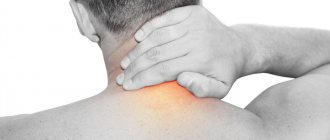
Thus, with cervical osteochondrosis, headaches are observed, aggravated by turning the neck, dizziness, flickering “spots” in the eyes, fainting, and others. Sometimes a headache is accompanied by nausea or vomiting, sensations of noise in the head, ringing in the ears, visual disturbances in the form of flickering, pain in the eye, and perhaps a sensation of a foreign body in the throat.
With lumbar osteochondrosis - acute pain (“shooting”) in the back and below, numbness of the legs, disturbances in the functioning of the genitourinary organs. Osteochondrosis of the thoracic spine is characterized by pain in the interscapular region. Often the pain in the chest becomes girdling and is felt in the area of the ribs.
Features of diagnostic measures
Anyone can seek qualified medical help if one or more symptoms of the disease develop.
To make a diagnosis, you must first consult a therapist. Who will give a referral to a highly specialized specialist (rheumatologist/orthopedist/surgeon).
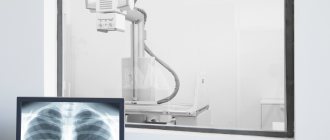
The first stage of diagnosis is the collection of anamnesis and medical history of the patient. After this, instrumental research methods are prescribed, which include such medical procedures as:
- radiography;
- ultrasonography;
- computed/magnetic resonance imaging.
In order to exclude the possibility of concomitant diseases, the patient is also prescribed laboratory tests of blood and urine.
In addition, it is worth noting that to determine the existing degree of development of pathological processes, diagnostic methods such as an immunogram and a test for rheumatoid factor are used.
Causes of osteochondrosis.
Osteochondrosis is a consequence of biological aging of the body. As the intervertebral disc wears out, collapses and flattens over time, it loses its shock-absorbing function. The intervertebral space narrows, and the radicular nerves and blood vessels become pinched in it. This leads to the above-mentioned unpleasant symptoms.
Other causes of this disease include:
- sedentary, sedentary lifestyle;
- excessive loads (lifting objects with a large mass, physical overload);
- autoimmune diseases;
- metabolic disorders with critical excess body weight;
- imbalance of calcium in the body;
- pregnancy (provokes osteochondrosis in women due to depletion of the body in calcium and inadequate distribution of increasing body weight relative to the vertical axis);
- spinal injuries;
- flat feet or daily wearing of shoes with too high heels;
- household or professional lifestyle features (long standing, sitting on uncomfortable furniture, sleeping on an uncomfortable mattress, etc.).
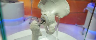
A common consequence of osteochondrosis is the formation of a herniated intervertebral disc, which may require surgical intervention. Depending on the degree of complication, symptoms can range from mild to severe.
Gymnastics for osteochondrosis of the knee joint
If you have ACS, you should not completely avoid physical activity. Physical education is needed in order to maintain muscle tone and ligaments, but at the same time not to overload the joints themselves. They need to be developed gradually, and this can be done with the help of certain exercises. In the case of knee joints, static loads are indicated.
Exercises:
- Lie on your back, straighten your leg and hold it there for a few seconds.
- In a similar position, pull your socks towards you and away from you.
- Lying down imitates riding a bicycle. 5-30 times depending on physical fitness.
- Sit on a chair, raise your leg, extend it, hold it for 30 seconds, lower it back, repeat with the other leg.
- In a sitting position, bend towards straight legs.
Remember! Gymnastics for osteochondrosis of the knee joint should last no more than 15-20 minutes a day. Try to avoid sudden movements, do 5-10-15 repetitions with rest breaks.
Diagnostics.
Most often, the diagnosis of “osteochondrosis” is made by a neurologist. At the initial examination, the doctor conducts an examination in connection with the patient's complaints of pain or limited mobility of the spine. The patient's spine is examined in standing, sitting and lying positions, in states of rest and movement.
Feeling the spine allows you to supplement the examination data (presence or absence of deformation), determine the location, degree and nature of pain. When palpated, tension in the muscles located next to the spine is noted. The patient is asked to bend or squat to determine the range of motion in various parts.
The final diagnosis is made by a neurologist based on a physical examination, as well as the results of radiography, CT or MRI. With the help of these examinations, the level of damage is determined, the diagnosis is specified, and hidden pathologies are revealed. After diagnosis, the attending physician determines treatment tactics and selects the most effective treatment method.
How does osteochondrosis of the knee joint develop?
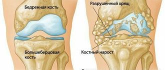
The knee joint accounts for the majority of both statistical and dynamic loads when maintaining the body in an upright position, moving, and carrying heavy loads. Changes begin with thinning of the cartilage tissue and a decrease in the lubricating fluid that provides gliding during mobility. A deficiency of useful and nutritional elements, poor blood circulation, frequent and heavy exercise, or lack of activity - all this triggers the process of destruction of cartilage and stops the production of synovial fluid.
The cartilage tissue fades and peels off, causing the joint to become inflamed and limiting range of motion. The fact is that such important substances as glucosamine and chondroitin are no longer synthesized in the body or their dose is too small to serve the body’s needs. Because of this, the cartilage cannot recover on its own; it continues to lose strength and elasticity, and at the same time, its basic functions. At this time, the first noticeable symptoms of osteochondrosis of the knee joint appear.
Among the forms of ACS, the following diseases are distinguished:
- Koenig or osteochondrosis dissecans.
Mostly typical for young men (from 16 to 30 years old). The cartilage gradually separates and can completely peel off and expose the bones. If not treated in time, disability cannot be ruled out. - Larson-Johansson or ossification of the patella.
The disease is rare and difficult to diagnose. Can appear at any age. - Osgurd-Schlatter or proliferation of the tibial tuberosity.
With the disease, large growths form. It is often detected in adolescents during the period of bone formation and growth. Risk factor – participation in professional sports. For example, football.
Do not forget! At the initial stage, any form of ACS is treated much faster.
Treatment of osteochondrosis.
The first stage of treatment is pain elimination. For this, the patient is prescribed nonsteroidal anti-inflammatory drugs (NSAIDs), for example, Movalis, Voltaren. It should be noted that these medications are not recommended to be taken without a doctor’s prescription, since most of them have a detrimental effect on the gastrointestinal tract.
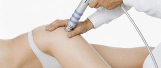
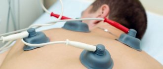
Further treatment of osteochondrosis can be conservative or surgical, depending on the severity and advanced stage of the disease. As a medical treatment, the patient is also prescribed B vitamins and muscle relaxants, for example Mydocalm, to relieve muscle hypertonicity and relieve pain. In some situations, the doctor may recommend physiotherapy.
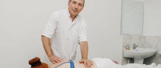
After the pain has been relieved, that is, the symptoms of the disease have subsided, the patient is recommended physical therapy and massage. Physical therapy exercises help strengthen the muscular corset of the spinal column, develop the flexibility of the ligamentous apparatus, thereby increasing the amplitude of movements. By strengthening the muscles, decompression of the nerve roots occurs, as a result of which blood circulation and metabolism in the intervertebral discs are normalized. As a result, pain in the spine is significantly reduced. Therapeutic massage, in turn, relieves muscle spasms. Controversial treatments that are not traditional medicine include manual therapy, osteopathy and reflexology.
It is also worth noting that osteochondrosis, like any chronic disease, has periods of exacerbation and remission. Sometimes pain appears depending on the time of year or weather conditions. Knowing the characteristics of your body, you can prevent exacerbations in advance.
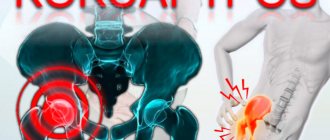
Degrees of osteochondrosis of the hip joint
Today, it is customary to distinguish three degrees of development of osteochondrosis of the hip joint. Each of them has its own characteristics and requires a specific treatment plan, which can only be determined by the attending physician, taking into account the individual characteristics of the patient and the clinical picture of the disease.
Osteochondrosis of the hip joint 1st degree
Osteochondrosis of the hip joint of the 1st degree is quite difficult to diagnose due to mild or absent symptoms.
During this period, dystrophic changes begin, which can be determined using x-ray examination (visually noticeable minor growths along the edge of the pelvic cavity).
Osteochondrosis of the hip joint 2nd degree
With osteochondrosis of the hip joint of 2nd degree, dystrophic changes expand in area and affect bone tissue.
The head of the femur undergoes deformation, the cartilage tissue becomes thinner, bone growths form, and the area of inflammation expands its boundaries.
The patient experiences a change in gait and lameness.
Osteochondrosis of the hip joint, grade 3
With osteochondrosis of the hip joint of the 3rd degree, degenerative-dystrophic changes lead to the complete destruction of the cartilage tissue of the joint; individual cartilaginous remains are observed between the tibia and pelvic bone.
During the examination, replacement of cartilaginous tissue with bone growths is noted. Inflammation and visually noticeable swelling are diagnosed.
Prevention of osteochondrosis.
The main recommendation for those who want to maintain the health of the osteoarticular system is an orthopedic regimen. It is important to learn how to sit correctly, stand correctly, lift and carry heavy objects, and sleep. For example, the patient is advised to avoid upholstered furniture, including for sleeping. The work chair should have lumbar support. It is not recommended to stand in one position for a long time; it is advisable to periodically warm up. It is not recommended to lift and carry things weighing more than 15 kilograms. Every morning it is recommended to do short exercises for 15-30 minutes to pre-warm up the muscles.
Back to articles
Nutritional Features
Compliance with the basics of dietary nutrition during complex treatment helps to reduce the load on the affected lower limb and relieve inflammation, improving the nutrition of the affected tissues.
It is recommended to expand the daily diet and enrich it with such foods as:
- nuts and seeds;
- Fish and seafood;
- dairy and fermented milk products;
- citrus;
- whole grain food products.
If you want to achieve the most positive result in the treatment of osteochondrosis of the hip joint, you should limit the consumption of strong tea/coffee, as well as instant foods and bakery products. Bad habits (drinking alcohol and smoking) should be abandoned altogether.
Possible causes of pain radiating to the leg
Most often, the cause of radiating pain in the leg is orthopedic diseases or neurological disorders. The first group includes:
- osteochondrosis;
- hernia;
- spondylosis;
- osteoporosis;
- osteomyelitis;
- prolapse;
- spinal column deformity (scoliosis).
To the second:
- damage to the lumbar roots;
- pinching;
- benign neoplasms;
- inflammation of the sciatic nerve.
- Other causes of pain radiating to the leg include:
- nagging or shooting pain down to the toes;
- diseases of internal organs;
- spinal and lumbar injuries.