The stoller method for diagnosing and classifying meniscal injuries is a modern method based on MRI of the knee joint. New and still expensive, it allows you to obtain maximum information about changes in cartilage tissue and select treatment that will be most effective in a particular case.
Damage to the meniscus can be traumatic or degenerative in nature. Both damages are determined accurately by stoller. Depending on the degree of injury, the patient will be prescribed conservative or surgical treatment. Other methods of detecting pathology cannot accurately reflect the level of cartilage damage.
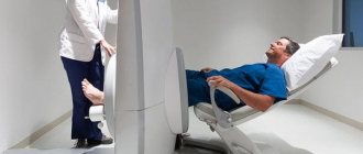
MRI principle for Stoller classification
MRI principle for Stoller classification To diagnose the slightest changes in the integrity of the cartilaginous structures of the knee joint, magnetic resonance imaging is often used.
The essence of this method of visualizing the internal structures of the knee is layer-by-layer scanning of tissue in a magnetic field. This leads to the development of the nuclear resonance effect, which occurs when protons of the nuclei of hydrogen atoms that are part of almost all organic compounds in the body are excited. In a state of magnetic resonance, protons of the nuclei of hydrogen atoms release energy, which is recorded using a special sensor, which makes it possible to construct an image through digital processing.
Basic principle of classification
Damage to the meniscus according to Stoller is classified based on the severity of the destruction of cartilage tissue. The separation is based on the appearance of an increased signal intensity during scanning using MR imaging, which indicates the development of degenerative processes causing damage to the meniscus. According to Stoller, classification involves the use of a criterion of signal intensity, as well as its localization and prevalence.
How to prepare for the procedure
Special preparation is not required before MRI with further classification of the condition of the Stoller meniscus. The study is subject to the same contraindications as MRI, and primarily the presence in the body of metal elements and various electrical stimulators and insulin pumps, the operation of which can be affected by the magnetic field of the device.
There will be no pain or discomfort during the examination. The only thing that the patient needs to take into account is that the procedure is lengthy. If the patient is very nervous before the examination, it is recommended that he take valerian or another herbal sedative.
The mechanism of pathology development
Mechanism of development of pathology
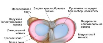
The medial cartilaginous meniscus is fixed more rigidly with the help of ligaments, therefore, when exposed to excessive mechanical force (rotation of the thigh with a fixed shin, a direct blow to the knee area or a fall on it), a violation of integrity occurs. In the case of the development of a degenerative-dystrophic process associated with the destruction of cartilaginous structures against the background of a violation of their nutrition, or inflammation, a decrease in strength occurs. In this case, a violation of integrity can occur even without the impact of excessive functional loads on the joint.
Pathological changes associated with the development of degenerative-dystrophic processes in the knee mainly develop in older people (gonarthrosis). Pathological damage to the meniscus of the knee joint develops more often. Stoller classification, based on objective visualization of changes, allows the doctor to select adequate treatment.
Causes of damage
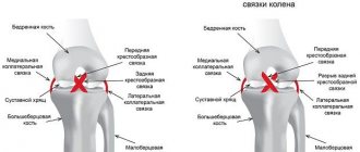
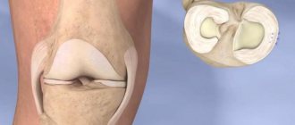
As is known, the menisci are a paired organ consisting of cartilage cells and located in the cavity of the bone joint. They are designed to provide protection and additional cushioning during movements, as well as to reduce unnecessary movement and reduce the intensity of friction in it.
The main cause of damage to the meniscus is an unexpected, sharp blow to the cup with sliding, a fall of this area onto steps or other ribbed surface, which are accompanied by a sudden turn of the tibia outward, as well as inward.
The pathological condition is often diagnosed in representatives of sports professions, in particular football players.
Damage to the meniscus of the knee joint is very common in athletes
This type of injury often affects people who spend a lot of time standing.
Rupture or partial tear of the cartilage pad is sometimes the result of its degenerative degeneration, provoked by a number of factors, including:
- chronic intoxication;
- rheumatoid process;
- frequent microtrauma of the menisci;
- gouty lesion of the knee joint.
Degenerative damage to the meniscus is a long-term process. As it progresses, a person's articular surfaces slowly erode, and the functioning of the knee joint is disrupted. Such changes without adequate therapy lead to inevitable disability and complete loss of the ability to make full movements in the joint.
Stoller degree of meniscus damage
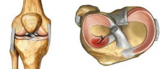
Due to the presence of certain anatomical features, damage to the posterior horn of the medial meniscus develops more often. According to Stoller, there are 4 degrees of destruction:
- Grade 0 – no changes were detected in the cartilage tissue during the study, which indicates its normal condition.
- Grade 1 meniscal injury – Stoller classification involves identifying a localized focal signal of increased intensity that does not reach the surface of the cartilage.
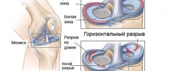
- Stoller grade 2 meniscal damage includes the determination of a linear signal of increased intensity, which also does not reach the surface of the cartilaginous structure. Horizontal damage to the meniscus of grade 2 according to Stoller indicates a partial violation of the integrity of the cartilage tissue without disturbing the general anatomical structure (there is no complete separation of part of the cartilage yet).
- Stoller grade 3 meniscus damage - this degree of severity of changes is characterized by the determination of a linear signal of increased intensity that reaches the surface of the cartilage tissue. Such changes indicate a violation of the anatomical structure in the form of avulsion. Separately, there is a grade 3a meniscus injury according to Stoller, in which part of the cartilaginous structure of the knee joint is displaced (complete tear with displacement).
Determining the degree of pathological or traumatic changes in cartilaginous structures according to Stoller is possible only using the visual examination technique of magnetic resonance imaging.
How are meniscus injuries diagnosed?
Diagnosis is primarily based on clinical manifestations during orthopedic maneuvers. The presence of pain on the inside of the knee with hyperflexion, hyperextension and external rotation of the bent knee at 90° is a sign of damage to the medial meniscus. While the appearance of pain during hyperflexion or internal rotation of the leg bent at 70° and 90° is a sign of a lateral meniscus tear.
As for instrumental diagnostics, it is carried out using magnetic resonance imaging. This often helps identify degenerative changes.
X-ray examination
X-ray examination
X-ray examination. Includes taking x-rays of the knee in frontal and lateral projections. They make it possible to identify gross changes, therefore this method of visual examination is mainly used in the initial stages of diagnosis (in case of traumatic injuries, radiography is the first research method prescribed by a doctor in a trauma center).
CT scan. An imaging method based on layer-by-layer scanning of tissues performed using X-rays in a special installation. The resulting series of images is processed on a computer, which makes it possible to determine the slightest changes in tissues at different depths. The method has a fairly high resolution, but is not suitable for determining the degree of change according to Stoller.
Ultrasound. Tissue visualization occurs using a sound wave, which is recorded when reflected from media with different densities. This diagnostic research technique allows us to identify inflammatory signs, in particular an increase in the volume of synovial fluid in the joint cavity.
Arthroscopy. A modern invasive research technique in which the internal structures of the joint are examined using an arthroscope inserted into its cavity (a special tube with a video camera and lighting). Arthroscopy is used to perform surgical procedures.
Diagnostic highlights
How to determine meniscal damage? Currently, there are many instrumental techniques that allow confirming such a diagnosis.
In the process of diagnosing the disease, doctors prescribe the following examinations to the patient:
- MRI, which allows you to accurately determine the localization of the pathological process, its severity and the presence of complications;
- X-ray examination of the knee joint in several projections, which is relatively informative;
- ultrasound diagnostics with tissue visualization, which allows you to determine signs of the inflammatory process and assess the amount of synovial fluid;
- arthroscopy, which makes it possible not only to diagnose disorders, but also to correct them.
Read more about hardware methods for diagnosing joint pathologies in this article...
Clinical signs of damage classification according to Stoller
Pathological or traumatic changes in the medial cartilage of the knee are more often recorded. Symptoms of changes depend to a certain extent on their severity:
- Zero degree determines the absence of any changes in cartilaginous structures without the development of clinical symptoms.
- Damage to the medial meniscus, grade 1 according to Stoller - minimal changes lead to the appearance of unexpressed pain in the knee joint, which usually intensifies in the evening, after prolonged standing or walking. Also, when bending the knee, a characteristic crunch may appear, indicating the development of a degenerative-dystrophic process.
- Damage to the medial meniscus, grade 2 according to Stoller - more severe disorders are manifested by pain, the intensity of which increases during movements of the knee or after prolonged standing. Against the background of clicking and crunching, inflammatory changes may appear, including redness of the skin and swelling of the soft tissues.
- Damage to the medial meniscus grade 3 according to Stoller - a violation of the anatomical structure of the cartilage of the knee is accompanied by severe pain and limitation of movements in it. Damage to the internal meniscus, grade 3a according to Stoller, can be accompanied by severe acute pain and blocking of the ability to move in the joint.
Injury to the internal meniscus of grade 3 according to Stoller often has a traumatic origin, and therefore is characterized by the acute development of clinical manifestations.
Treatment of a torn meniscus of the knee joint
First aid for a knee injury that is accompanied by severe pain is immobilization (using a splint or splint) and cooling (using an ice pack or sports cooler). Then the victim must be taken to a traumatologist as soon as possible to receive adequate treatment. This approach significantly reduces the risk of complications from a knee meniscus tear.
Meniscus tears are divided into:
- separation at the point of attachment to the joint (the damage can heal with bed rest and immobilization of the limb with a splint);
- rupture in the central part (requires surgical treatment);
- flap rupture (the most severe damage).
The choice of treatment strategy for a tear in the meniscus of the knee joint is also influenced by which meniscus is damaged. The medial (internal) meniscus is much less well supplied with blood vessels, so its rupture requires longer treatment and the use of auxiliary drugs (for example, intra-articular administration of glucocorticoids).
After an accurate diagnosis, the doctor chooses the direction of treatment - conservative or surgical. The first is to fight inflammation, accelerate the regeneration of cartilage tissue, and relieve pain. The limb is immobilized or protected with a compression orthosis; if necessary, a puncture is performed (removal of fluid accumulated in the joint, be it blood or effusion), and ice compresses are applied (about 20 minutes a day).
Surgical treatment for a knee meniscus tear involves arthroscopy.
Immobilization of the knee - first aid for a torn meniscus
Physiotherapy for a torn meniscus
Physiotherapy is especially advisable during the rehabilitation period after a torn meniscus of the knee joint. It is designed to strengthen the muscles of the thigh and lower leg, naturally reducing the load on the knee joint, and also fully restore mobility in the joint. This is facilitated by:
- mechanotherapy;
- kinesiotherapy;
- ultra-high frequency therapy (UHF);
- electromyostimulation;
- massage;
- taping.
These types of physiotherapy are used to relieve spasm, restore atrophied muscles, and improve innervation.
When treating a knee meniscus tear, only anti-inflammatory techniques are usually used, for example:
- physiotherapy;
- medicinal electrophoresis;
- cryotherapy and thermotherapy;
- ultrasound.
They are also used to relieve swelling before meniscus surgery.
Drug treatment of a torn meniscus of the knee joint
In the conservative treatment of a rupture of the meniscus of the knee joint, as well as after surgery, patients are prescribed non-steroidal anti-inflammatory drugs to relieve swelling and pain, and improve soft tissue trophism. eliminating “inflammatory” enzymes that destroy articular cartilage.
If NSAIDs do not provide sufficient effect and do not reach the meniscus well, the doctor may decide on intra-articular injections of corticosteroid hormones and hyaluronates. This procedure allows you to relieve pain and inflammation within 15-20 minutes after administering the medicine.
In case of poor blood circulation and injuries of the medial meniscus, blood microcirculation correctors are additionally connected.
Chondroprotectors play almost the central role in the treatment of meniscal tears of the knee joint. These are preparations enriched with natural polymers - components of cartilage tissue and synovial fluid. Chondroprotectors accelerate the regeneration of cartilage tissue, serve as a prevention of complications (from joints and ligaments), and ensure fusion of the meniscus with minimal scarring of the tear. They help relieve inflammation, improve nutrition of the joint and improve the shock-absorbing function of cartilage.
It is especially important to take chondroprotectors for degenerative meniscus tears - when the cartilage tissue is already severely worn out, has a low potential for recovery and needs to be recharged. In this case, chondroprotectors should be taken for life, in annual courses of 3-6 months.
One of the most effective chondroprotectors is the drug Artracam.
Surgery
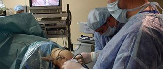
Modern surgical treatment for knee meniscus tears is usually performed in the form of arthroscopy. Since arthroscopy is a diagnostic procedure, minimally invasive surgery during its process can be performed immediately after clarifying the location and extent of the meniscus tear. The surgery begins with the insertion of a microscopic camera into a tiny incision (1.5-3 cm) and takes only 30 minutes.
After the examination, the doctor makes a decision on stitching, resection or removal of the meniscus. Stitching the edges with bioabsorbable suture material is usually indicated for 1st and 2nd degree tears of the meniscus of the knee joint - after this operation, rest for the affected limb is required for 3-4 weeks. Stitching is especially effective for lateral tears of the meniscus of the knee joint, as well as for fresh injuries.
Resection involves partial removal of a fragment of the meniscus that blocks movement in the joint, or severely damaged tissue, followed by grinding of the edges. If most of the meniscus is preserved, the patient does not feel any discomfort or changes in sensations after surgery. After 3 weeks of rehabilitation, athletes can return to training.
Complete removal (meniscectomy) is performed in exceptional cases and only for grade 3 meniscal tears. Removal is recommended for old ruptures or complex injuries, when reconstruction is impossible, as well as for severe damage to the meniscus, excessive looseness of cartilage tissue (arthrosis and other chronic diseases). Rehabilitation after such an operation is more complex, and the shock-absorbing characteristics of the joint worsen (patients are recommended to take lifelong chondroprotectors and be monitored by a rheumatologist). However, with arthroscopic removal of the meniscus, the patient can step on the affected leg on the day of surgery - using a walking stick or crutch. If medical recommendations are followed, motor activity does not suffer after meniscectomy.
Recovery time after arthroscopy of the knee meniscus varies from person to person. Typically patients require:
- 3 days before discharge from hospital;
- 2-4 weeks for recovery at home.
In this case, disability (depending on the profession) lasts from 2 to 6 weeks. Driving is allowed 6 weeks after surgery.
Gymnastics and exercises for meniscus tears
Performing gymnastic exercises for a torn meniscus of the knee joint is allowed only during the recovery period, after complete removal of inflammation and with the permission of the attending physician. The physical therapy instructor (PT) selects the following groups of exercises for the patient;
- training the muscular-ligamentous apparatus to strengthen the knees (sit down/stand up from a chair, shift your body weight first to one side, then to the other, step-up exercises);
- strength training (mainly for the thigh muscles) and flexibility training;
- training for coordination of movements and maintaining balance, which prevent re-injury of the knees and help to work the deep muscles;
- workouts to maintain general tone.
The exact list of exercises is selected by a specialist taking into account the general health, constitution, physical fitness, age of the patient and other factors. Rehabilitation after surgery involves a gradual increase in loads.
Rehabilitation for meniscus injuries
This is a very important stage in the treatment of meniscus injuries (if the outer meniscus is damaged, then rehabilitation will be faster) and restoring the functionality of the knee.
In the following table you will find a description of the different stages of rehabilitation and physical therapy after meniscus injuries.
(first 4 weeks)
Characterized by persistent pain and effusion in the knee joint.
The joint is protected with braces that are worn day and night.
- reduction of pain and effusion;
- restoration of the range of joint mobility;
- preventing the development of restrictions in the patella;
- muscle control;
- restoration of postural stability;
- Improved hip and ankle strength and flexibility.
How to do it:
- application of cold and compression against pain and effusion (medication with Voltaren is also often used);
- when walking with the help of crutches, they create a partial load on the knee;
- exercises of continuous passive movement from a sitting position;
- exercises to develop the quadriceps muscle;
- development of the ability to balance in a vertical position;
- stretching the hamstrings and leg flexors.
(from 4 to 10 weeks)
Effusion and pain are minimal. The knee can be fully extended and extended to 120°.
- restoration of normal mobility of the knee joint;
- improving flexibility, strength and endurance of the entire lower limb.
How to do it:
- control of knee mobility (especially extensor muscles) by using a bandage;
- regaining mobility through stretching exercises;
- strengthening muscles using exercise bikes, soft resistance machines;
- regaining the ability to maintain balance (for example, going up and down stairs);
- increasing the range of mobility of the leg and rectus femoris.
(between 9 and 12 weeks)
There is no pain or effusion, the range of motion of the joint is fully restored, and muscle strength reaches 60-80% of what it was before the injury.
- preparation for return to full functional activity;
- teaching the patient behavior that preserves the meniscus.
Read also: Knee meniscus treatment without surgery
How to do it:
- exercises that simulate functional activity;
- increasing the duration and intensity of exercises that increase muscle strength;
- after 5-6 months you can completely return to the standard training program (in the case of athletes).
Always remember that the rehabilitation process is very long and requires complete dedication from the patient , especially in the early stages when you have to endure pain.
