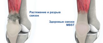
The ankle joint is a movable articulation of the lower ends of the tibia and fibula with the talus bone of the foot. The ankle experiences intense loads, so the joint is strengthened by ligaments.
Types of ankle ligament injuries:
- stretching;
- partial tears;
- ankle ligament rupture;
- tearing of the ligament at its insertion, often along with a small piece of bone.
At CELT you can get a consultation with a traumatologist-orthopedic specialist.
- Initial consultation – 3,000
- Repeated consultation – 2,000
Make an appointment
Diagnostics
When you have an ankle injury, the first thing you need to do is make sure that the bone is not damaged. To exclude a fracture, an x-ray of the foot and an ultrasound examination (ultrasound) are performed.
For mild to moderate injuries, other additional diagnostics are usually not required. During the examination, the doctor will assess the condition of the joint and prescribe appropriate treatment.
If a serious injury is suspected, the doctor will order a magnetic resonance imaging scan. This study allows you to obtain clear layer-by-layer sections of the ankle, and, if necessary, to recreate a three-dimensional model.
Inversion injuries of the capsular-ligamentous apparatus of the ankle joint in children and adolescents
Introduction. Injuries to the ankle joint account for up to 25% of the total number of musculoskeletal injuries in the general population [1]. In children, this percentage increases to 35% [2]. Among injuries to the soft tissue structures of the ankle joint, injuries to the lateral group of the capsular-ligamentous apparatus (inversion injuries), which account for 70-75% of ankle joint injuries, deserve special attention [3]. In the available literature, we found a few studies devoted to damage to the capsular-ligamentous apparatus of the ankle joint in children and adolescents [4-7].
A study of foreign publications showed that more than 40% of injuries to the lateral ligaments of the ankle joint lead to chronic instability and the development of degenerative processes in the joint [8, 9]. Loss of the supporting function of the ligaments predisposes the patient to repeated ligament ruptures or other injuries resulting from much lesser trauma, which has led to discussion and detailed study of this problem.
According to our observations, in most cases, when children come to the emergency room with damage to the ankle joint, an examination and radiographs are performed in standard projections. In the absence of bone damage, a diagnosis is made: ankle sprain. Such patients are recommended to fix the limb with an elastic bandage and apply warming ointments locally. However, with this treatment method, constant mobility in the joint leads to the formation of a stretched scar, which increases the risk of repeated injuries [10]. The term “sprain” makes doctors, patients and their parents less vigilant.
The problem of the pathogenesis of ankle ligament injuries has been studied for a long time. Early scientific papers described the possibility of stretching collagen fibers due to their folding. Research has shown that the extensibility of ligaments is their property [11]. A.A. Zavarzin experimentally measured the elastic modulus of collagen fibers, which amounted to 2.6-8.8 kg/mm² [18]. Therefore, with increasing load, one should expect ligament rupture rather than stretching. Based on this, the term “sprain” is incorrect. Therefore, for practical purposes, it is more appropriate to make a diagnosis of partial or complete ligament damage [12].
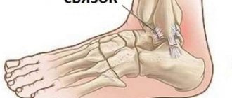
Among injuries to the external ligaments, 90% are injuries to the anterior talofibular ligament (65% of them are isolated, and 25% are combined with damage to the calcaneofibular ligament). The posterior talofibular ligament (or the third component of the external collateral ligament) is resistant to posterior displacement of the talus and is therefore rarely injured except in cases of complete foot dislocation [10].
In foreign guidelines, the “gold standard” in diagnosing ankle ligament injuries is the magnetic resonance imaging method with a sensitivity of 90% and a specificity of 83% [10]. However, the low prevalence of magnetic resonance imaging scanners and the high cost of the study significantly limit the use of this method when searching for injuries to the ankle joint. In addition, in young children this diagnosis can only be carried out under anesthesia.
The most accessible method for visualizing ankle ligament injuries is ultrasound. The sensitivity of the method according to the literature is 92%, specificity is 64% [13]. As a non-invasive, relatively cheap and widely available research method, it is of high value for visualizing the capsular ligamentous apparatus [14].
When studying and analyzing the information available in the literature, we noted many different methods of treating ankle ligament injuries. However, the tactics of treating ligamentous injuries remains controversial and this problem has not been solved to date. A number of authors recommend starting with conservative treatment methods in all cases of ankle ligament injuries [15]. In case of partial ligament damage, some authors recommend immobilization, while others oppose it [16].
Kannus P. writes in his publication that to understand the principles of treatment of ankle ligament injuries, it is necessary to know the stages of regeneration and the conditions that need to be created for the healing of the ligament [16]. The reparative process for damage to the ankle ligaments occurs in 3 phases:
1st phase. Inflammation (1-7 days). During this phase, capillary walls thicken, plasma and fibrin sweat, and inflammatory cells migrate to the site of damage. During this phase, it is necessary to ensure rest through immobilization and an elevated position. Cold has a beneficial effect. Lack of immobilization increases the inflammatory phase, which leads to the formation of large amounts of scar tissue.
Phase 2. Proliferative (7-21 days). During this phase, inflammatory cells carry out phagocytosis of damaged tissues. Fibroblasts produce type 1 collagen (mature collagen). In the middle of this phase, the formation of type III collagen (immature collagen) begins. At this stage of treatment, it is necessary to limit movement in the ankle joint to prevent excessive production of type III collagen, which contributes to the formation of a soft, stretched scar and chronic sprains.
Phase 3. Remodeling (21 days to 12 months). Collagen maturation. At this stage, controlled gradual loading will promote the correct orientation of the collagen fibers. Therapeutic exercise to develop movements in the ankle joint and strengthen weakened periarticular muscles in combination with a complex of physiotherapeutic treatment to improve blood supply, reduce swelling and pain will avoid the harmful effects of immobilization on muscles and articular cartilage. Treatment will continue until the collagen matrix matures so that full return to activity is possible 4 to 8 weeks after injury.
The purpose of the study is to develop a diagnostic algorithm and improve the results of treatment of children and adolescents with inversion injuries of the capsular-ligamentous apparatus of the ankle joint.
Materials and methods. An analysis of case histories and own clinical material of 53 patients with injuries to the lateral ligaments of the ankle joint was carried out. Depending on age, patients were divided into groups of preschool, primary school, senior school and youth. There were 23 girls with ankle ligament injuries, and 30 boys (Table 1).
Table 1. Distribution of patients by gender and age
The average time for patients to go to a medical facility after an injury is 1-7 days. Before admission to CITO, all patients were treated in medical institutions in Moscow. In 15% of patients the injury was recurrent.
We have developed an algorithm for examining patients with ankle joint injuries:
1. Taking an anamnesis.
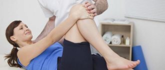
2. Examination, including diagnostic stress tests (inversion, pronation, compression test, anterior drawer test).
3. X-ray of the ankle joint.
4. Ultrasonographic examination of the ankle joint.
The following anatomical structures were examined during ultrasound examination:
1. Anterior section: ligaments of the tibiofibular joint, tendons of the long extensor digitorum and the first finger.
2. Lateral section: tendons of the peroneal muscle group, joint capsule, anterior talofibular ligament, calcaneofibular ligament.
3. Medial section: tendons of the tibialis posterior muscle, flexor digitorum longus and flexor digitorum longus, deltoid ligament.
4. Posterior section: posterior talofibular ligament, Achilles tendon.
To assess the degree of damage to the capsular-ligamentous apparatus of the ankle joint, we used the classification developed by S. Trevino [17]
1st degree – fiber separation of the ligament.
2nd degree – partial damage to the ligament.
3rd degree – total damage to the ligament.
Ultrasound examination assessed the amount of effusion in the joint, the presence of intra-articular bodies, the thickness of the joint capsule, and determined the integrity and echogenicity of the capsule and joint ligaments.
Treatment of ankle ligament damage was carried out in 3 stages.
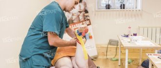
Stage 1 – plaster immobilization (or immobilization with modern rigid fixation orthoses) for up to 3 weeks. Loading on the injured limb was excluded. During immobilization, gymnastics was prescribed for the healthy limb, isometric gymnastics for the injured limb, and magnetic therapy.
Stage 2 - after the cessation of immobilization, active lightweight exercises are carried out to develop movements in the ankle joint, a special set of exercises to strengthen the periarticular muscles, special attention is paid to the peroneal muscle group, as the main stabilizers of the ankle joint. Exercises that cause trauma and stretching of the scar - adduction and supination - are excluded. An orthopedic regimen is prescribed - limiting the load on the injured leg for 1 week. To prevent scar trauma, we recommend that patients wear a special bandage or high lace-up boot that limits lateral movements of the foot and wear arch supports. The following physiotherapeutic measures are used: phonophoresis with hydrocortisone and lidase, magnetic therapy, warm foot baths with sea salt.
Stage 3 – consolidation. Therapeutic physical education according to the methodology of the final period. The procedures are performed in a sitting or standing position. Walking in place, on toes, on heels, in a straight line, sideways, with turns, going up and down ramps, stairs, etc. is used. During this period, exercises on leg exercise machines are prescribed. At the same time, we include running and jumping in the set of exercises.
Results and discussion. An examination of 7 patients with a referral diagnosis made at the emergency room: a closed fracture of the lateral malleolus revealed damage to the capsular-ligamentous apparatus of the ankle joint in the lateral part. The bone structures were intact. According to the examination, 45.3% of patients had damage to the capsular-ligamentous apparatus of grade I, 41.5% had grade II, and 13.2% had grade III (Table 2).
Table 2. Distribution depending on the severity of ankle ligament injuries
In accordance with the results obtained, damage to the anterior talofibular ligament was diagnosed in more than half of the cases (77.4%). The calcaneofibular ligament was in second place in terms of frequency of injuries (22.6%). No damage to the posterior talofibular ligament was detected in the examined patients. Analysis of the degree of damage showed a significant predominance of partial ligament damage (86.8%) over complete ligament rupture (13.2%) (Table 3).
Table 3. Distribution of inversion injuries to ankle ligaments
Objective assessment of the treatment effectiveness of patients was carried out after 3 weeks, 6 weeks, 6 and 12 months using the AOFAS questionnaire.
86.8% of patients had excellent treatment results, and 13.2% had good results.
Here is a clinical example. Patient K., 10 years old. During figure skating training, I sprained my right leg at the ankle joint. At the time of injury, she noted a click. When visiting the emergency room: examination, radiographs of the ankle joint. A diagnosis was made: sprain of the right ankle joint. Soft tissue immobilization of the ankle joint was performed using a figure-of-eight bandage, and local warming ointments were prescribed. The girl could not train due to severe pain in the joint. I contacted the CITO on the 3rd day after the injury. Examined. Ultrasound examination revealed partial damage to the anterior talofibular ligament, partial damage to the calcaneofibular ligament and joint capsule (Fig. 1). Plaster immobilization was performed for 3 weeks. After removing the plaster splint, physical therapy was carried out according to the proposed method, as well as a set of physiotherapeutic measures. Joint function was restored after 5 weeks. The girl returned to playing sports.
Rice. 1. Sonograms of the lateral ankle joint: 1a – the arrow indicates the anechoic area of the joint capsule, indicating a rupture of the joint capsule; 1b – the arrow indicates a hypoechoic area corresponding to partial damage to the anterior talofibular ligament; there is a partial tear of the ligament with a detached cortical layer from the lateral malleolus; 1c – a hypoechoic area is indicated, corresponding to partial damage to the calcaneofibular ligament.
conclusions
1. Considering the high frequency of relapses of inversion injuries of the capsular-ligamentous apparatus of the ankle joint in children and adolescents, examination and treatment of such patients requires a special approach.
2. The data obtained during the examination of patients indicate the need to introduce into everyday practice the ultrasonographic diagnostic method for traumatic injuries of the ankle joint in order to visualize damage to the capsular ligamentous apparatus in children and adolescents. The use of classical radiography alone as a visualization method in many cases leads to diagnostic errors and to the wrong choice of treatment method.
3. Ultrasonography, as a relatively cheap and widely available research method, is of high value both for visualizing the capsular-ligamentous apparatus and for assessing the degree of its damage. This is of great importance for the prognosis of the disease and determining further treatment tactics.
4. In case of damage to the ankle joint ligaments in children, in order to form a full-fledged scar, it is necessary to carry out short-term rigid immobilization of the joint (up to 3 weeks)
5. Comprehensive treatment of ankle ligament injuries helps prevent recurrence of injuries and the development of instability in the joint.
Our doctors
Poltavsky Dmitry Ilyich
Traumatologist-orthopedist
Experience 28 years
Make an appointment
Marina Vitaly Semenovich
Traumatologist-orthopedist, head of the minimally invasive traumatology and orthopedics service
Experience 36 years
Make an appointment
Zubikov Vladimir Sergeevich
Traumatologist-orthopedist, Doctor of Medical Sciences, doctor of the highest category, professor
44 years of experience
Make an appointment
Samilenko Igor Grigorievich
Traumatologist - orthopedist, doctor of the highest category
24 years of experience
Make an appointment
Treatment
Treatment for a sprained ankle is simple and is considered a minor injury. It is important to provide first aid to the victim in a timely and correct manner. Cold is applied to the joint area, and then it is fixed with an elastic bandage. After this, you need to contact a traumatologist to clarify the diagnosis.
Moderate injuries include partial ligament tears. The victim must be taken to the emergency room as quickly as possible. The doctor performs anesthesia with an anesthetic solution and applies a fixing bandage. After some time, physiotherapy, massage, and then therapeutic exercises are prescribed.
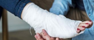
Ligament tears and avulsions are classified as more severe injuries. Most often, after pain relief, the traumatologist applies a plaster splint, which must be worn for 6 to 8 weeks. After removing the plaster, physiotherapy, massage, and therapeutic exercises are prescribed. Restoration of performance after injuries to the ligamentous apparatus of the ankle joint can take 1.5 months.
Experienced specialists at the multidisciplinary CELT clinic provide high-quality medical services to patients with ankle injuries and other types of injuries.
Physiotherapeutic procedures
Physiotherapy for the treatment of ankle ligament injuries is always prescribed. Their therapeutic effectiveness is no lower than that of pharmacological drugs. For grade 1 and 2 ligament injuries, physiotherapeutic procedures are recommended already on the 3-4th day of treatment. And after surgery they are carried out during the rehabilitation period. The traumatologist selects physical procedures individually for each patient. It takes into account:
- severity of injury;
- rate of tissue regeneration;
- a history of chronic cardiovascular and renal pathologies.
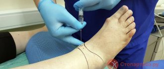
At the initial stage of treatment, electrophoresis is used. A sterile swab soaked in the drug solution is applied to the damaged area, and metal plates are placed on top. Under the influence of electrical impulses, drug molecules penetrate into the deepest tissues. During the procedure, glucocorticosteroids, analgesics, and non-steroidal anti-inflammatory drugs are used. For better fusion of ligaments torn from the bone base, solutions of chondroprotectors are prescribed.
In addition to electrophoresis, the following physiotherapeutic procedures can be performed during the rehabilitation period:
- ultrasound therapy . Used to improve microcirculation in damaged tissues and accelerate lymph outflow. After it is carried out, more complete absorption of medications occurs - ointments, gels, balms. Drugs are better accumulated and distributed in ligaments and tendons;
- UHF therapy . Accelerates reparative processes, stops the inflammatory process. And due to the expansion of blood vessels, the metabolism of substances improves;
- paraffin therapy . Physiotherapy helps to quickly eliminate severe swelling and even long-standing extensive hematomas. And improving microcirculation replenishes the supply of oxygen, nutritional and biologically active compounds in damaged tissues.
Magnetic therapy is an indispensable physiotherapeutic measure for ankle injuries. After it is carried out, the outflow of blood and lymph improves, and inflammation decreases. More complete absorption of medicinal substances from ointments and gels was also noted.
Orthopedics and traumatology services at CELT
The administration of CELT JSC regularly updates the price list posted on the clinic’s website. However, in order to avoid possible misunderstandings, we ask you to clarify the cost of services by phone: +7
| Service name | Price in rubles |
| Appointment with a surgical doctor (primary, for complex programs) | 3 000 |
| Ultrasound of two symmetrical joints (except hip) | 4 000 |
| MRI of the ankle (1 joint) | 7 000 |
All services
Make an appointment through the application or by calling +7 +7 We work every day:
- Monday—Friday: 8.00—20.00
- Saturday: 8.00–18.00
- Sunday is a day off
The nearest metro and MCC stations to the clinic:
- Highway of Enthusiasts or Perovo
- Partisan
- Enthusiast Highway
Driving directions
Reconstructive surgeries
Reconstructive surgeries are used to treat chronic ligament tears that are accompanied by ankle instability. The integrity of the ligaments is restored surgically, after which the joint is fixed with an Ilizarov apparatus. If the injury is less than 5 months old, surgery is often performed by arthroscopy. But old ruptures require open reconstruction.
The integrity of the ligaments can be restored:
- local tissues;
- allotissues;
- synthetic endoprostheses.
Plastic surgery with local tissue is associated with additional trauma to the donor site, so it is rarely performed. To restore ligaments, synthetic tissue or allotendon is often used.