Physiotherapy
The patient’s task is to ensure adequate elasticity of the heel (plantar) fascia. Do this in different ways. The following therapeutic exercises will increase stretching, reduce pain, and help protect the plantar fascia from microtears.
- Position yourself next to the wall at a distance of approximately 30–40 cm (at the length of your thigh, since you will have to squat).
- Place your palms on the wall at shoulder level, as if you were about to do a push-up.
- Place your feet in a line, one after the other. The patient must be in the back. If both hurt, the exercise must be repeated twice, changing legs.
- Sit down, your heels are firmly on the floor and do not come off. The depth of the squat is until you feel how the bottom of the lower leg of the outer leg stretches.
- Stay in this position for 10–15 seconds.
- Repeat this warm-up 5 times.
Read also: Therapeutic exercises for arthrosis of the shoulder joint
The next exercise continues to warm up the calf muscles, while simultaneously working the plantar fascia itself, stretching it a little and preparing it for further action.
- Place a low bench, about 7–10 cm high, at a short distance from the wall. It is necessary for someone to press one edge of it with their foot - the one closest to the wall.
- Stand with half your foot on the edge of the bench. Place your hands on the wall as in the first exercise. Lower your heels below the level of the bench.
- Press your whole body against the wall, feel how the muscles of your lower leg tighten.
- Return to the starting position and rise onto your toes. Lower yourself back down, but your heels should be level with the bench you're standing on. Repeat the movement 15 times.
- Make it more difficult by lifting one leg. Perform calf raises on one leg, then on the other. 15 times each.
The next and final exercise is aimed at stretching and normalizing the plantar fascia itself. Remember, if you feel severe pain, stop and rest. If the pain does not go away, take a long break. Return to exercise when it becomes easier.
- Sit on the floor, back straight, legs extended and slightly apart.
- Bend forward and grab your toes and the arch of your foot with your hands.
- Pull your hands towards you.
- When you can no longer stretch your foot, hold for 10–15 seconds.
- Then return to the starting position and repeat with the other leg.
You can use a strong band instead of your hands if your flexibility does not allow you to reach your feet with your hands.
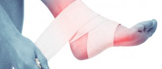
After therapeutic exercises for the feet, perform taping - fixation of the foot in order to reduce the load on the plantar fascia.
Using orthotics for plantar fasciitis
Arch support with custom or standard orthotics is another strategy that can protect your feet during treatment.
It is unclear whether there is a significant difference between custom orthotics and standard orthotics such as SuperFeet Green or Powerstep when it comes to treating plantar fasciitis.
While they may not be ideal for your feet, standard insoles are not as expensive and are easily available, whereas with custom insoles you will have to wait at least a few weeks for them to be made.
Avoid using soft gel arch pads as they will likely do nothing to help treat your injury.
Choose your casual and athletic shoes carefully when you have plantar fasciitis.
Thus, many runners find that wearing casual shoes with more arch support (including Birkenstocks and other brands of shoes with cork soles) improves their symptoms.
Calf and foot stretches and the use of a night splint for plantar fasciitis were also studied.
All three treatments are designed to stretch the calf muscle/Achilles tendon/plantar fascia complex, which helps reduce the tensile stress placed on the arch of the foot.
Causes
The very first changes that occur in the fascia are associated with the onset of the inflammatory process, which is localized in the area of the heel bone. Plantar fasciitis develops after the fascia is unable to cope on its own with micro-tears due to mechanical injuries and tension. After some time, dystrophic transformations develop in this part: the fascia loses its elasticity, becomes denser, and there are no reactions to stretching. As a result of the deposition of calcium salts, a bone growth called a heel spur is formed.
The disease develops due to prolonged and severe overload of the ligaments, so most often it is diagnosed in middle-aged and elderly people. Weightlifters, track and field athletes, and dancers are also at risk.
Among the main reasons provoking negative changes in the fascia are the following:
- excess body weight;
- work that requires constant standing;
- flat feet or high arches;
- a foot with an incorrect position, where there is an outward rotation - pronation. At the same time, shoes wear out faster from the inside;
- degenerative and inflammatory leg diseases, for example, arthritis or arthrosis;
- wearing narrow and uncomfortable shoes without heels and with them;
- obliterating atherosclerosis, which provokes disruption of normal blood supply in the legs;
- osteochondrosis in the lumbar spine.
Causes of plantar fasciitis
Some causes of plantar fasciitis include:
- tight calf muscles or Achilles tendons (tight tissue that connects the calf muscles to the heel bone);
- prolonged walking, standing or running, especially on hard surfaces;
- wearing shoes that don't fit;
- wearing shoes that do not provide the necessary support for the foot;
- weakening of foot tissues with age;
- flat feet, high arches, or uneven gait;
- overweight;
- pregnancy or hormonal changes; under the influence of hormones, ligaments and tissues may become more mobile than usual.
to come back to the beginning
Traditional medicine
Traditional methods for eliminating the pathological course in the fascia are not inferior in effectiveness to drugs from pharmacy chains. In addition, homemade medicines are several times cheaper.
Alternative medicine suggests the use of ointments and compresses to eliminate the pathology of fasciitis. It is effective to use ointments as a lotion to eliminate plantar pain.
To make one of these you will need:
- Egg.
- Butter – 200 grams.
- Vinegar – 2 tablespoons.
The egg is broken and vinegar is poured on top. The butter is melted, but not brought to a boil, and sent to the prepared products. There is no need to mix the mixture. The mass is covered with a lid and left for 3 days. Afterwards everything is mixed and used for its intended purpose.
The fabric is soaked in the prepared product, applied to the plantar area, then gauze is applied and socks are put on. Fasciitis should be treated before the ointment is completely used.
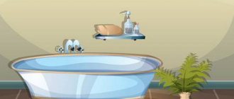
Baths
Before using creams and lotions to cure fasciitis, traditional medicine recommends taking baths.
Sea salt
The following traditional medicines are used for the procedure:
- Coarse sea salt – 2 tablespoons. Keep the heels in the bath for 15 minutes.
- Iodine – 10 drops and salt – 3 tablespoons. Manipulate in 3 liters of water with the addition of these products for 10 minutes. After the bath, the plantar ligament is lubricated with iodine.
- Use vinegar 9%, turpentine, vodka in equal volume. The products are mixed and heated using a water bath. The affected leg is dipped into the mixture and held until it cools. Afterwards, the solution is heated again and the procedure for treating the plantar zone is repeated.
Compresses
The treatment course using traditional medicine for the plantar ligament includes the use of compresses.
You can prepare night lotions using the following folk remedies:
- Using the herb cinquefoil to cure fasciitis. Chopped root - 2 tablespoons, pour ¼ cup of water, leave to stand under a table lamp. When 2 hours have passed, the softened roots are ground into a paste-like mass and applied to the sole, secured with a bandage. A bag is put on top, then a sock. The lotion is placed in the plantar area for 10 o'clock.
- Elderberry infusion. The berries are placed in a 0.5 container and filled with alcohol. You need to let the infusion brew in the dark. You need to use a compress daily to relieve fasciitis.
Therapeutic gymnastics by Bubnovsky for heel spurs
Doctor of Medical Sciences, Professor Sergei Mikhailovich Bubnovsky develops original training complexes for patients with various pathologies of the musculoskeletal system. In particular, he proposed a number of exercises that strengthen the arch of the foot and stretch the plantar aponeurosis.
- We lie down flat, extend our arms parallel to the body, spread our legs to shoulder width. Simultaneously bend both big toes towards you, and then pull up.
- We move our thumbs inward, moving them away from their neighbors as much as possible.
- We forcefully squeeze all the toes on our feet at the same time, and then fan them out.
- We alternately raise our legs above the floor and carry out rotational movements in the ankle joints clockwise and counterclockwise.
This complex is convenient to perform in the morning, before getting out of bed. It is also suitable as a short warm-up during the working day.
A heel spur is not a dangerous disease, but it can cause a lot of discomfort and significantly complicate life. To prevent this from happening, start training when the first signs of pathology appear. And the best option is to exercise preventively if at the end of the working day you experience heaviness and aching in your feet.
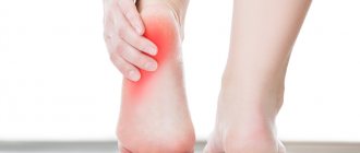
How do you know if you have plantar fasciitis?
The most obvious sign is the so-called “pain of the first step.”
If you experience a sharp, stabbing pain at the base of your heel immediately after getting out of bed in the morning, you can unfortunately be sure that the diagnosis has been made.
How common is plantar fasciitis?
Plantar fasciitis accounts for about 8% of all running injuries, and is common among runners of all levels, but affects even ordinary overweight people who spend the workday on their feet.
Runners commonly experience injuries due to the impact that is an integral component of running, but don't discount your shoes or daily living habits if you're dealing with plantar fasciitis.
Women's shoes are especially hard on the arch, but men's shoes with hard soles and no arch support can also pose problems.
Although it is quite possible to find shoes suitable for wearing with plantar fasciitis (both men's and women's).
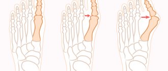
Hallux valgus (hallus valgus)
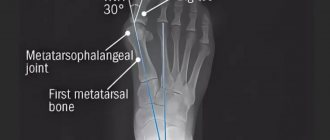
In this case, lateral deviation of the big toe occurs at the metatarsophalangeal joint. Apparently, wearing pointed-toed shoes with heels contributes to this deformation of the finger. In this case, there is increased pressure from shoes on the metatarsophalangeal joint of the big toe, which contributes to the formation of bursitis in this area. Arthritis in the named joint develops secondarily.
Pain can be relieved by soft pads in the bunion area and plastic “pegs” inserted between the first and second toes, but severe deformity requires surgical intervention. Various types of operations are used.
Thus, the medial part of the metatarsal head can be trimmed or a Keller operation can be performed, in which the proximal half of the proximal phalanx is removed (excisional arthroplasty), and the toe is formed into a flail shape.
Other surgical interventions used include osteotomy with metatarsal bone transfer, Mayo procedure (arthroplasty with excision of the distal head of the metatarsal bone) and arthrodesis of the metatarsophalangeal joint.
Taping the arch of the foot to limit the load
Several studies have confirmed the effectiveness of the arch taping method invented by Ralph W. Dye. Although there are several ways of taping using this method, even the basic version is effective.
The magnitude of the effect, however, is small, so arch taping is only part of the rehabilitation plan.
Taping instructions. The tapes that cover the sole in a vertical plane (in the photo - below left and right) should be stretched quite tightly and should always be glued from the outside to the inside.
Causes and symptoms
A common misconception concerns the causes of the development of pathology. Many people believe that the disease is the lot of older people, but this is a completely wrong judgment.
Plantar and nodular fasciitis occurs in people with flat feet (therefore it is important to correct the situation from childhood), excess weight, athletes (incorrectly calculated loads on the legs, excessive stretching of the calf muscle, overload, and so on). Also, the disease can occur due to a sharp change in physical activity, appear due to heavy physical work and due to injuries
The main symptom is a sharp pain in the heel bone during exercise, just when walking, which is felt more in the morning. The disease is diagnosed by examining the patient by a doctor and performing x-rays. If there is no spur as such, then it could be other diseases (rheumatoid arthritis, and other inflammatory processes, pain in the heels).
In any case, before starting treatment for heel fasciitis with folk remedies or other, even the most harmless, methods, you need to consult a doctor for diagnosis and treatment. After all, incorrect treatment, insufficient treatment, or attempts to get rid of a non-existent pathology lead to serious complications.
Next, we’ll talk about which recipes are the most effective. We will also consider a set of exercises for the feet, which will help cope with the disease faster, or prevent its development (you can do it for preventive purposes).
Symptoms of plantar fasciitis
The plantar fascia is a thick fibrous band that runs from the base of the heel to the heads of the metatarsal bones.
It has several branches, any of which can be damaged, but by far the most susceptible area of the plantar fascia to damage is the base of the inner bundle of fibers, right at the base of the heel.
Plantar fasciitis is most painful at the beginning of a run, but the pain gradually goes away as you warm up.
The pain may return at the end of your run and will be more severe if you run in shoes with insufficient arch support or in bare feet.
Your arch or heel may also hurt after a long day on your feet, especially if you wear uncomfortable, hard-soled shoes.
Prognosis for Sharp syndrome
Despite its complex range of symptoms, Sharpe syndrome is usually not too serious. Most people can improve their health with medications and lifestyle changes.
Your healthcare provider will need to develop a long-term treatment protocol that will best suit your symptoms.
During the course of the disease, various complications and long-term consequences may occur. If the disease remains untreated, mixed diseases of the connective tissue of the skin and polyarthritis with painful changes and vascular diseases may appear.
Anatomical structure of the plantar fascia
The plantar fascia plays a significant role in maintaining the normal shape of the foot. On the dorsum of the foot, the fascia is fixed to individual bone points. In the area where the muscles are located, it is divided into two leaves, which form a bed for the superficial muscles of the dorsum of the foot. The deep leaf isolates the interosseous muscles from the digital extensors.
On the plantar side of the foot, the fascia is much thicker compared to the dorsal side. In the middle part, the fascia has even greater compaction and consists of fibrous bundles. It is called the plantar aponeurosis. In the distal section, those bundles that form the aponeurosis are called transverse bundles. A significant part of the fibers of the plantar aponeurosis breaks up into five bundles. In the area of the calcaneal tubercle, some of them are a continuation of the tendon of the triceps surae muscle. With increased physical activity, micro-tears occur in the aponeurosis. In the case when the loads are not prolonged, the connective tissue regenerates. However, with regular physical activity, the aponeurosis does not have time to recover. The result is plantar fasciitis.
Yoga therapy for heel spurs
Yoga for heel spurs helps to completely relieve pain and also neutralizes areas of irritation.
Elements of yoga are performed after a thorough, comprehensive warm-up of the body muscles, since it is based on the development of flexibility, the level of which varies from person to person. Perform movements of the toes, rotation and movement of the feet, alternate bending of the legs with the heel placed behind the outer side of the pelvis.
Tibetan gymnastics contains the main exercise - Virasana. To do this, get down on your knees and move your pelvis back until you sit with your heel on the floor. If you feel pain in your knees, stop and put the folded blanket back and try to lower yourself again. Adjust the height of the mat until you sit completely without excessive pain in your knees. The exercise is performed in a completely relaxed state and in a fixed position while sitting on the floor, the execution time is three minutes. Yoga classes contain a set of exercises to treat heels.Finish your yoga with simplified Sarvagasana. To do this, lie on the floor, lift your legs vertically up and lean your elbows against the wall, and place your straightened arms behind your head on the floor.
Fasciitis: diagnosis
Based on the symptoms of plantar fasciitis, a preliminary diagnosis can already be made. At the initial appointment, the doctor carefully collects anamnesis, listens to the patient’s complaints, conducts a visual examination, palpating the foot in order to establish the localization of the pain syndrome. In the absence of a heel spur, along with heel pain, a differential diagnosis with systemic inflammatory pathologies is necessary. These include: rheumatoid arthritis, Reiter's syndrome and tarsal tunnel syndrome.
In addition to visual and tactile examination, the patient is often prescribed an X-ray examination. This is a simple diagnostic method that has virtually no contraindications and does not require lengthy preparation. The resulting x-ray image clearly shows the ossification of the ligament. In addition, instrumental examination makes it possible to distinguish pathology from a bone fracture and other possible ailments that provoke the appearance of painful sensations.
Treatment of the disease
Drug therapy involves the use of special agents. The medication course is based on the use of drugs that are classified as NSAIDs. They are available in the form of gels, tablets and ointments.
You also need special gymnastics, which you can watch in the video:
Traditional medicine
The most common representatives of this class are the following drugs:
- Ortofen;
- Nurofen;
- Diclofenac;
- Voltaren.
If a non-steroidal anti-inflammatory drug does not bring a positive result, corticosteroid drugs should be used. Give injections or take tablets only as directed by your doctor. If you do not adhere to certain regimens for taking these medications, they will provoke heel bone disorders. This reaction often becomes the cause of various complications.
Physiotherapy
In addition to the internal impact on the pathological process, it is also necessary to use certain procedures. These measures help relieve the inflammatory reaction from the fascia and ensure normalization of metabolism in the affected joint.
We list some of the prescribed procedures:
- ultrasound;
- therapy with a device that provides shock wave effects.
You can even be treated with mud applications with natural bioactive components. Laser may be prescribed to treat the disease.
Inflammation of tissues, as well as pain relief, are relieved using the following procedures:
- applying ice;
- massage manipulations;
- use of Botox;
- organizing warm baths;
- gymnastics;
- rubbing with warming ointments against inflammation;
- taking analgesics - Ibuprofen, Naproxen.
Treatment with folk remedies
As an additional tool for influencing the affected heel, treatment should be organized at home, using folk advice. You can make a compress of honey or buy bile. An effective way to combat plantar fasciitis is:
- hot potatoes that need to be mashed;
- grated horseradish;
- thoroughly crushed garlic;
- petrolatum.
A tincture of red elderberry, which should be used for rubbing, is recognized as a useful product for this pathology. To prepare it, you need to take the berries, pour alcohol into them and leave for 2 weeks in a place where there are no ultraviolet rays. A tincture of sunflower pulp has a similar effect.
People's experience shows the effectiveness of such measures - an adult is able to feel relief 8 months after treatment. However, in order to cure the pathology correctly and without complications, it is necessary to include the entire range of health procedures.
If, after using ointment, Dimexide, patch, massage, training, Bubnovsky exercises and taking pills, the patient’s condition does not improve, he should immediately contact a doctor, who will probably prescribe surgery.
Last resort
In advanced cases, the doctor recommends surgery. Part of the fascia on the sole is cut off, reducing its tension. This action relieves pain, although it can accelerate the development of flat feet.
After surgery, a person is recommended to use elastic stickers to help reduce heel stress.
Treatment of plantar fasciitis
Although plantar fasciitis usually resolves without causing long-term problems, it can last anywhere from 6 to 18 months.
You may need to take medicine to reduce pain and swelling. Your doctor will tell you which of these drugs is best for you. You should always take your medications as directed by your doctor.
The most common medications taken for this condition are nonsteroidal anti-inflammatory drugs (NSAIDs). Examples of NSAIDs:
- ibuprofen (Advil® and Motrin®);
- naproxen sodium (Aleve®);
- naproxen (Naprosyn®).
NSAIDs should be taken with food. Check with your doctor before taking NSAIDs if you:
- have previously consulted a doctor due to diseases of the stomach and intestines, liver or kidneys;
- have previously consulted a doctor due to bleeding disorders;
- are taking aspirin or other medicine that prevents blood clotting;
- are taking corticosteroids, which are steroid hormones produced by the adrenal cortex (the outer part of the adrenal glands).
For more information, read Common Medications Containing Aspirin and Other Nonsteroidal Anti-inflammatory Drugs (NSAIDs) (
Your doctor may also suggest applying tape to the arch of your foot or placing special support products in your shoes, such as heel insoles, orthotics (such as braces or splints), or arch supports. You may also need physical therapy.
If these treatments do not help, contact your doctor. You may be offered other methods, such as corticosteroid injections or surgery.
to come back to the beginning
Leg exercises
With regular exercise, you can not only strengthen your leg muscles, but also overcome heel fasciitis. The exercises are simple and even a person who is far from any sport can cope with them. We suggest considering several exercises:
- Go to the wall, lean on your hands. Place one leg forward, move the other back as far as possible, but do not lift your heels off the floor. Have you felt a stretch and the heel of your leg, when pulled back, can barely stay on the floor? Everything is correct! Hold your body in this position for 30 seconds. Switch legs, repeat 10 times for each.
- This exercise is especially good to perform in the morning, when the muscles are relaxed. Before you go on errands, do tasks that will help you stretch them, and the pain will not be so intense. If you do exercises every day, you will get better quickly. Take a tin can (0.5 l) with lemonade, you can also use a golf ball. Place the object under your foot, pressing lightly with it. Start rolling from toe to heel and back. It will take 50 circles for each leg.
- Sit on a straight, hard surface with your legs extended. Use your hands to reach your toes (at first you can bend your knees a little, then a stretch will appear) and pull them towards you. Straighten up, releasing your fingers, then repeat, and so on 10-20 times.
Causes and types of pathology
Plantar fasciitis is often caused by injuries to the plantar fascia. If we talk about other factors in the occurrence of the disease, we can highlight:
- Staying in one position for a long time. Office workers are at risk. People standing behind the conveyor also have increased load on their feet. Athletes are highly susceptible to the disease. These could be lightweight and weightlifters or bodybuilders.
- Flat feet. Due to a flat arch, uneven pressure is created on the foot. In this case, the inflamed area “suffers”.
- Overweight. In obese people, the load on the foot increases many times.
- Shoes without arch supports. This type includes sneakers. Frequent wearing of flip-flops or flip-flops also puts excessive stress on the fascia.
- Increased load on the foot. Plantar fasciitis is common among professional athletes and people whose work activities involve carrying heavy objects. Long walking can also lead to illness. With this pathology, the stairs are sometimes an insurmountable obstacle.
- Congenital weakness of the soles of the feet.
- Disorders of metabolic processes and blood circulation of the foot. This condition can occur in people suffering from diseases such as gout, diabetes or atherosclerosis.
- Age-related changes in the tissues of the sole.
- Infectious process. With diseases such as chlamydia or gonorrhea, the spur forms covertly, and it may appear long after infection. Necrotizing fasciitis is caused by bacteria that enter the human body through wounds or insect bites.
People over 50 are more likely to suffer from plantar fasciitis.
With age, the structure of the fascia changes. It becomes thicker and less elastic. As the pathology progresses, inflammation of the fascia occurs, especially in the place where it attaches to the heel bone. Swelling and pain appear.
The classification of plantar fasciitis includes five types:
- Distal. Pain in this form manifests itself throughout the entire foot. The strongest sensations occur in the area of the toes.
- Proximal. This type is characterized by the inflammatory nature of the pain present in the heel area.
- Front. With this form, the pad of the finger hurts, and then the pain spreads to the heel bone itself.
- Nodal. It is also called Ledderhose disease. Nodules form on the aponeurosis of the sole. This leads to its thickening.
- Bilateral. With this form, the fascia on both feet becomes inflamed.
There are several types of plantar fasciitis depending on the location of the lesion.
How to distinguish fasciitis from other foot diseases?
Fasciitis
is an inflammatory process that occurs as a response to the constant death of fascial cells due to multiple microtraumas.
The body cannot cope with the dissolution and removal of dead destroyed cells and swelling and inflammation begin. Characteristic features:
- “starting” pain in the heel, which appears not as a result of the load, but before it, when the legs have been at rest for a long time - in the morning or after a long sitting;
- the pain intensifies with increasing load - during long walking, running and long standing;
- swelling of the arch of the foot;
- decreased mobility (contracture with foot deformity);
- in some cases - lameness.
In the evening, the leg hurts much more as a result of the day's stress.
The pain syndrome most often manifests itself in the sole area, but can radiate to the toes and lower leg.
Symptoms of plantar fasciitis can be similar to those of other foot diseases, so it is important to get the correct diagnosis early.
An orthopedic surgeon treats inflammation of the plantar fascia
or
surgeon
.
Therapeutic gymnastics is a component of complex treatment of heel spurs
You should not expect that physical therapy will save you from heel spurs in just a week. To notice a positive effect, you need to constantly train, gradually increasing the load. However, you must understand that therapeutic exercises are not a panacea for heel spurs. Gymnastics is one of the measures in the complex treatment of the disease, advisable in the initial stages of the development of plantar fasciitis to eliminate pain.
The patient also needs to take non-steroidal medications, massage, orthotics, physiotherapy, arch supports, heel pads, ready-made or custom insoles, gels and ointments for external use, weight loss, limiting physical activity, wearing comfortable shoes (women are recommended to wear shoes with stable heels with a height of 3 to 5 cm). You can also take warm baths with sea salt or use orthopedic patches. They will improve blood flow and nutrition of foot tissues.
Read also: Cervicothoracic osteochondrosis exercises
If proven methods of treating heel spurs are unsuccessful within six months, the patient is prescribed surgery. During surgery, the plantar fascia is first incised at its attachment to the heel bone, then the spur is removed.
Additional video about gymnastics for heel spurs (in English):
Characteristics of the disease
Plantar fasciitis is an inflammatory process in the area of attachment of the plantar aponeurosis and all surrounding perifascial structures to the periosteum of the calcaneal tubercle. Inflammation develops as a result of increased extensibility of the fascia, which leads to chronic microtrauma with ruptures of the aponeurosis and the subsequent development of the degenerative process. Synonyms for fasciitis are heel spur, subcalcaneal pain, painful heel syndrome, calcaneodynia.
Anatomy and function of aponeurosis
The plantar aponeurosis runs from the medial tubercle of the calcaneus to the proximal phalanges of the toes. Normally, the thickness of the plantar aponeurosis, according to ultrasound, ranges from 3 to 8 mm. The plantar fascia stabilizes the foot through the “winch effect,” which is when the heel is pulled in two directions when you push off. It is pulled upward by the Achilles tendon, and pulled forward by the plantar aponeurosis, which, when supported by the fingers, wraps around the heads of the metatarsal bones like the gate of a winch. The aponeurosis is a passive stabilizer of the arch of the foot. It supports the arch of the foot, is involved in toe flexion, and reduces stress on the lateral edge of the foot.
Causes of fasciitis
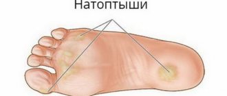
1. Excessive pronation of the foot when rolling. With valgus of the hindfoot, as well as with a flat arch, there is a decrease in supination at the beginning of the roll and an increase in pronation in the middle and end of the roll. Pronation causes a redistribution of the load along the foot, reducing the load on the lateral edge of the foot and increasing the load on the medial edge and aponeurosis. During excessive pronation, the tension of the aponeurosis increases, which leads to its stretching at the site of attachment to the tubercle of the calcaneus. Constant pronation causes chronic microtrauma of the aponeurosis. As plantar fascia insufficiency progresses, the load on the metatarsal heads increases with increasing pressure underneath them, which leads to the formation of calluses of the skin in the forefoot. 2. Hollow foot. With a cavus foot, its deformation is relatively severe. In a person with a cavus foot, there is an increase in the angle of inclination of the metatarsal bones due to increased tension of the plantar aponeurosis and thickening of the plantar aponeurosis. With a hollow foot, there is a lack of support on the lateral edge of the foot, a decrease in the area of support of the foot, which leads to a decrease in the ability to distribute the load during push-off from the support. The load on the middle section is reduced, and the load on the anterior and posterior sections is increased. As a result of a decrease in the ability to distribute pressure over the surface of the foot, secondary thickening of the plantar fascia develops. A small foot support area causes high average pressure under the foot, which contributes to discomfort in the foot.
Factors contributing to the progression of fasciitis:
1. High body weight, obesity. 2. Night sleep with flexion of the ankle joint under the influence of the weight of the forefoot and hypertonicity of the calf muscle, which leads to contraction of the plantar aponeurosis and the gradual development of its contracture. In the morning after waking up, stretching of the aponeurosis during the first steps is painful. The situation is aggravated in patients with joint hypermobility. 3. With fasciitis, clinical symptoms can be caused by a complex of changes, which include: inflammation, neuritis of the first branch of the lateral plantar nerve, perineural fibrosis of the nerve that innervates the muscle that abducts the 5th finger, calcaneal spur, injury to the heel area, periostitis, traumatic rupture of the aponeurosis , seronegative arthritis.
Symptoms of fasciitis
Manifestations of fasciitis include pain in the arch of the foot, pain in the lateral part of the foot, swelling of the proximal part of the foot, weakness of the foot muscles, and lameness. With fasciitis, the pain syndrome has its own characteristics. Unpleasant sensations occur in the morning after waking up when the patient takes his first steps. The pain is localized along the inner surface of the heel and intensifies when the fingers are straightened. If unpleasant sensations appear in the morning, they completely disappear within a day. In addition to morning pain, a feeling of discomfort in the foot may occur when moving after prolonged sitting. The severity of pain decreases when the legs are warmed.
Diagnostics
With fasciitis, as a result of prolonged irritation in the area of fixation of the ligament to the heel tubercle, an inflammatory reaction occurs, which is accompanied by pain on the plantar surface of the foot. Ultrasound shows thickening of the plantar fascia. The difference in the thickness of the fascia on the diseased and healthy feet reaches 4 mm. Fasciitis is differentiated from calcaneal nerve neuropathy. Patients with neuropathy complain of pain in the posterior surface of the heel 4-5 cm in front of its posterior edge in the area of the transition of the lateral surface to the plantar surface from the medial malleolus to the edge of the heel. The EMG of the calcaneal nerve appears unchanged.
Treatment
The main method of treating fasciitis is conservative. Conservative treatment is effective in 89% of patients. The most successful is complex treatment, which includes several methods. Each individual method provides less treatment effect than their combined use.
Table 1 Frequency of use and effectiveness of treatment methods for fasciitis
| Treatment method | Frequency of application (%) |
| Peace | 70 |
| NSAIDs | 69 |
| Orthoses | 63 |
| Modified shoes | 48 |
| Heel brace | 45 |
| Corticosteroids | 41 |
| Thermal treatments | 29 |
| Cold treatments | 27 |
| Shock wave therapy | 24 |
1. Rest Rest means staying indoors and walking within the home for hygienic needs. Rest is resorted to during an acute attack of the disease, mainly in unemployed people, throughout the day, in combination with physical exercise and physiotherapeutic procedures to prepare for other types of treatment. 2. Cold procedures Carry out at home. An ice pack is applied to the plantar surface of the foot. 3. Physical education with exercises for stretching the foot Do stretching of the triceps muscle by passive extension in the ankle joint with the knee extended. The soleus muscle is stretched by passively extending the ankle joint with the knee bent. 4. Heel brace Reduces the degree of tension in the Achilles tendon and plantar aponeurosis, making walking easier. The braid is inserted into shoes, or tucked under the heel. 5. Design of the orthosis The requirements for the orthosis are to limit the pronation of the foot to normalize the distribution of the load along the foot. 6. Cushioning of loads in the orthosis According to the degree of depreciation, in descending order, the material for orthoses is distributed as follows: silicone, rubber, felt, plastic. The orthosis is indicated for people whose work involves walking or prolonged standing for more than 8 hours. The best properties are provided by a complex orthosis made of several materials, which reduces the impact load and limits excessive pronation of the foot. 7. Special shoes For constant stretching of the aponeurosis, rocker shoes or shoes with rocking soles are used. In shoes, the thickness of the sole in the front section is less than in the back. Due to the difference in height, the foot rolls from heel to toe relatively easier during the roll, which relieves the metatarsophalangeal joints from excessive extension. Ordinary shoes are modified to give them orthopedic function. To do this, a steel tire with a curved toe end is placed in the sole. It corrects the foot and provides the necessary unloading of the toes. 8. Ankle splint A splint is placed on the ankle and foot at night. The splint achieves 5º flexion of the foot in the ankle joint in relation to the shin and 30º extension of the fingers in the metatarsophalangeal joints in relation to the metatarsus. The splint eliminates contracture of the plantar aponeurosis and Achilles tendon and ensures painless first steps after sleep. The effect of treatment is achieved within 1 month. 9. NSAIDs
Table 2. Use of NSAIDs for foot pain
| A drug | Dose | Action | Contraindications |
| Ibuprofen | 600-800 mg | Suppress the synthesis of prostaglandins, reduce the activity of cyclooxygenase, relieve the inflammatory response and pain | Peptic ulcer of the stomach and intestines, renal and liver failure, decreased blood clotting, hypersensitivity to the drug, hypertension, pregnancy |
| Ketoprofen | 25-50 mg | ||
| Naproxen | 500 mg |
10. Steroid drugs In outpatient practice, steroid injections into the area of the heel tubercle are given in approximately half of patients with plantar fasciitis. An injection of diprospan (betamethasone) is used as a steroid drug at a dose of 6 mg per 1 ml of lidocaine. The injection is made into the heel tubercle on the inside of the foot. A single injection provides a therapeutic effect in 41% of patients for a period of 6 to 8 weeks. When treated with a course of hydrocortisone injections, an analgesic effect is observed in 68% of patients for a period of more than one year. When using steroids, complications are possible in approximately 1 case. Complications such as atrophy of adipose tissue on the plantar surface of the foot or rupture of the plantar aponeurosis occur. When the aponeurosis ruptures, there is a decrease in pain in the heel and an increase in pain along the outer edge of the foot, which lasts for a short period. After rupture of the fascia, there occurs a weakening of the passive stabilization of the foot, its destabilization, redistribution of the load along the foot, stretching of the structures along the inner edge of the foot, compression of the structures along its outer edge, increased load on the ligaments of the midfoot and the tendon of the posterior tibial muscle. Closure of the calcaneocuboid joint develops with pain along the outer edge of the foot. The insufficiency of the plantar aponeurosis is compensated by other structures. With rigid ligaments, compensation occurs relatively faster, and with elastic ligaments relatively slower. With increased ligament extensibility and hypermobility, the stabilizer defect is compensated worse and fascial rupture is accompanied by prolonged pain in the foot.
Combined conservative treatment
The optimal treatment method for plantar fasciitis is a combination of several methods of influencing the pathological process. During the day, this means wearing an insole or rocker shoe and doing stretching exercises, and at night, this means sleeping in a splint.
Surgery
Surgery is performed in less than 10% of patients with plantar fasciitis. The indication is severe pain in the heel, disabling the patient. The following operations are used to treat fasciitis: heel spur removal, fasciectomy, fasciotomy, neurolysis, neurectomy, calcaneal osteotomy, calcaneal tunnelization, arthroscopic plantar fasciotomy. Fasciotomy Release of the plantar fascia is performed using endoscopic techniques. Access to the fascia is made on the plantar surface of the foot 1 cm distal to the medial edge of the calcaneal tubercle. The optics are directed from the inside out. A blade is inserted and the fascia is cut in the direction from the medial to the lateral edge. To achieve an analgesic effect, it is enough to cross the medial and central parts of the fascia along 4/5, leaving approximately 1/5 of the diameter of the fascia. Neurolysis A 3 cm long incision along the inner surface of the heel, or an oblique incision 2 cm from the edge of the medial malleolus. The branches of the medial calcaneal nerve and the muscle that abducts 1 finger are distinguished. The superficial and deep fascia are separated and released. Under the deep fascia there is a nerve that goes to the muscle that abducts the 5th finger. The nerve may be pressed between the fascia and the superior edge of the muscle. The fascia is cut along the medial edge at the site of its attachment to the calcaneal tubercle to 1 cm in width and depth. If the nerve going to the muscle that abducts the 5th finger is injured by a heel spur, then the nerve is decompressed by resection of the spur. Decompression of the nerve going to the muscle that abducts the 5th finger and partial resection of the plantar aponeurosis gives an analgesic effect in 75% of cases.
Mitskevich Viktor Aleksandrovich Orthopedist-traumatologist, Doctor of Medical Sciences
Physiotherapy
When and how to treat trochanteritis of the hip joint with exercise therapy, the doctor decides. Typically, a course of therapeutic exercises is prescribed after the acute symptoms of the disease have been relieved, that is, when the pain completely disappears. Special gentle exercises provided by the course help to activate blood flow and strengthen the thigh muscles. However, they must be performed with the permission of a doctor, under the supervision of a specialist, so as not to harm the joint.
As a type of exercise therapy, post-isometric relaxation can be prescribed. This technique involves performing passive stretching of the external ligaments and muscles in certain positions of the patient’s body. Stretching them will help overload the hip joint and related structures. A regular stretching program to maximize the length of the muscles and ligaments is essential for proper hip function. Exercises are carried out together with a doctor, for 20 minutes every other day. For tangible results, a minimum of 10 sessions are required.
Prevention measures
Proper care and careful handling will help you avoid problems with sunglasses. Strictly follow the manufacturer's recommendations, the basic rules and glasses will serve you for a long time:
- store only in a special hard case of the appropriate size;
- subject to regular cleaning with soap suds or special wipes, sprays;
- additionally purchase and apply protective films;
- do not use acetone-containing compounds or abrasive substances;
- do not place lenses face down on hard surfaces;
- Do not expose to too high or low temperatures, do not leave in the open sun or car panels;
- remove the frame, holding the temple with both hands;
- use glasses only for their intended purpose (do not, for example, wear them as a hairband);
- Avoid contact with the product with varnishes, air fresheners, and other chemicals.
Recommended clinics
EUROPEAN CLINIC OF SPORTS TRAUMATOLOGY AND ORTHOPEDICS (EMC Orlovsky)
Around the clock. Russia, Moscow, Orlovsky lane, 7 +7
Interpretation of PET CT results from another health care facility – RUB 5,700. Remote telemonitoring of health status – 5800 rub. Doctor's appointment/Consultation:
- orthopedist-traumatologist – 10,700 rubles.
- surgeon 10,700 rub.
- Lecture by a specialist doctor – 35,000 rubles.
- Foreign trauma surgeon – 9,600 rubles.
Russian-Israeli Medical
Mon-Fri: 09:00 – 19:00 Russia, Moscow, 2nd Tverskoy-Yamskoy lane, building 10 +7,,,
- Admission to Ph.D. for joint replacement – 3000 rubles.
- Appointment with a professor of joint replacement – 5,000 rubles.
- Admission to Ph.D. for arthroscopy of joints – 3000 rubles.
- Removal of intra-articular bodies – 24,000 rubles.
- Reconstruction of feet – 97,000 rubles.
- Hip replacement – 410,000 rubles.
- Endoprosthetics of the knee joint – 400,000 rubles.
Treatment options for plantar fasciitis
Inflammation of the fascia is predominantly treated conservatively - in almost 80% of cases. Injections of non-hormonal or hormonal anti-inflammatory drugs will help relieve severe pain. Physiotherapy increases the chances of a speedy recovery. If conservative methods are ineffective, surgery is performed.
Exercises
It is best to use an anti-stress ball - it has something like soft spikes on its surface. The ball needs to be rolled with the sole on the floor with slight pressure to activate blood circulation and relieve pain, and most importantly, to work the plantar fascia.
You can also perform exercises on a special massager to stretch the fascia. It is purchased in specialized stores; outwardly it consists of several balls located side by side, which need to be dynamically rolled with the heel and foot. This treatment method also works for heel bumps.
Video: Exercises, massage and taping for fasciitis.
Medicines
To treat plantar fasciitis of the heel, it is better to use non-steroidal anti-inflammatory drugs based on naproxen, ibuprofen or aspirin. These are effective painkillers and anti-inflammatory medications for spurs. It is better to choose drugs in the form of creams or gels that are applied directly to the affected area.
Folk remedies
Pine-salt baths relieve pain and fatigue.
Folk remedies for plantar fasciitis are effective at the initial stage of the disease. What the populists recommend:
- Contrast baths, in which chamomile decoction is added to a bowl of hot water. The feet are held in each container for approximately 30 seconds. After the water has cooled, the feet are lubricated with spur cream and massaged.
- Treatment with a strong saline solution helps to activate blood flow and reduce pain. Add 300 g of salt per liter of hot water and stir until completely dissolved. Baths are made until the solution cools completely. The course of therapy is two weeks.
- Dried lilac baths will help relieve pain and swelling in the foot. The flowers are infused for ten days with vodka in a ratio of 1 to 10, after which they drink 50 ml of the product three times a day, and rub the heels with the same folk remedy for spurs in the evenings.
Physiotherapy
Physiotherapeutic methods for treating fasciitis include:
- mud applications - warming the foot with a sulfide-silt mixture, the course consists of 10 procedures;
- Ultrasound therapy – heating the tissues of the foot using ultrasound on the spur, which, with the simultaneous use of therapeutic mixtures, allows you to achieve the effect in 2-5 procedures;
- laser treatment, which helps to activate blood circulation, relieve swelling and pain, and accelerate the recovery of fasciitis tissue;
- shock wave therapy – exposure of the problem area to acoustic waves allows you to quickly get rid of plantar fasciitis;
- interstitial electrical stimulation;
- cryotherapy;
- magnetotherapy.
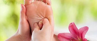
Massage
For plantar fasciitis, you can do self-massage at home and use the services of a professional chiropractor. With the help of massage, blood circulation is activated, inflammation goes away faster, the muscles of the foot and leg are strengthened, which prevents relapses of fasciitis in the future.
The direction of massage for fasciitis is from the toes to the heel and back.
Orthopedic products
For people suffering from plantar fasciitis, boot models have been developed that are used at night. It is inconvenient to use them during the day, but at night the foot in the boot takes a gentle position and rests, and in the morning when stepping on the foot, patients do not feel pain. To prevent injury to the fascia when walking, use heel pads.
Additional Treatments for Fasciitis
If you've tried all of the above treatments to relieve arch or heel pain but find yourself going a little further, there is something else you can do.
Massage for plantar fasciitis
Massaging the plantar fascia tissue is an approach that has recently become increasingly popular among runners.
Using a golf ball or other hard round object, you can knead your arch in the same way you would knead your quadriceps or calf muscles with a foam roller.
More aggressive soft tissue techniques such as the Active Release Technique or the Graston Technique are also popular.
However, all of these types of massage have not been scientifically proven to be effective, so while many runners do find them very helpful, there is no guarantee that they will work for you.
If you decide to roll the ball of your foot or perform any other soft tissue manipulation, applying ice after the procedure is a very good idea.
Steroid injections into the foot to relieve heel pain
Corticosteroid injections are a common second-line treatment among podiatrists.
Although some studies have shown that they can help, scientists urge caution as the success rate with them is quite low and there is a risk of complete tearing of the plantar fascia.
Administering a corticosteroid such as dexamethasone through the skin via electrophoresis may be more beneficial and have a lower risk of complications than direct injection.
But in any case, you should discuss the need to use electrophoresis with an orthopedist or podiatrist.
New, alternative treatment for plantar fasciitis
Cases of chronic plantar fasciitis can be especially challenging.
Two new treatments, extracorporeal shock wave therapy (ESWT) and PRP therapy (platelet-rich plasma injections), are showing good promise in treating recalcitrant fasciitis, especially in runners.
Due to the relatively recent development of these therapies, they may not be readily available, but their effectiveness has not yet been sufficiently studied.