Price list Doctors clinic
X-rays of joints and bones are widely used to diagnose pathologies of these structures. This is one of the fastest and most informative examination methods. X-rays are used when degenerative or inflammatory diseases are suspected, as well as when assessing the consequences of injuries. The images help make an accurate diagnosis and subsequently monitor the effectiveness of the prescribed treatment. The price of x-rays of the hip joints and bones is more affordable than the cost of complex studies (CT, MRI, etc.), while the method allows you to obtain accurate information about the condition of the tissues in a short time.
Indications and contraindications
X-rays of bones and joints are prescribed by a doctor if the following diseases and consequences of injuries are suspected:
- cracks/fractures;
- dislocation, sprain;
- ligament or tendon rupture;
- tumors, cysts;
- inflammatory diseases of the joints;
- arthrosis;
- osteoporosis;
- osteophyte, etc.
A specialist may suspect diseases of bone tissue and joints based on symptoms such as pain, redness and swelling, limited joint mobility, and local fever. The resulting injury is also a reason to take an x-ray - it is important to assess the condition of the bone tissue, exclude serious damage and take timely measures if they are detected.
A relative contraindication to radiography is pregnancy, but there are cases when performing such a diagnostic procedure is justified. A mental illness in which the patient cannot maintain a still body position may also be a contraindication.
Causes of injuries and diseases of the wrist joint
When falling, as a rule, a person puts his arms forward to protect the body and face from impact, so the wrist joint is most often injured when falling on an outstretched arm with emphasis on the palm and hand. Bones are easily damaged by mechanical stress and body weight. As a result of unsuccessful falls, falls from a height, accidents and other accidents, a person receives injuries of varying degrees of severity, including damage to the wrist joint.
In addition, patients suffering from osteoporosis are susceptible to wrist fractures. This disease causes increased bone fragility and decreased bone density, which leads to frequent injuries.
In addition to mechanical damage, the causes of diseases of the wrist joint can be improper exercise, aging of the body, poor nutrition, excessive muscle strain, physical inactivity, etc.
You can have a quick and high-quality x-ray examination of the wrist joint in St. Petersburg in the trauma department of the Medi clinic network. It is equipped with the latest Italian digital Clinomat device, multi-mode and practically safe, allowing for a complete x-ray diagnosis of the human body in the shortest possible time. The device allows Medicenter specialists to examine images in various processing modes, quickly and accurately establish a diagnosis and prescribe competent and effective treatment.
Indications for x-ray examination of the wrist joint:
- fractures of the radius, Galeazzi fracture, wrist bones;
- wrist sprains;
- bruises, cracks;
- acute pain;
- swelling, hematomas;
- tumor, degenerative-inflammatory processes;
- developmental anomalies of the wrist joint, etc.
In addition, x-rays of the wrist joint are prescribed if the patient complains of stiffness in wrist movement, crunching, pain and other unpleasant symptoms. Radiography of the wrist joint is also necessary during the treatment period to monitor its effectiveness, or before surgery.
Contraindications to x-ray examination of the wrist joint
When prescribing an x-ray examination, each case is considered individually. As a rule, doctors try to avoid x-rays of the wrist joint if the patient is pregnant or the patient’s general serious condition. In these cases, it is recommended to replace x-rays with diagnostic methods such as ultrasound and MRI.
Preparation and procedure
No special preparation is required for the study. The patient is placed on a special table, the groin area is protected with a lead apron. For children, additional protection is used for the thyroid gland and eyes; the baby is completely covered with an apron, leaving only a limb exposed.
An image of a joint or bone can be taken in one or more projections. To obtain accurate data, it is important to remain still. The study takes just minutes. The doctor sees the condition of the tissues immediately, and a conclusion with a transcript is issued after the procedure.
Excess body weight can become a hindrance - fat cells distort X-rays. This results in the picture not being clear enough. In this case, the attending physician will suggest alternative examinations. Sometimes it is possible to solve the problem by taking pictures from several projections. Some diseases and injuries require only a direct projection, others require a lateral projection, on both sides. Occasionally they resort to a combination of several options. This allows you to get the most accurate picture of the condition of the bone tissue.
How are x-rays taken of the bones and joints of the arms and legs?
An X-ray of the bones of the legs and arms takes on average no more than five to ten minutes and does not require the use of painkillers, since it is an absolutely painless and safe procedure.
Tubular bones are most often examined. To obtain high-quality images, two perpendicular projections are used. The main condition for the patient is to remain motionless during the shooting. The procedure includes the following:
- The limb being examined must be freed from clothing and any metal objects;
- The limb is placed in the center of the X-ray cassette and the rays are directed at it at the appropriate angle;
- The radiologist takes pictures in such a way that adjacent joints are also clearly visible. This approach allows for high-quality visualization of the entire bone and ensures that adjacent joints and bones are not damaged.
If it is necessary to conduct a functional study, the patient is asked to flex/extend the joint of the area being examined or provide a load on the limb being diagnosed. There are often cases when, due to illness or injury, the patient cannot achieve the required position of the arm or leg. Then CELT radiologists use special projections, not forgetting to maintain the perpendicular position of the cassette relative to the X-ray emitter.
How safe is the examination?
Patients who need to have joint x-rays often ask whether the procedure is harmful. The harm from radiation during such diagnostics is minimal if we are talking about the use of modern equipment. Using an advanced X-ray machine allows you to receive a radiation dose comparable to the amount of natural radiation from TV or an airplane flight. So, with an X-ray of the knee joint, the dose is 0.001 mSv - almost the same amount a person receives every day in the course of life. Therefore, the image can be repeated, for example, after 2–4 weeks, to assess the effectiveness of the prescribed treatment or the dynamics of tissue healing.
You can get an x-ray of your joints in Moscow at the Family Doctor clinic. We use advanced equipment with minimal radiation. The photo is taken quickly, which reduces the burden on the body.
What will an x-ray show?
Radiography of bones and joints is widely used in orthopedics and traumatology. Despite the variety of other research methods (ultrasound, MRI, etc.), it is the x-ray that remains the leading method for determining injuries to the musculoskeletal system. Without it, rapid diagnosis of closed and open fractures, cracks, subluxations, dislocations, and ligamentous injuries is unthinkable. X-rays are also performed after reposition, that is, stabilization of the fracture.
The image also allows you to detect birth defects and anomalies (dysplasia). These include, for example, arthrosis. This is an acquired disease, which is characterized by pathological changes in tissue. With its help, the doctor can detect violations of the conformity of articular surfaces, growth of bone tissue along the edge of the cartilage, areas of calcium deposition, etc. Diagnosis is carried out if infectious and inflammatory diseases (arthritis, etc.) or oncological pathologies (tumors and neoplasms) are suspected.
The images are interpreted by a radiologist. He studies the picture and draws up a written report, which is subsequently sent to the attending physician.
Features of joint diagnostics
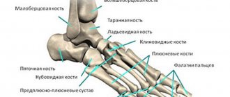
Foot.
The picture is taken in several projections. Based on the results of the examination, the doctor gets an accurate picture of all the bone and joint formations of the feet. The method allows you to detect injuries, degenerative changes, diagnose deformities, flat feet, heel spurs, synovitis, etc.
Ankle joint.
This joint experiences enormous loads and is therefore susceptible to disease. The most common pathologies are injuries, arthrosis, inflammatory diseases of the joint and joint capsule. To make a diagnosis, the image is taken in three projections.
Knee-joint.
A common reason for investigation is injuries: meniscus tear, dislocation, condyle fracture. Also, using an x-ray, a doctor can diagnose arthrosis, arthritis, hemorrhages in the joint and other diseases. This diagnosis makes it possible to examine both the joint and part of the fibula, tibia, and femur.
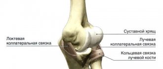
Elbow joint.
Injuries to this joint occur quite often, and they are especially common among athletes. Fractures of the shoulder, radius, processes of the ulna, and dislocations of the bones of the forearm may occur. Often there are cases of bursitis - inflammation of the synovial bursa.
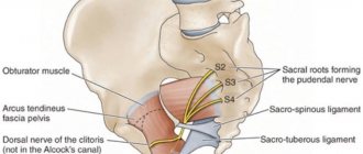
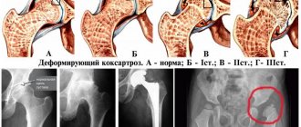
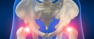
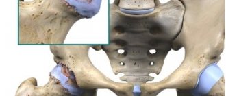
Hip joint.
X-ray allows you to diagnose arthritis, necrosis of the femoral head, neck fracture, tumors, changes in tissue structure, coxarthrosis, etc. X-ray of joints is affordable. This is one of the few types of radiography that requires preparation: to increase the accuracy of the image and eliminate unnecessary shadows, you must have a bowel movement before the examination. This can be done naturally, using a laxative or an enema. When choosing a method, it is better to consult a doctor. Also, if possible, it is better to avoid foods that increase gas formation 2-3 days before the procedure. Such preparation measures will also be required when planning an x-ray of the pelvic bones.
Shoulder joint.
An X-ray may be required if glenohumeral periarthritis is suspected, as well as tumors, inflammatory and degenerative processes, fractures and dislocations. The image also allows you to visualize the collarbone and scapula.
Temporomandibular joint.
Radiography of this joint is often used in dental practice and maxillofacial surgery. It allows you to diagnose joint diseases and injuries. The data obtained is also used by the doctor to identify malocclusions. An image of the jaws allows one to draw conclusions about the volume and structure of bone tissue; this information is necessary when planning orthopedic treatment and dental implantation.
The procedure for performing an X-ray examination of the wrist joint
Radiography of the wrist joint is performed in two projections. The image itself should simultaneously show the lower thirds of the bones of the forearm, the bones of the wrist, the area of the wrist joint, and parts of the metacarpal bones, because Wrist injuries are often confused with wrist injuries.
The entire procedure takes about 5 minutes, and the radiation dose is very small. After completing the X-ray examination, the patient receives an image and its description and is sent for consultation and further treatment to the appropriate specialist.
Conditions for accurate diagnosis: where to get an x-ray
To obtain reliable information about the structures being studied, it is necessary to remain still. The doctor will tell you at what point to “freeze”. It takes a few seconds to take a photo. The lack of preparation required also simplifies the examination. However, it is important to remember that the study of the pelvic bones and hip joint can be complicated due to the accumulation of gases and/or feces in the intestines. In an emergency situation, such as in the case of injury, the doctor performs the test without thorough preparation.
There are many institutions in Moscow where you can get x-rays of your joints. When choosing a clinic, pay attention to the equipment. Modern X-ray machines have maximum resolution. This increases the diagnostic value of the procedure and eliminates the need to take a series of images.
Our clinics in St. Petersburg
Structural subdivision of Polikarpov Alley Polikarpov 6k2 Primorsky district
- Pionerskaya
- Specific
- Commandant's
Structural subdivision of Zhukov Marshal Zhukov Ave. 28k2 Kirovsky district
- Avtovo
- Avenue of Veterans
- Leninsky Prospekt
Structural subdivision Devyatkino Okhtinskaya alley 18 Vsevolozhsk district
- Devyatkino
- Civil Prospect
- Academic
For detailed information and to make an appointment, you can call +7 (812) 640-55-25
Make an appointment
We draw your attention to the schedule of technological breaks in the CT and X-ray rooms.
X-ray, as one of the most informative and visual diagnostic studies, helps to identify various types of damage, developmental pathologies, injuries, inflammatory processes of the wrist joint, the condition of bones, cartilage, soft tissues of the lower part of the forearm, wrist. The most common diseases in this area of the upper limb are arthritis, arthrosis, tendinitis, necrosis, tunnel syndrome, trauma, and osteochondropathy.