Hernias of the thoracic (syn. thoracic) spine, in contrast to lumbar and cervical hernias, are a rather rare pathological phenomenon. According to statistics, pathology with such uncommon localization in the general group of intervertebral hernias occupies no more than 1%, in the structure of all diseases of the spinal column - 0.5%. But, despite the infrequent development of lesions in the chest, it is characterized by a very wide variability of symptoms and the severity of consequences.
The thoracic intervertebral lesion is located in the central part of the spine. This section occupies the entire area from the collar zone to the zone of lumbar lordosis, providing innervation to vital structural units of the body. Nerve spinal fibers in the corresponding area innervate the upper limbs, the respiratory center (trachea, lungs and bronchi), the solar plexus, and the anterior wall of the chest. In addition, neural connections are made with the esophagus, liver, biliary tract, duodenum, large/small intestine, spleen, urinary system, groin area and fallopian tubes.
The protrusion is visible on MRI images (indicated by arrow)
Thoracic intervertebral hernias often cause pinching of nerve roots, provoking not only noticeable painful discomfort along the affected nerve, but also, in advanced situations, serious dysfunction of the listed organs and systems. Experts characterize discogenic pathogenesis in this part of the spinal system as clinically unfavorable, with a high degree of disabling risk. Therefore, in order to avoid dangerous complications, it requires early diagnosis and timely treatment. People aged 20-45 years are most susceptible to the disease. The risk category includes athletes, hairdressers, welders, seamstresses and tailors, office workers, programmers, and vehicle drivers.
Therapeutic measures for such a complex diagnosis must be adequate to the true clinical picture! Otherwise, a person will be subject to unbearable suffering for life from illiterate therapy that does not bring a single drop of benefit or expected relief.
Valuable information conveying the whole truth about the treatment of intervertebral hernias of the thoracic region will be found in full further in the article.
What is a thoracic disc herniation?
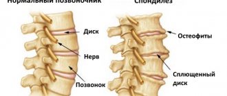
An intervertebral hernia is a protrusion of an intervertebral disc (the shock-absorbing pad between the vertebrae) outside the spinal column or into the spinal canal. In the area of the thoracic spine, disc herniation is rare, since this is the least mobile part. The 12 thoracic vertebrae (Th1 – Th12) connect to the ribs and sternum, forming a stable structure.
According to statistics, among all intervertebral hernias, this pathology in the thoracic region is no more than 1%. Young and middle-aged people are more likely to get sick. But since this area innervates the internal organs, as well as the upper and lower extremities, a herniated disc in the thoracic region can lead to serious complications. According to the International Classification of Diseases, 10th revision (ICD-10), disc herniation of the thoracic spine belongs to the section “Involvement of intervertebral discs in other parts,” code M51.
Disc herniations are best treated in the initial stages, so if you experience pain in the chest, which is often mistaken for diseases of the internal organs, but cannot be treated for six months or more, you need to contact a neurologist and check the condition of the spine.
How to treat a thoracic hernia?
In most situations, therapy is effective without the use of surgical interventions. It is very important to consult a doctor in a timely manner and strictly follow all his recommendations. The following methods are used in the treatment of thoracic hernia:
- Taking medications. Non-steroidal anti-inflammatory drugs, painkillers, muscle relaxants, hormonal drugs, and chondroprotectors are usually prescribed. Medicines are used orally, externally, administered in the form of injections and blockades.
- Physiotherapeutic treatment. When the acute inflammatory process is stopped, physiotherapeutic procedures are prescribed.
- Massage.
- Manual therapy.
- Physiotherapy.
- Folk remedies.
Causes and mechanism of development of thoracic intervertebral hernia
If the cause of disc herniation in the cervical and lumbar spine is most often osteochondrosis, then thoracic disc herniation most often develops against the background of injuries, poor posture, heavy physical activity, or, conversely, a sedentary lifestyle. Osteochondrosis is also important, especially if there is a hereditary predisposition to the early development of this disease.
Under the influence of various reasons, the intervertebral disc (an elastic fibrocartilaginous plate containing an elastic nucleus pulposus) wears out, becomes thinner, cracks, and the nucleus pulposus extends beyond the spine, compressing the roots of the spinal nerves and the spinal cord.
People at risk for thoracic intervertebral hernia include:
- those engaged in active heavy physical labor with lifting weights - loaders, athletes, circus performers;
- overweight, leading a sedentary lifestyle - office workers, IT specialists, etc.;
- spending a long time behind the wheel - sitting position plus shaking (shaking is the main factor);
- those who spend a lot of time standing - cooks, hairdressers, salespeople, etc.;
- with a family history, if close relatives had similar problems with the spine.
Intervertebral hernia of the thoracic region
Psychosomatics
Sometimes the cause of back pain is the severity of psychological problems. In such cases they talk about psychosomatics. The mental state of men and women, what they constantly think about, what torments them, also affects the physical body.
The spine is the support of our body, so its condition reflects all our mental troubles. Feelings such as resentment, envy, some mistakes in the past accumulate inside, do not allow the back to straighten, cause aching and sharp pain, and impaired sensitivity. A person who confidently moves through life, without resentment or thoughts about his own mistakes in the past, does not have such problems. Soreness in different parts of the back may indicate the following problems:
- upper half of the back
- from the shoulders to the middle of the shoulder blades - disappointment in life, lack of interest due to the fact that I had to mind my own business; - lower half of the back
- from the middle of the shoulder blades to the lower ribs - inability to make any decision due to existing obstacles or weakness of character.
Spinal hernia of the thoracic region means the maximum concentration of problems that cannot be solved. Most often they are associated with mistakes of the past, to which a person attaches too much importance. To get rid of the pain of a spinal hernia, you need to recognize and forgive yourself for these mistakes. This is not so simple; a person will need the help of a psychotherapist or psychologist.
Characteristics of pain syndrome
The clinical picture of the disease is determined by which part of the spinal column the hernia is localized in:
- If it is located in the upper spine, painful sensations occur in the lower neck, in the area between the shoulder blades. The pain becomes more intense when turning the head or moving the limbs. The irradiation of pain is the heart, shoulder blades, shoulder girdle or the entire limb. In advanced situations, when there is no treatment and the disease continues to progress, neurological symptoms arise - paresthesia, tingling, paresis, deterioration in the sensitivity of nerve fibers.
- If the hernia is localized in the middle section, there are often no neurological symptoms. A girdle pain appears. When moving, coughing, sneezing, or trying to take a deep breath, the pain becomes severe.
- If the hernia is located in the lower part, pain appears below the ribs. It radiates to the lower back, stomach, and becomes more intense during movements, coughing and sneezing.
Thanks to such symptoms, the doctor can guess in which part of the thoracic region the hernia is located. His assumption can be confirmed by radiography, computed tomography or magnetic resonance imaging.
Depending on the severity of the clinical picture, the stage at which the pathological process is located is established:
- At the first stage, moderate pain occurs and is felt in the damaged area. They are aching or stabbing in nature. In the morning after waking up the pain is more intense. During physical activity, when turning the body, lumbago may occur - acute pain.
- In the second stage, the pain becomes more pronounced. Their appearance does not depend on the phase of the day and whether there is physical activity or the person is resting. To make the pain a little less, you need a long rest. At this stage, complications such as loss of sensation in the limbs and muscle weakness occur.
- At the third stage, severe pain of an excruciating nature appears. You can get rid of it only by taking a strong painkiller. Since blood circulation in the affected area is impaired, the skin becomes thinner and becomes excessively pale. Fragility of the nail plates, paresis or paralysis occurs.
Symptoms of a herniated thoracic spine
Symptoms of a herniated thoracic spine can be divided into initial and obvious.
Initial symptoms
The first signs of a thoracic vertebral hernia are erased; not all patients pay attention to them in time. This:
- a feeling of fatigue in the back, especially when standing for a long time;
- discomfort in the back when turning the body;
- mild chest tenderness or short-term shortness of breath;
- a feeling of crawling on the back, arms, legs.
Obvious symptoms
As changes in the spine increase and a hernia forms, the disease can manifest itself in the form of a variety of symptoms similar to diseases of the internal organs, masquerading as them, but sometimes actually causing disorders in these organs. It can be:
- aching pain in the heart area, sometimes they are acute and short-term in nature - a person has been treated for heart disease for a long time without much success; unlike angina attacks, these pains are not relieved by nitroglycerin; women may be bothered by painful sensations in the mammary glands, more often this is a symptom of damage to the upper thoracic vertebrae Th1 - Th4;
- periodic shortness of breath, difficulty in inhaling or exhaling, painful inhalation or exhalation, cough make one think about diseases of the bronchopulmonary system; attacks of girdle pain are very similar to symptoms of pancreatic disease; all these are signs of damage to the middle thoracic vertebrae Th5 – Th8;
- painful attacks in the stomach or intestines are often mistaken for a stomach ulcer, and in the projection of the gallbladder - for signs of cholecystitis; these pains do not decrease from taking antispasmodics (No-shpy, Papaverine), which is typical for any pain associated with the digestive organs. Such symptoms occur when the lower thoracic vertebrae are affected Th9 - Th
Overt symptoms often appear after a disc bulge
Characteristic signs of a herniated thoracic spine also include:
- symptoms of intercostal neuralgia - burning pain attacks along the intercostal nerves; the patient cannot take a deep breath, turn around, or sneeze;
- aching, debilitating back pain that gets worse when standing or lifting heavy objects;
- disturbance of sensitivity in the back, abdomen, upper and lower extremities in the form of its complete absence, crawling sensation, etc.;
- gait disturbance, uncertain movements.
What types of intervertebral hernias are most difficult to treat?
4 stages of treatment for intervertebral hernia
Dangerous symptoms
In the absence of adequate treatment, fecal and urinary incontinence and disorders of the reproductive system may develop:
- in men
- impotence with erectile dysfunction and ejaculation; - in women
- infertility.
Very dangerous symptoms occur when the spinal cord is compressed by a herniated protrusion. If this happened in the upper part of the thoracic region, then the upper limbs may suffer, paresis and paralysis develop in them, if in the area of Th-12, which gives branches to the lumbar plexus, the lower limbs suffer.
If paresis (temporary disturbance of movements) or loss of sensitivity or dysfunction of the pelvic organs appears, you should immediately contact a neurologist.
Difficulties in diagnosis
Many symptoms of a herniated disc coincide with the manifestations of a number of other diseases. It is not necessarily the back that hurts and not necessarily exactly where the protrusion is located. For example, a patient may think that his stomach or heart hurts, but in fact the pain is caused by pressure on the nerve roots and a negative effect on all organs. Diagnosis is difficult and requires experience and in-depth knowledge to determine the source of the problem.
Experienced diagnosticians of the Central Clinical Hospital of the Russian Academy of Sciences recommend that patients with complaints of pain in the abdominal cavity and heart also undergo spinal diagnostics. A hernia has dangerous consequences; its manifestations can begin with slight discomfort and develop into paralysis.
Stages of thoracic intervertebral hernia
A herniation of the thoracic spine occurs in several stages:
- Initial
– changes in the intervertebral discs are degenerative-dystrophic in nature. They become thinner and lose their shock-absorbing properties. Short-term unpleasant pain appears in the thoracic region of the back. - Protrusion
– the elastic core of the disc shifts to the side; the intervertebral disc can partially extend beyond the spinal column and impinge on the spinal nerves. But the fibrocartilaginous ring surrounding it has not yet been torn. The pain intensifies; if the roots of the spinal nerves are pinched, severe sudden pain may appear along the nerves, as well as simulating diseases of the internal organs. - Rupture of the fibrous ring
- exit of the nucleus pulposus from it, compression of the nerve roots and spinal cord. A complete picture of a vertebral hernia develops with all the characteristic symptoms. - Sequestration
is the separation from the nucleus pulposus of individual sequesters that can enter the spinal canal and compress the spinal cord tissue. At this stage, dysfunction of the pelvic organs, sexual disorders, paresis and paralysis may appear.
Based on the size of the protrusion, thoracic vertebral hernias are divided into:
- small – from 1 to 6 mm;
- medium – from 6 to 8 mm;
- large - more than 9 mm.
When the fibrous ring ruptures, characteristic symptoms of a hernia appear
How does a hernia develop?
The protrusion does not appear immediately. It is preceded by a number of pathological changes that can be eliminated without causing complications in the form of a hernia.
- The process begins with the disc weakening due to certain processes in the tissues. This may be a consequence of aging or regular injury.
- Protrusion. This stage is characterized by a significant protrusion of the disc contents into the canal of the spinal column. The disorder does not cause compression and is practically not felt by the patient except for slight pain.
- Extrusion. The semi-fluid core of the disk extends beyond the retaining ring, and the connection with the disk is maintained.
- Sequestration. The core is completely separated from the disk and extends beyond it. This is the last stage of hernia formation.
Possible complications
A herniated disc in the thoracic spine is rare, but can cause very serious complications if it is not treated promptly with adequate therapy. A feature of the thoracic spinal canal is its small width, so complications can develop already at the stage of prolapse. As you move into the next stages, the risk of their development will increase. This:
- paralysis and paresis, up to complete immobilization;
- fecal and urinary incontinence; reproductive dysfunction;
- severe disorders of internal organs;
- destruction of bone tissue of the affected vertebrae and dysfunction of the entire spine, spread of the pathological process to other discs.
Causes
Most often, people aged 25–40 years old experience disc herniations in the thoracic or thoracic spine, especially those who spend a lot of time in a sitting position and also regularly experience severe physical activity. Therefore, they are most often diagnosed in professional athletes, welders, seamstresses, office employees, programmers, and drivers.
The main cause of the formation of a hernial defect of the intervertebral disc is osteochondrosis, i.e. degenerative changes in its tissues. This may be a consequence of age-related changes, a sedentary lifestyle, excessive exercise, obesity, autoimmune diseases and a number of other factors.
In any case, the disc, which is a cartilaginous fibrocartilaginous formation, begins to experience increased stress, which leads to disruption of its nutrition and tissue wear. Gradually it becomes dehydrated, becomes thinner and ceases to cope with its natural functions. As a result, the pressure on the vertebrae increases, which causes an increase in pressure inside the disc.
Since its nutrition is disturbed, the fibers that form its outer shell, called the annulus fibrosus, lose their natural elasticity and firmness and micro-tears appear in them under the influence of pressure from the internal contents of the disc (nucleus pulposus). As a result, deformation of the disc shape already occurs, which is called protrusion. In this state, the nucleus pulposus penetrates into the thickness of the fibrous ring. This is accompanied by an inflammatory process and pain.
If at this stage you do not intervene in the pathological process and do not eliminate the destructive factors acting, the disc will continue to deform, since the processes of destruction proceed much faster than regeneration. Ultimately, under the pressure of the nucleus pulposus, the fibrous ring will finally rupture and the contents of the disc will have a free exit into the spinal canal or the anterior part of the spine, which is extremely rare. Thus, posterior or dorsal hernias are formed, which pose a serious threat.
In the absence of treatment, an already formed hernia can enter the final stage of its development, i.e., the prolapsed part of the nucleus pulposus (sequestrum) will separate from it. This is the most dangerous condition, since this piece of cartilage can move freely along the spinal canal, pinching, injuring the spinal roots, descending into the area where the cauda equina passes, and thus provoke irreversible consequences, including complete paralysis.
Classification of intervertebral hernias according to the direction of hernial protrusion
The hernial protrusion can move in different directions. Based on this feature, hernias are divided into:
- Dorsal (posterior) are the most dangerous types of hernias. They wedge into the spinal cord, causing severe complications. Sequestered hernias are especially dangerous, when part of the protrusion breaks off and wanders along the spinal canal. The following subtypes of posterior hernias are distinguished:
- median (average) - can be very large, from 5 to 15 mm;
- paramedian (median-lateral) - found more often than median ones, but not as large.
- Ventral (anterior) hernias are the safest, since the protrusion goes in the opposite direction from the spinal cord.
- Foramial - wedged into the hole from which the roots of the spinal nerves emerge, always accompanied by severe pain;
- Lateral (lateral - left-sided, right-sided) are the most common type of hernia.
- Schmorl's hernia (vertical) - a protrusion penetrates into a higher or lower located vertebra. It is asymptomatic, but can sometimes cause vertebral destruction.
Sometimes the following types of hernias are distinguished:
- local - the release of the hernial mass occupies no more than a quarter of the disc circumference;
- diffuse (widespread) - the exit occupies up to half the circumference of the disk;
- sequestered - part of the hernial mass is separated and penetrates the spinal cord.
Read about other types of hernias in this
.
Each type of intervertebral hernia has serious complications, so you should not delay treatment.
See how easy it is to get rid of a hernia in 10 sessions
Pain due to a herniated thoracic spine
Painful symptoms of a herniated thoracic spine and their treatment are the main problem of the attending physician. The pain is most often localized in the back between the shoulder blades in the area of the affected vertebra. They can be aching and constant. Their intensity is less in the morning, then it can increase during the day, especially when lifting heavy objects or carelessly turning the body.
When the roots of the spinal nerves are pinched, acute burning pain appears in the thoracic spine along the intercostal nerves. Painful sensations in the chest and abdomen are disguised as cardiac, pulmonary pain, and also those associated with diseases of the digestive system.
How to relieve pain
You can eliminate pain in the back due to a herniated thoracic disc with the help of painkillers, as well as medications that eliminate increased tone of the spinal muscles. Various drugs from the group of non-steroidal anti-inflammatory drugs (NSAIDs) are prescribed - diclofenac, nimesulide (Nise), ketorolac (Ketorol, Ketanov), etc. For severe pain, drugs from this group are administered as injections or rectal suppositories. Moderate pain is relieved by taking painkillers orally. At the same time, external agents are used in the form of ointments, creams or gels.
To relieve tension in the spinal muscles, which increases and maintains pain, Mydocalm is prescribed. Inflammation and swelling can be treated with glucocorticoid hormones.
When the roots of the spinal nerves are pinched, when the pain is very severe, paravertebral blockades are performed - anesthetic solutions (novocaine or lidocaine) are injected pointwise into the muscle at the exit site of the spinal nerve.
If the pain syndrome is not relieved by anything, an epidural block is performed - an anesthetic is injected into the epidural space - the gap between the dura mater and the bone of the spinal canal.
How to prevent a hernia
There are several rules that, if followed, can minimize the risk of bulging disc tissue. The following will help keep your spine healthy for as long as possible:
- Swimming, yoga, gymnastics.
- Frequent long walks.
- Correct posture, well-organized workplace during sedentary work.
- Warm-up during the day and morning exercises.
- Refusal of junk food, spicy and fried foods.
- A varied menu rich in vegetables and healthy fats.
- Quitting bad habits, especially smoking.
- Visit a doctor in a timely manner if complaints occur or for preventive purposes.
Diagnostics
Diagnosis of thoracic intervertebral hernia
It is not easy to identify a thoracic intervertebral hernia: its symptoms cannot always be distinguished from the symptoms of other diseases. Therefore, to clarify the diagnosis, it is necessary to conduct additional instrumental studies:
- magnetic resonance imaging (MRI)
is the most reliable way, you can see changes in soft tissues, including a hernia, assess its size, direction and the risk of developing severe complications; - computed tomography (CT)
is also a good method, but in this case a hernia can be diagnosed by indirect signs - changes in the inert structure; soft tissues will not be visible on CT; - radiography
- assessment of the condition of the spine can be carried out only by indirect signs - the condition of bone tissue and the height of the space between the vertebrae, which indicates thinning of the disc and the possible presence of a hernia.
Only a doctor can establish the correct diagnosis of a thoracic vertebral hernia; it is impossible to do this on your own, at home.
Establishing diagnosis
if the patient’s symptoms indicate the presence of a hernia, an examination, instrumental examination, and the use of a number of diagnostic tests are necessary. An MRI or CT scan of the spine will help you see the full picture of the disease. These methods not only help to visualize the protrusion and its exact location, but also make it possible to assess the condition of the surrounding tissues and the width of the spinal canal. When performing tomography, a specialist can change the step at which the slice is made in order to get into the desired area. In addition, tomography does not harm the body and has virtually no contraindications.
Using computer diagnostics, concomitant pathologies are also identified, which play a role in the further treatment of the hernia. If conventional diagnostics do not provide the necessary information, an examination with a contrast agent may be used. It is rarely prescribed, as it has a number of contraindications and can negatively affect the patient’s health.
Treatment of herniated thoracic spine
Adequate comprehensive treatment for a herniated thoracic spine can only be prescribed after a thorough examination of the patient. Complex conservative therapy includes:
- drug treatment;
- physiotherapeutic procedures;
- medical massage;
- manual therapy;
- reflexology;
- physiotherapy;
- kinesitherapy, taping;
- hirudotherapy;
- folk remedies.
Drug treatment
During treatment, first of all, they try to relieve pain, using the entire arsenal of painkillers:
- Injections:
- intramuscular administration of drugs from the NSAID group; if there is tissue swelling, glucocorticoid hormones are prescribed;
- intramuscular administration of drugs that restore cartilage tissue - glucosamine and chondroitin (Dona, Structum).
- Blockades – for severe pain, paravertebral and epidural blockades are performed.
- Tablets (capsules) - all of the listed groups of drugs are also available in oral form. Back muscle spasms should be treated with Mydocalm. Chondroprotectors for hernias include Dona, Sustilak, and Teraflex. B vitamins with a neuroprotective effect are also prescribed, which have a positive effect on the state of the central and peripheral nervous system - Milgamma, Neuromultvit, etc.;
- Ointments:
- NSAIDs – Voltaren, Pentalgin, Favtum-gel, Nise, Diclofenac;
- chondroprotectors – Chondroxide, Chondroitin, Teraflex.
Physiotherapeutic procedures
Physiotherapy is prescribed at any stage of treatment:
- Electrophoresis and phonophoresis with analgesic medicinal solutions are prescribed for severe pain. If inflammation and swelling predominate, electrophoresis with hydrocortisone is prescribed. The duration of treatment depends on the patient's condition.
- Diadynamic currents (DDT) – have an analgesic effect in case of disc herniation, improve blood circulation and tissue nutrition; course of treatment – up to 10 procedures.
- Shock wave therapy (SWT) – relieves muscle spasm, improves blood circulation and tissue nutrition. A course of treatment for a hernia will require 15 procedures, which are carried out every three days. A good effect is obtained when conducting annual courses.
Massage
Massage helps improve blood circulation in case of thoracic disc herniation
Massage for a thoracic disc herniation is part of a comprehensive treatment. Procedures are given to reduce the intensity of pain using gentle methods. The load is increased gradually, focusing on the patient’s condition: his pain should not increase.
Massage helps improve blood circulation and nutrition of the affected tissues of the spine, relieves tension from the back muscles, improves the general condition and mood of the patient. It should only be carried out by a medical professional who has the appropriate specialist certificate.
Manual therapy
Manual therapy techniques allow a specialist to treat a disease with his hands. The technique came to official medicine from folk medicine. It is currently accepted all over the world. An experienced manual therapist (he must be certified as a specialist) can relieve a pinched nerve and even completely cure a herniated disc.
Reflexology
This method comes from Ancient China. In case of thoracic disc herniation, the pain can be significantly reduced and even completely removed after the first session. After a course of reflexology (RT), inflammation and tissue swelling are relieved, blood circulation improves, and the cartilage tissue of the discs begins to recover. Several courses of acupuncture, acupressure or cauterization of acupuncture points with wormwood cigarettes can completely eliminate pain and return the patient to normal life.
Physiotherapy
For a patient with a thoracic disc herniation, movement is necessary, but it must be very careful. Complexes of physical therapy (physical therapy) are prescribed by a doctor and the first classes are conducted under the supervision of an instructor. The following exercises for a herniated thoracic spine are recommended to be performed at home:
- Sit on a chair, back straight. Shine on your shoulder blades, fix this state and hold it for 5-10 seconds at first, after several sessions gradually increase the time and bring it to 30 seconds. Relax, repeat 5 times. In the future, the number of approaches can be increased, focusing on your condition (lack of pain).
- Sit on a chair, back straight, arms crossed over your chest and touching your shoulder joints. Stand up, back straight, sit down, back also straight. Repeat 5 times. Over time you can reach up to 15 times.
- Stand next to the wall, feet shoulder-width apart, hands resting on the wall. Bring your body closer to the wall, then return to the starting position. Repeat 5 – 6 times, then gradually reach 20 – 30 times.
All these exercises can be included in exercise therapy courses, and then they can be transferred to daily exercises. Exercises for intervertebral hernia should not be done in case of severe pain, as well as in case of any acute or exacerbation of chronic diseases.
For spinal herniation of the thoracic disc, swimming and walking in the fresh air are recommended. Cycling, tennis, and motorsports are contraindicated.
Kinesitherapy, taping
Kinesitherapy is a separate area of exercise therapy. Many of the individualized exercises are performed using passive movements and are performed by a therapist or instructor. This allows patients with a hernia, without straining, to restore normal muscle tone, relieve tension, and activate blood circulation and metabolism. Kinesitherapy courses are increasingly being included as part of the complex treatment of hernias.
Taping is one of the methods of kinesitherapy. During treatment, the doctor applies elastic bands that secure certain areas of the body in the back area, preventing the occurrence of overexertion and additional injury. The method is new, but has already managed to prove itself from the best side, therefore it is often used in the treatment of vertebral hernias.
Taping helps patients with hernia restore normal muscle tone
Hirudotherapy
Treatment with leech bites. Their saliva contains substances that improve blood circulation and metabolic processes, restoring the normal state of cartilage tissue. There is also evidence that leeches promote the resorption of segments of the nucleus pulposus that have entered the spinal canal.
We use non-surgical hernia treatment techniques
Read more about our unique technique
Folk remedies at home
Folk remedies for a herniated disc in the thoracic spine should be treated with caution. It is very important not to cause harm with such treatment. Therefore, before starting the procedures, you should definitely consult your doctor. Here are some recipes:
- salt lotions; dissolve 2.5 tablespoons of salt in 500 ml of warm water, moisten a cotton napkin, squeeze lightly, apply to your back and leave until dry; relieves swelling and pain; treatment can be carried out daily;
- Peel a hard-boiled, well-warmed (but not hot!) egg and roll it over your back in the painful area; this treatment perfectly warms the tissues and improves the patient’s condition;
- ointment from aspen buds; mix a tablespoon of crushed aspen buds with four tablespoons of Vaseline, grind and gently rub into painful areas; treatment should be carried out before bedtime.
The truth about treatment: analysis of the effectiveness of non-surgical techniques
Do medications help?
Medicines are a group of first-line symptomatic drugs prescribed by a specialist at the time of exacerbation of the disease. The medicinal caste is headed by painkillers for internal, local, intramuscular injection use from a number of NSAIDs - Diclofenac, Ibuprofen, Miloxicam, Nise, Ketoprofen, etc. In combination with them, muscle relaxants (Mydocalm, Sirdalud, etc.) are usually prescribed to relieve muscle tension. If the pathology has acquired a chronically aggressive nature, tormenting with constant unbearable pain, the doctor may prescribe intravertebral blockades. Therapeutic drug blockades are injections into the spine based on steroid hormones with the known anesthetics Lidocaine or Novocaine.
All of these medications are intended to relieve painful manifestations and reduce inflammation in the problem area. They have absolutely no effect on eliminating osteochondrosis and reducing the size of the existing protrusion. Their therapeutic effect consists only of a temporary “dulling” of pain and providing an anti-edematous effect around the hernia, while the hernia itself does not change in size and still continues to grow.
In the last, penultimate stages, often even the strongest painkillers turn out to be ineffective or do not help at all. Domestic specialists are very fond of prescribing chondroprotectors, which are intended to prevent degenerative changes in the spine. But they often keep silent about the fact that it is possible (not always!) to achieve improvement of intracellular metabolism and prevent the further spread of degenerations through the use of chondroprotectors only at the stage of protrusion.
If the patient does not notice any benefit from the prescribed medications in the near future, or the pain returns again and again after their effect ends, this indicates a complex case that requires a radical revision of treatment tactics. Drug-induced disease, stage 3-4. it is simply pointless to treat. Only a surgical operation aimed at radically eliminating the cause (excision of the hernia), and not at temporarily suppressing symptoms, will help here.
Long-term use of pain medications for months/years, while maintaining or aggravating the underlying pathology, additionally leads to the emergence of new health problems. Due to prolonged use of drugs, stomach and duodenal ulcers, problems with hematopoiesis, autoimmune pathologies, liver and kidney diseases can develop.
The effect of gymnastic exercises
Special physical exercises are developed individually for each individual patient, taking into account the characteristics of the hernial protrusion (according to MRI) and the general condition of the patient. There is no single exercise therapy program for everyone! In addition, it is allowed to exercise only at the stage of remission of the disease, starting with minimal load and range of motion with a very careful increase in them.
Gymnastic exercises are aimed at increasing blood supply and nutrition in problem areas, relieving spasm and peridiscal edema, and beneficially distributing the load on the spine. Proper movement exercises gently strengthen and stretch muscles, improve mobility and endurance of the spinal structure, thereby reducing the number and severity of relapses. However, just like any conservative tactics, therapeutic exercises are not able to regenerate already damaged disc tissue and “deflate” the hernia.
Degenerative-dystrophic destruction of the fibrocartilaginous element, due to physiology, does not have a reversibility mechanism. But physical therapy can effectively resist the emergence or progression of degenerative processes in the thoracic intervertebral segments. The effectiveness of gymnastic exercises has been proven and scientifically substantiated, but people with an early form of pathology can count on better results.
Patients with a medium-sized or large hernia cannot use exercise therapy without the recommendation of a doctor and the vigilant supervision of a rehabilitation instructor over the performance of each exercise. Otherwise, a sharp deterioration in the clinical picture may follow due to greater disc displacement and pinched nerves, an increase in the degree of spinal canal stenosis, sequestral detachment, and damage to the spinal substance. In this situation, the patient will not achieve a delay in surgical intervention, but its emergency implementation for vital indications.
Impact of massage
Massage tactics, by increasing blood circulation, help improve cellular nutrition within the massaged part of the body, eliminate congestion, muscle relaxation, and prevent atrophy of the back muscles. Massage and all kinds of manual techniques for this diagnosis are recommended for a narrow group of people. For the most part, they are suitable for patients with mild forms of protrusions, as well as small hernias not complicated by neurogenic disorders. For such an audience, this type of therapy, considered, again, as preventive, can be highly effective and at the same time safe.
But you shouldn’t naively believe those who tell stories about the “absorbing” effect that massage supposedly has. Alas, no, it will not resolve the hernia, nor will it correct the shape of the distorted disc. If surgery is indicated, sooner or later it will still have to be done. According to statistics, 6-12 months after unsuccessful testing of all existing conservative tactics, including massage, people consciously go for surgery.
It is forbidden to use massage or manual therapy during an exacerbation of the disease and without agreeing on the possibility of their use with the treating doctor. Under no circumstances should you undergo such sessions with specialists of dubious qualifications. It is necessary to realize that the slightest technical error in the massage process can erase the drop of benefit for which the patient went for the procedure with a sea of irreparable conservative problems. You should be wary of any tactics for realigning the vertebrae due to the high risk of developing spinal instability. Only a few people are professionally proficient in safe reduction techniques, but we still need to look for them in the domestic environment of orthopedists and neurologists.
Physiotherapy
Physiotherapy is prescribed at any stage of the disease, but outside the acute phase. Serves to increase blood flow in the affected area, reduce inflammation and normalize metabolic processes, regulate muscle tone. However, local activation of restoration processes through physical influence is too difficult with such a complex pathogenesis, so not everyone feels the positive effect of the procedures. Moreover, about 70% of patients note, on the contrary, a deterioration in their health and an increase in painful phenomena.
Physiotherapy together with physical therapy is of particular clinical value after a neurosurgical hernia removal procedure. After an ectomy, sessions of EHF, magnetic therapy, laser treatment, electrical myostimulation and other methods will bring the greatest benefit in the restoration of the operated spine, in the regeneration of nerve tissue freed from compression. After cutting off the hernia, the root cause of pain and impaired conduction functions is completely eliminated. But for a successful complete recovery after surgery, high-quality rehabilitation is required, where physiotherapy and therapeutic exercises are its fundamental methods. At the postoperative stages, they have no equal; they fully cope with their therapeutic tasks.
Surgery
Most experts believe that surgery is a last resort; treatment of intervertebral hernias is best done conservatively. And yet, surgical treatment has indications: severe pain that cannot be relieved by conservative methods;
- severe pain that is not relieved by conservative treatment;
- the appearance of paresis, paralysis and significant sensory impairment;
- fecal and urinary incontinence.
The following types of operations are carried out:
- discectomy – removal of a disc followed by its replacement with an implant;
- laminectomy - removal of vertebral arches that compress the nerve roots of the spinal nerves;
- Nucleoplasty – removal of the disc core that extends beyond the spinal canal.
Surgery always carries a risk of complications with no guarantee of complete recovery. Therefore, you should not delay contacting a doctor; most patients with a herniated disc in the thoracic spine can be helped by conservative treatment methods.
Who will treat you?
The Innovative Medical Center employs doctors of the highest category, doctors and candidates of medical sciences, who have accumulated extensive experience in the treatment of intervertebral hernias and have already helped many patients:
Bogdanov Vadim Yuryevich – chief physician, orthopedist-traumatologist. Completed internships in Germany to perform complex arthroscopic interventions, successful experience in performing thousands of operations. Read more about the doctor.
Ronami Valery Guseinovich is a professor, doctor of medical sciences, neurologist, reflexologist and chiropractor with 40 years of experience. Read more about the doctor.
Approach to treating the disease in our clinic
The Paramita clinic has developed its own approach to the treatment of vertebral hernias. It includes:
- thorough examination of the patient using the most modern instrumental and laboratory tests, including MRI;
- development of an individual treatment plan for each patient;
- inclusion in the complex treatment of the most modern Western techniques for treating a specific organ and traditional Eastern techniques aimed at restoring the health of the body as a whole.
This approach to treatment allows us to help each patient, eliminate pain, prevent relapses of the disease and restore their joy of life. Our patients know that Paramita will always be able to help them.
Detailed information about treatment can be found on our website.
FAQ
Hernia of the thoracic spine - what not to do?
Lifting heavy objects, driving or standing for long periods of time.
Is rehabilitation necessary after surgery to remove a herniated thoracic spine?
Necessarily. Come to our clinic, we have extensive experience in postoperative rehabilitation of hernias.
Is exercise necessary for herniated thoracic spine?
Exercise is necessary, but it must be done in consultation with a doctor and it is better if you spend the first exercises under his supervision.
Literature:
- Krotenkov P.V., Kiselyov A.M., Sherman L.A. MRI tomography in the diagnosis and treatment of herniated thoracic intervertebral discs. (Russian) // Bulletin of radiology and radiology: scientific article. - Radiation diagnostics, 2007. - No. 4. - P. 53-57. — ISSN 0042-4676.
- Arts MP, Brand R., van den Akker ME, Koes BW, Bartels RH, Peul WC; LeidenThe Hague Spine Intervention Prognostic Study Group (SIPS). Tubular diskectomy versus conventional microdiskectomy for sciatica: a randomized controlled trial // JAMA.- 2009.-V.302.-N.2.-P.149-158.
Themes
Intervertebral hernia, Spine, Pain, Treatment without surgery Date of publication: 07/10/2020 Date of update: 03/04/2021
Reader rating
Rating: 5 / 5 (2)
