- Home >
- Services >
- Neurology Clinic >
- Osteoarthritis of the cervical spine
Do you suffer from aching and dull pain in the neck?
Has it become difficult to turn and tilt your head? Do you occasionally experience cervical subluxations? Don't put off visiting a specialist! Perhaps the cause of these symptoms is osteoarthritis of the cervical spine - a pathology that causes inevitable degenerative changes and threatens with dangerous complications.
Causes of cervical osteoarthritis
Osteoarthritis is a pathological process that gradually affects the cartilage tissue separating the vertebrae. The causes of the development of osteoarthritis of the cervical spine may be:
- Sedentary lifestyle, excess weight
- Incorrect posture and uncomfortable back position at work, school and at home
- Frequent and excessive physical activity
- Back injuries in the cervical region
- Other pathologies: Poliomyelitis
- Flat feet
- Mineral metabolism disorders
Unfortunately, modern lifestyle largely influences the development of this pathology, which often occurs in adults and older people. It is all the more important to identify the disease at the earliest stage. To do this, you need to consult a doctor at the first symptoms of the disease!
At the MART clinic on Vasilyevsky Island
- Experienced doctors (including those practicing in the USA and Europe)
- Prices affordable for everyone
- Expert level diagnostics (MRI, ultrasound, tests)
- Daily 8:00 — 22:00
Make an appointment
Causes of spinal osteochondrosis
Osteoarthritis of the spine can develop under the influence of the following provoking factors:
- Heavy physical activity combined with monotonous movements (lifting weights during sports or work);
- Static loads on the cervical spine during sedentary work;
- Sedentary lifestyle;
- Overweight.
With osteochondrosis of the spine, the facet (facet) joints are primarily affected. Their synovial capsule is richly innervated by articular nerves, which are branches of the posterior rami of the spinal nerves, and small accessory nerves from the muscular branches. Due to their vertical orientation, the facet joints offer very little resistance to compressive forces, especially during flexion. During extension, the facet joints account for 15 to 25% of the compressive forces. They can increase with disc degeneration and narrowing of the intervertebral space.
Osteoarthritis of the spine of any location develops if functional overload occurs. It is more often observed in older people, since they have less anatomical and functional reserves and irregularities in the shape of the spine are more common. In a normal spinal motion segment, which includes a three-joint complex, 70-88% of gravity forces occur in its anterior sections, 12-30% - in the posterior, mainly intervertebral joints.
When the position of the articular facets changes, a redistribution of gravity forces occurs within the spinal motion segment. The mechanical load on the cartilage surfaces increases. When discs are damaged, the weight load gradually transfers to the intervertebral joints. It reaches from 47 to 70%. Due to overload, the following changes sequentially occur in them:
- Synovitis with accumulation of synovial fluid between facets%
- Degeneration of articular cartilage;
- Stretching of the joint capsule and subluxations in them.
Continued degeneration due to weight-bearing and rotational overloads, repeated microtraumas, leads to fibrosis around the joints and the formation of subperiosteal osteophytes. They increase the size of the upper and lower facets, which take on a pear shape. Over time, the joints sharply degenerate and almost completely lose cartilage.
The degeneration process quite often occurs asymmetrically. This is manifested by uneven loads on the facet joints. Due to a combination of changes in the disc and facet joints, there is a sharp restriction of movements in the corresponding motor segment of the spine. The intertransverse, interspinous and rotator muscles, under the influence of impulses from the affected spinal segment, reflexively tense, which leads to the formation of muscular-tonic syndrome.
The development of spinal osteoarthritis in young people is usually preceded by trauma, increased mobility of spinal segments, or congenital skeletal abnormalities:
- Nonfusion of the arches of the lumbar vertebrae;
- Sacralization is the fusion of the first sacral and fifth lumbar vertebrae;
- Lumbarization – loss of the relationship of the first sacral vertebra with the sacrum and the formation of an additional lumbar vertebra;
- Violation of articular tropism - asymmetrical arrangement of the facet joints.
Osteoarthritis of the spine is rare in young people.
Symptoms of osteoarthritis of the cervical spine
How to suspect that you have cervical osteoarthritis? Check for the following symptoms:
- Pain syndrome in the neck: Aching and dull pain
- Often occurs in the morning
- The pain gradually spreads to the arms and shoulder blades
Common situation? Make an appointment with a neurologist today! Remember: the sooner you detect pathology, the higher the chance of a successful and quick recovery!
Diagnosis of spinal osteoarthritis
Vertebrologists at the Yusupov Hospital diagnose the disease using a clinical examination of patients and additional examination methods. Upon examination, smoothness of the cervical or lumbar lordosis (curvature of the spine facing forward), rotation or curvature of the spine in the cervicothoracic or lumbosacral regions is visible. On the sore side, there is tension in the paravertebral muscles and quadratus dorsi muscles. Local tenderness may be detected over the affected joint. When palpated, muscle tension around the intervertebral joint is determined. Sometimes in chronic cases, neurologists detect some weakness of the erector spinae and popliteal muscles.
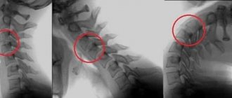
X-ray examination and computed tomography reveal an increase in intervertebral joints and the presence of bone growths (osteophytes) on them. Using radionuclide scintigraphy in active osteoarthritis, accumulation of the isotope in the intervertebral joints is detected. The final diagnosis is made after a diagnostic periarticular block with local anesthetic. Reduction of back pain after blockade confirms the diagnosis of spinal osteoarthritis.
Diagnosis of cervical osteoarthritis
Comprehensive and timely diagnosis is the key to successful treatment. Therefore, the MART Clinic approaches diagnostic measures with special attention and responsibility. So, to diagnose cervical osteoarthritis, the following is used:
- Neurological examination
- Blood chemistry
- MRI of the cervical spine
The high qualifications of our specialists and expert-class equipment allow us to obtain the most accurate disease data in the shortest possible time!
If you have any questions, ask our specialist! Ask a Question
Arthrosis of the shoulder joint - symptoms
As with most types of osteoarthritis, pain is the main symptom. Pain usually occurs when moving the joint. Sometimes a person may experience pain while sleeping. Another symptom is limited movement. It can be easily noticed when attempting maximum flexion/extension of the shoulder. A doctor can diagnose shoulder arthrosis based on this particular symptom. He or she may hold your hand and use movements to check the joint for restriction of movement. Sometimes clicking and cracking noises may occur when moving. Another sign that your doctor will look for first is atrophy of the muscle around the shoulder due to lack of use and weakening. For diagnosis you may need:
- X-ray
- Blood test (to detect rheumatoid arthritis and rule out other diseases)
- Joint synovial fluid analysis
- MRI
Treatment of cervical osteoarthritis at the MART Clinic
To successfully treat cervical osteoarthritis, the MART Clinic uses an integrated approach aimed at solving the problem rather than masking the symptoms. Thus, our experienced neurologists select an individual course of treatment, which may include:
- Drug treatment
- Physiotherapy
- Massage and manual therapy
- Exercise therapy
- Surgical intervention and others
Help yourself get better! Make an appointment with a neurologist at the MART Clinic today!
Make an appointment with a neurologist at the MART medical center in St. Petersburg (see map) by calling 8 or leave a request on the website.
Diagnostics
Diagnosis of osteoarthritis (deforming arthrosis) based on a combination of clinical and instrumental data. In modern medicine, there are certain diagnostic criteria for this disease, both clinical and instrumental. The clinical criteria are: The presence of pain in the joints, mainly at the end of the day or at the beginning of the night; pain, as a rule, also occurs during physical activity and regresses after a little rest and joint deformation is visually determined (including due to Heberden’s and Bourchard’s nodes). X-ray manifestations of osteoarthrosis are: decreased joint space, the presence of sclerotic changes in the bone tissue, the presence of bone growths (osteophytes). Certain features are characteristic of some forms of osteoarthrosis (coxarthrosis). The following clinical manifestations are characteristic of coxarthrosis: decreased range of motion in the hip joint (decreased external rotation) and pain during internal rotation; stiffness in the joint in the morning for no more than an hour; patients over 50 years of age.
Laboratory examinations show abnormalities in the presence of complications (for example, with synovitis, the ESR may increase and in biochemical parameters with synovitis, the amount of fibrin seromucoid sialic acids increases).
X-ray research methods usually provide sufficient information about the presence of degenerative changes in the joint. Depending on the x-ray picture, the classification of osteoarthritis is carried out:
- 0 absence of radiological signs of osteoarthritis
- Stage 1 – cystic restructuring of the bone tissue structure, the appearance of small osteophytes, signs of linear osteosclerosis
- Stage 2 osteosclerosis, more pronounced and signs of narrowing of the joint space appear.
- Stage 3: severe osteosclerosis, osteophytes become large, and the joint space narrows significantly.
- Stage 4 osteophytes are more massive, the joint space is practically not visualized, the deformation of the epiphyses of the bones is flattened.
X-ray examination of joints.
To diagnose osteoarthritis, an analysis of a biopsy of synovial fluid is often prescribed, which allows one to determine the presence of signs of degeneration or an inflammatory process.
MRI and CT are prescribed when detailed visualization of morphological changes and differential diagnosis with other joint diseases is necessary
Types and degrees
Each type of degenerative process in the tissues of the spine has its own name. Thus, cervical arthrosis is called cervicoarthrosis, lesions in the lumbar region are called lumbarthrosis. Uncovertebral pathological process is a type of cervicoarthrosis.
There are 5 types of pathological process:
- Primary - developed on a previously healthy joint.
- Secondary – a consequence of other diseases of the articular apparatus of the cervical spine.
- Local – 1 joint is affected.
- Multiple – several joints are involved in the pathological process.
- Deforming - at the point of contact of bone structures, deformation of cartilage tissue, tendons, and articular surfaces develops. There is severe pain.
The disease is characterized by a long, persistent course. Doctors distinguish 3 stages of development:
- At the first stage, the symptoms do not bother the patient much. The changes affect the articular cartilage. He experiences nutritional deficiencies and becomes exhausted. Other vertebral structures are not affected.
- The hyaline cartilage is destroyed. Osteophytes are clearly visible on x-rays. Treatment is difficult, but possible.
- Ankylosis of the joints develops. Movement of the head without turning the body is impossible; neurological symptoms are observed. Conservative therapy does not bring results.
Treatment
To cure this deformity of the cervical area, drug and non-drug therapy is used. Outpatient treatment conditions are indicated for patients with chronic features of the disease. If symptoms temporarily disappear, the doctor prescribes a sanitary-resort treatment method.
Drug therapy
The list of medications that doctors prescribe is wide; they are divided into:
- anti-inflammatory (non-steroidal drugs);
- antispasmodics (nosh-pa, actovegin);
- protective (chondroprotectors);
- relaxing (muscle relaxants);
- vitamin complex (vitamin B);
- hormonal (glucocorticosteroids);
- to normalize blood circulation (Trental, Reopoliglyukin);
- local medications (Finalgon ointment, Nicoflex).
Non-drug treatment of spondyloarthrosis of the uncovertebral joints of the cervical spine is considered to be the methods described below.
Manual therapy
This technique is effective for treating deformed areas of the neck, because with the help of manipulations, the specialist relaxes the muscles, eliminates pathological inflammation of the tissue structure of blood vessels, neurons and the vertebral region. But this practice can be used after the inflammation is relieved.
Massage
This method of complex neck treatment will provide muscle relaxation and prevent muscle spasms. The doctor prescribes massage manipulations on the collar area if the patient does not have a pathological exacerbation.


