Spondyloarthritis of the cervical spine is known as cervicoarthrosis. This pathology is one of the forms of a disease such as osteoarthritis, which is characterized by degenerative-dystrophic changes in the structures of the facet joint (ligaments, cartilage, muscles located around the joint, capsule and bone). Spondyloarthrosis of the cervical spine is characterized by osteochondral growths in the joint area, which often cause a decrease in the distance between the vertebrae and constant irritation of the spinal cord roots. Often the growths put pressure on the artery, which has its negative consequences.
In this article you can learn about the symptoms and treatment of spondyloarthrosis of the cervical spine. If you find yourself with similar clinical manifestations, immediately contact the CELT multidisciplinary clinic!
Treatment
The treatment plan for spondyloarthrosis of the cervical spine is drawn up by CELT doctors based on a number of individual indications. In most cases, conservative techniques are used, which include:
- taking medications: non-steroidal anti-inflammatory drugs, chondroprotectors, muscle relaxants, B vitamins;
- physiotherapy: professional massage, therapeutic exercises, magnetic therapy, electrophoresis;
- wearing special corrective corsets.
As for surgical intervention, our specialists resort to it only in extreme cases - when there are appropriate indications:
- spinal stenosis;
- paresis or paralysis after spinal cord injury;
- dysfunction of the pelvic organs due to compression of the spinal cord;
- lack of effect from conservative treatment;
- spinal instability.
Do you want to be sure that the treatment will be successful? Contact the CELT clinic! We employ highly qualified doctors who have extensive experience in successfully treating this pathology!
Make an appointment through the application or by calling +7 +7 We work every day:
- Monday—Friday: 8.00—20.00
- Saturday: 8.00–18.00
- Sunday is a day off
The nearest metro and MCC stations to the clinic:
- Highway of Enthusiasts or Perovo
- Partisan
- Enthusiast Highway
Driving directions
Stages of development of deforming spondyloarthrosis of the cervical spine
There are 4 stages in the development of spondyloarthrosis.
- Stage I. Dystrophy of the hyaline cartilage covering the articular surfaces of the vertebrae begins. There are no symptoms, sometimes with increased load the patient feels discomfort in the neck area.
- Stage II. Due to a decrease in the production of joint fluid, the nutrition of the cartilage is disrupted, it begins to thin out, the elasticity of the ligaments is weakened, and the range of motion in the affected cervical segment decreases. The patient experiences a feeling of stiffness and moderate pain in the neck in the morning, which soon passes.
- Stage III. Spondylolisthesis develops - one of the damaged vertebrae moves backward, and osteophytes - spiky bone growths - grow. Osteophytes narrow the intervertebral foramina, irritating the spinal nerve roots passing through them, causing the patient to experience constant excruciating pain.
- Stage IV. Complete destruction of cartilage, in its place massive bone growths are found on the articular surfaces.
Lumbar
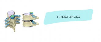
Lumbar spondyloarthrosis is more common. Painful signs that occur in the lower back radiate to the buttocks and thighs, but do not fall lower, as happens with a hernia. Movements are constrained, it becomes impossible to tilt or turn. After being in a horizontal position for a long time, it is very painful to sit down.
Spondyloarthrosis of the lumbosacral spine develops slowly. Articular cartilage loses its elasticity, causing the joint to become thinner and begin to deteriorate. Osteophytes grow, which can subsequently lead to joint immobilization and disability. Pain attacks in the lower back and stiffness lead to changes in gait and the formation of flat feet.
Causes of the occurrence and development of spondyloarthrosis
Spondyloarthrosis can be caused by congenital spinal abnormalities, spinal injuries and chronic microtraumas. Spondyloarthrosis can also be caused by poor posture and metabolic disorders (especially in old age). Constant static loads that cause overload of the spinal column (sedentary work - for example, at a computer) can cause spondyloarthrosis.
Another cause of spondyloarthrosis is prolonged physical activity (professional sports, weightlifting, etc.). In addition, the cause of this disease may be flat feet (of various types). With this disease, gait disturbance occurs and, accordingly, improper distribution of the load on the spine in a vertical position, which in turn leads to spondyloarthrosis.
Main excitatory factors
Together with age-related changes of involutional origin, the progression of spondyloarthrosis in the cervical area is associated with developmental disorders (incompletely formed arches, the presence of asymmetry) and damage. An important reason for athletes, people engaged in heavy physical labor and who need to stay in one position for a long time is non-physiological stress. Scientists report the likelihood of a genetic predisposition, the presence of autoimmune abnormalities, hormonal imbalances and metabolic disorders.
Physical examination
First, the doctor finds out the presence of symptoms, their intensity, location, and history of the disease. A neurological examination is then performed, which includes examining reflexes, testing muscle strength, and determining sensory deficits and range of motion in the neck. A doctor may perform a gait analysis to determine how much damage to the spinal cord is present.
If a doctor suspects cervical spondylosis, he will order imaging tests and neurophysiological studies to verify the diagnosis.
How to get rid of cervical spondyloarthrosis?
The maximum effectiveness of medical therapy appears only in the first stage of the disease, when there are no ankyloses in the joints. Doctors prescribe the use of pharmacological drugs aimed at eliminating the inflammatory response. Additionally you will need to take:
- Chondroprotective agents.
- Vitamin complexes, in particular group B, which provide a blockade.
The patient is prescribed treatment with acupuncture sessions, classes to learn exercises that help strengthen the muscle area of the neck, and seminars on proper breathing. Based on the indications, the patient is prescribed massage and manual therapy. However, you should attend such sessions with extreme caution and contact a qualified worker only on the recommendation of the attending physician and there are no signs of inflammation. In the case of self-medication or incorrectly selected therapy, the patient may experience serious consequences that will provoke an exacerbation of spondyloarthrosis, a rapid increase in pain and the formation of neurological symptoms. Medical techniques should be used in short courses simultaneously with the use of anti-inflammatory drugs.
The operation is prescribed quite rarely in the presence of dynamic disorders caused by displacement of the endings and excessive pressure on the nerve parts of the osteophytes.
Spinal gymnastics
Orthopedists and traumatologists emphasize that regular performance of special therapeutic and preventive exercises plays a leading role in spinal arthrosis. An individually developed set of exercises for a particular department can significantly improve blood flow in the affected area, activate the nutrition of depleted tissues, establish the delivery of important substances for reparative and regenerative functions, normalize motor capabilities, and strengthen the muscles responsible for the functioning of the intervertebral joints.
Epidemiology
Cervical spondylogenic myelopathy is the most common cause of non-traumatic spastic paraparesis and quadriparesis. Almost 23.6% of patients with non-traumatic myelopathic symptoms had cervical spondylogenic myelopathy.
Cervical spondylosis is more common in men.
The results of studies using radiographic data showed that spondylous changes are most common in people over 40 years of age. After all, more than 70% of men and women suffer from spondylosis in adulthood, but radiographic changes are more pronounced in men than in women.
Causes of uncovertebral arthrosis
The main reasons for the development of this dystrophic process are: congenital (Olenik syndrome or occipitalization of the atlas), as well as acquired (spinal injuries, infectious diseases, such as polio, physical work with increased load).
Predisposing factors include: excess body weight, decreased physical activity, dislocation of the hip joint, flat feet.
Uncovertebral arthrosis, the main symptom of which is a decrease in the height of the cartilaginous plate between the vertebral bodies, is a dangerous disease.
The signs of uncovertebral arthrosis are quite specific: a decrease in the height of the cartilaginous plate, the growth of osteophytes (bone outgrowths) on adjacent vertebrae that meet each other, as well as ossification (calcification) of the spinal ligaments.
Visualization methods
- X-rays can be used to visualize bone spurs and other abnormalities.
- A CT scan can provide more detailed images of the neck.
- MRI scans, which create images using radio waves and a magnetic field, help doctors detect compression of nerve structures.
- Myelogram - This procedure uses contrast to be injected into specific areas of the spine. A CT scan or x-ray is then used to take more detailed images of these areas.
- EMG (ENMG) is used to test the functionality of nerve fibers, the method checks the electrical activity of the nerves and the conduction of impulses to the muscles.
- ENMG determines the speed and strength of the signals that the nerve sends. This allows you to determine the presence and level of damage to nerve fibers.
How to diagnose
Diagnosis is based on the patient’s complaints, information about the course of the disease, clinical examination data with an assessment of the neurological status, as well as the results of laboratory and instrumental examinations.
Radiography
allows you to determine the condition of the facet joints if pronounced structural changes have already occurred in them: thinning of the cartilage, osteophytes, narrowing of the joint space.
Computed tomography (CT)
is most informative for detecting pathologies of hard tissues - bones and cartilage, and records the initial signs of spondyloarthrosis.
Magnetic resonance imaging (MRI)
detects the slightest changes in cartilage, bones, ligaments and blood vessels of the vertebral joints.
Diagnostic methods
To clarify the correct conclusion, the attending physician prescribes the patient to undergo a series of examinations:
- Radiography - informative information can be obtained in case of severe deformations that occur in the facet areas.
- MRI scanning - visualizes accompanying changes localized in soft tissues.
- Computed tomography provides doctors with data on the presence of anomalies in the first stages of development.
- Dopplerography - allows you to check the state of cerebral circulation.
If the patient has neurological disorders, the physician writes a referral for consultation with a colleague in the neurological department.
When should you see a doctor?
If a person suddenly experiences numbness or tingling in the shoulder, arms or legs, or if the person has lost bowel or bladder control, then seek medical help as soon as possible!
If discomfort and pain begin to interfere with daily activities, it is necessary to consult a neurologist. Despite the fact that spondylosis is part of involutional changes, there are nevertheless various treatment methods that can improve well-being and reduce symptoms.
Classification
Doctors of the traumatology and orthopedic department distinguish the stages of spondyloarthrosis of the cervical area, focusing on pathomorphological and clinical indicators. These include:
- The asymptomatic form is the formation of the first changes in the joints.
- Initial form - the first signs of the disease appear (pain, discomfort, minimal decrease in the level of mobility).
- The average form is distinguished by the bright course of the lesion, which can be observed on radiographic examination (changes in the facet joints, bone growths form at the borders).
- Severe form - noticeable changes occur that affect the overall degree of motor activity of the neck; ankylosis, disruptions in the functioning of the blood supply to the brain and neurological disorders may be observed.
Spondyloarthrosis: treatment with the NANOPLAST forte therapeutic patch
In the treatment of spondyloarthrosis, the NANOPLAST forte therapeutic patch is very effective, it allows you to relieve pain and inflammation, improve blood circulation in the area affected by spondyloarthrosis, and reduce the dose of painkillers and anti-inflammatory drugs. In the therapeutic treatment of spondyloarthrosis , various agents are used, such as NSAIDs, analgesics, and hormones. All these remedies are effective, but if used for a long time they can cause harm to the body. Therefore, it is very important to minimize side effects and increase the effectiveness of treatment of spondyloarthrosis. A new generation drug can help with this - the pain-relieving anti-inflammatory medical patch NANOPLAST forte .
For spondyloarthrosis , the therapeutic plaster NANOPLAST forte is applied to the area of the affected joint of the spine, depending on the location - lower back, neck or chest. To relieve acute symptoms of spondyloarthrosis, use for 3 to 5 days. The duration of the course of treatment for spondyloarthrosis is from 9 days. It is usually recommended to use the patch in the morning for 12 hours, but it can also be used at night.
High efficiency, unique composition, long-term (up to 12 hours!) therapeutic effects, ease of use and affordable price make NANOPLAST forte the drug of choice in the treatment of spondyloarthrosis.
Read more about NANOPLAST forte
Symptoms of spondyloarthrosis
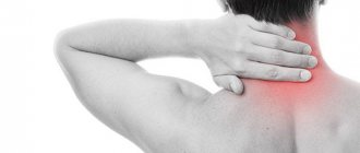
Pain is the main sign of damage to the articular processes of the vertebrae. In the early stages, it is periodic, wave-like in nature and worries mainly when moving. At rest, pain decreases or disappears completely. The pain syndrome is felt only in the damaged area and does not tend to spread. It does not radiate to the arms and legs, as with osteochondrosis and intervertebral hernias. There is no numbness or sensory disturbances. Spondyloarthrosis of the 2nd degree is accompanied by stiffness and limited mobility in the morning. It lasts about half an hour and gradually passes. If left untreated, symptoms become extremely severe. A pronounced stoop appears due to displacement of the vertebrae, pain and motor discomfort intensify.
Complications include compression of the nerve roots, which is manifested by sharp, burning pain that radiates - radiates - to different parts of the back and limbs. A consequence of the chronicity of the process may also be spinal canal stenosis.
Thoracic region
Spondyloarthrosis deformans of the thoracic region is characterized by discomfort and pain attacks in the chest area. Dull pain in the back when taking a deep breath. The upper girdle of the limbs is also affected, as a result of which the functions of the hands are impaired. The sources of pain are the affected facet joints. Performance decreases. Disorders in the thoracic region are much less common than, for example, spondyloarthrosis of the lumbar spine, since in this area the vertebrae are less susceptible to strong vibrations.