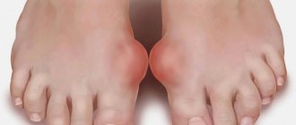
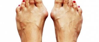
A painful foot deformity - a protruding bunion on the big toe - is a problem that is familiar to many women.
Why does this “bump” appear, is it possible to slow down its development, and how to remove the “ugly” growth that prevents you from wearing elegant shoes and spoils your gait? Modern medicine knows the answers to these questions.
A bunion on the big toe is not only a cosmetic problem. Foot deformation not only disfigures the leg, but also worsens health due to constant pain and inflammation. Choosing comfortable, but at the same time elegant, shoes turns into an impossible task.
“Bumps” on the legs: women’s retribution for the desire for beauty
What in everyday life we call a bunion on the big toe, in scientific language sounds like a hallux valgus deformity of the toe. There is no “bump” on the leg. It just seems that way. In fact, this is a deviation of the first metatarsal bone of the foot to the inside with a simultaneous inclination of the big toe to the outside.
If the problem is not addressed immediately, the situation will only get worse over time. Gradually the bone will stick out more and more. This not only disfigures the leg, but also causes severe pain when walking. And besides, it creates incredible difficulties when choosing shoes.
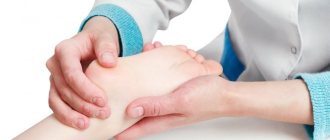
It happens that a bunion on the big toe is mistaken for arthrosis, gout or salt deposits. A traumatologist-orthopedist at the Komarova Clinic will help make a diagnosis based on a visual examination and x-ray of the feet, as well as a blood test.
Causes
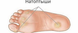
Many problems that arise in the legs are the result of abnormal pressure or friction. The easiest way to determine the presence of the consequences of pathological pressure is to examine the leg. The leg is a hard bone covered with skin. In most cases, symptoms develop gradually as the skin and soft tissues absorb the excess impact on the leg. Any protruding bone or injury aggravates the already existing consequences of the injury. The skin reacts to friction and pressure by forming a callus. The soft tissues located under the skin react to excess stress. Both the callus and the thickened soft tissue underneath the callus become painful and inflamed. Reducing pain helps to reduce pressure. Pressure can be reduced externally through looser shoes or internally through surgery and removal of excess tissue.
Why does a “bone” appear on the legs?
The most annoying thing is that women are primarily susceptible to this disease. According to statistics, women are 20 times more likely to experience thumb deformity than men. But we love to show off in elegant shoes with thin heels. According to doctors, women have congenital weakness of the musculo-ligamentous system. We should take care of our legs. And we, on the contrary, “climb” higher, onto stiletto heels, so the foot cannot withstand the load, flattens and becomes deformed.
The hereditary factor also plays a role: if your mother and grandmother had similar problems, then you are definitely at risk. Don't think that bumps on your feet are an age-related problem. Quite often, deformity of the big toe appears in young girls aged 12-15 years. In this case, after 25 years, when the body stops growing, the bone is surgically corrected.
Other reasons that provoke the formation of a bunion on the big toe include excessive loads: excess weight, long walking, previous diseases of the cartilage tissue of the joint, as well as flat feet.
How is the “bunion at the base of the big toe” formed?
So, what happens to the leg if additional stress is placed on it? A healthy foot has two arches: transverse and longitudinal. The arches act as natural shock absorbers, softening shaking when walking. If the load on the foot is distributed unevenly, then the muscles and ligaments gradually weaken, the arches sag and flatten. This is also reflected in the metatarsal bones of the foot, which in a normal anatomical structure should be parallel to each other.
If the foot is already flattened, has become wider, and we all still walk in tight narrow shoes, then the following happens. The first metatarsal bone gradually deviates from its correct position. Displacement of the finger and this bone leads to the formation of the so-called valgus angle. The head of the metatarsal bone is its apex, which thickens over time and transforms into that very bone that cannot be hidden.
Diagnosis and treatment of restless legs syndrome
It is customary to prescribe medications for RLS in cases where it significantly disrupts the patient’s vital functions, causing persistent sleep disturbance, and non-drug measures are not effective enough. In mild cases, you can limit yourself to taking sedatives of herbal origin or prescribing a placebo, which can give a good, but sometimes only temporary effect.
In more severe cases, it is necessary to choose a drug from four main groups: benzodiazepines, dopaminergic drugs, anticonvulsants, opioids [17].
Benzodiazepines accelerate the onset of sleep and reduce the frequency of awakenings associated with PDC, but have relatively little effect on the specific sensory and motor manifestations of RLS, as well as PDC. The most commonly used benzodiazepines are clonazepam (0.5–2 mg at night) or alprazolam (0.25–0.5 mg). With long-term use of benzodiazepines, there is a danger of developing tolerance with a gradual decrease in effect and the formation of drug dependence. The negative aspects of the action of benzodiazepines also include the possibility of developing or increasing drowsiness during the day, decreased libido, increased sleep apnea, episodes of confusion at night, as well as worsening cognitive impairment in the elderly. In this regard, benzodiazepines are currently used sporadically in mild or moderate cases - during periods of deterioration, and in severe cases requiring constant treatment, they are prescribed only when dopaminergic drugs are ineffective [16, 17].
Dopaminergic drugs (levodopa and dopamine receptor agonists) are the mainstays of treatment for RLS. They affect all the main manifestations of RLS, including the maximum concentration limit. Dopaminergic drugs are so effective in RLS that a positive reaction to them can serve as an additional criterion for diagnosing RLS, and its absence, as, for example, in Parkinson's disease, should be considered a basis for revising the diagnosis. The effect of dopaminergic drugs in RLS occurs in doses that are significantly lower than those used in Parkinson's disease. Apparently, dopaminergic drugs are equally effective in both primary and symptomatic variants of RLS [6].
Levodopa has been used for RLS since 1985, when its effectiveness in this category of patients was first shown. Currently, levodopa is prescribed in combination with DOPA decarboxylase inhibitors benserazide (Madopar) or carbidopa (Nakom, Sinemet). Treatment begins with 50 mg of levodopa (approximately 1/4 tablet of Madopar “250”), which the patient should take 1–2 hours before bedtime. If the effectiveness is insufficient, after a week the dose is increased to 100 mg, the maximum dose is 200 mg. Taking levodopa provides an adequate effect in 85% of patients. In many patients, it remains effective for many years, and in some patients its effective dose may remain stable and even decrease [10]. Levodopa medications are usually well tolerated by patients with RLS, and side effects (nausea, muscle cramps, tension headaches, irritability, dizziness, dry mouth) are usually mild and do not require discontinuation of the drug. Given the rapid onset of effect and the lack of need for dose titration, levodopa may be considered the treatment of choice for intermittent worsening symptoms.
However, with long-term use in a significant proportion of patients, the effectiveness of levodopa decreases, while the duration of action of a single dose is reduced to 2-3 hours, which may be followed by a rebound increase in the symptoms of RLS and PDC in the second half of the night. In this case, it is recommended to increase the dose of the drug or add a second dose immediately before bedtime or upon awakening at night. However, with an increase in the dose of levodopa, the rebound increase in symptoms may not be eliminated, but only shift to the early morning hours, and its intensity may increase. Experience shows that a more reasonable alternative in this situation is to switch to a sustained-release levodopa preparation (Madopar GSS). A slow-release drug that acts over 4 to 6 hours to ensure good sleep throughout the night and prevent morning rebound symptoms.
In approximately half of patients, during long-term treatment with levodopa, symptoms gradually begin to appear earlier (sometimes even during the day), becoming more intense and widespread (the so-called “augmentation”). The higher the dose of levodopa, the stronger the augmentation [14], so increasing the dose of levodopa in this situation only worsens the situation, completing a vicious circle. When using Madopar GSS as a basic therapy for RLS, rebound enhancement and augmentation are observed less frequently than when taking standard levodopa drugs. In this regard, Madopar GSS is now often used as a means of initial treatment of RLS (1-2 capsules 1-2 hours before bedtime). Sometimes it is reasonable to recommend to the patient 1 hour before bedtime 100 mg of levodopa as part of a standard drug or a soluble fast-acting drug, which provides a relatively rapid onset of effect, and 100 mg of levodopa as part of a slow-release drug (for example, 1 capsule of Madopar GSS). When augmentation develops, it is recommended to either replace levodopa with a dopamine receptor agonist, or add it to it (by reducing the dose of levodopa).
Dopamine receptor agonists (DRAs) have been used for RLS shortly after levodopa was shown to be effective, in 1988. Experience shows that the effectiveness of ADR for RLS is approximately equivalent to that of levodopa. ADRs can be considered as a means of choice if long-term daily medication is required. For RLS, both ergoline drugs (bromocriptine, cabergoline) and non-ergoline drugs (pramipexole, piribedil) are used [12, 14]. Non-ergoline drugs have the advantage of being free from side effects such as vasospastic reactions, pleuropulmonary, retroperitoneal fibrosis, and fibrosis of the heart valves. To avoid nausea, ADRs are taken immediately after meals and the dose is adjusted by slow titration. Pramipexole is initially prescribed at a dose of 0.125 mg, then gradually increased until the effect is achieved (usually no more than 1 mg). The effective dose of piribedil is 50–150 mg. For bromocriptine treatment, the starting dose is 1.25 mg and the effective dose ranges from 2.5 to 7.5 mg. Cabergoline treatment begins with 0.5 mg, and its effective dose is 1–2 mg. The indicated dose is usually prescribed once 1-2 hours before bedtime, but in severe cases, additional administration of the drug may be necessary in the early evening hours. Side effects when taking ADR include nausea, fatigue, headache, dizziness, and daytime sleepiness. Domperidone may be prescribed at the beginning of treatment to prevent nausea.
With long-term use of ADR, signs of augmentation are detected in approximately 25–30% of patients, but they are almost never as severe as with levodopa treatment. If one of the ADRs turns out to be ineffective, you can try replacing it with another drug from this group. It is important to note that dopaminergic drugs, while eliminating the symptoms of RLS, do not always lead to normalization of sleep, which requires the addition of a sedative drug (benzodiazepine or trazodone).
It should be noted that, probably due to the absence of denervation and the normal number of dopaminergic neurons, dopaminergic drugs are effective in RLS at doses significantly lower than those used in Parkinson's disease. Moreover, side effects such as dyskinesia, psychosis, impulsivity, and compulsive behavior (common in Parkinson's disease) are extremely rare in RLS.
In those few cases where the patient does not tolerate dopaminergic drugs well, and benzodiazepines are ineffective or cause intolerable side effects, they resort to anticonvulsants or opioids. Of the anticonvulsants, gabapentin is currently most often used, at a dose of 300 to 2700 mg/day [9]. The entire daily dose is usually prescribed once in the evening. Opioid drugs (codeine, 15–60 mg; dihydrocodeine, 60–120 mg, tramadol, 50–400 mg at night, etc.) can significantly reduce the symptoms of RLS and PDC, but the risk of developing drug dependence makes their use justified only in the most severe cases where all other treatment methods are ineffective. The treatment algorithm for RLS is shown in the figure.
For RLS, it is possible to use some other drugs (clonidine, folic acid, magnesium, vitamins E, B, C), but their effectiveness has not been confirmed in controlled studies [18]. In some patients, amantadine, baclofen, zolpidem are effective; beta blockers (for example, propranolol) can reduce symptoms, but sometimes cause them to worsen.
Treatment of RLS has to be carried out over a long period of time over many years, and therefore it is very important to follow a unified treatment strategy. Sometimes it is carried out only during the period of intensification of symptoms, but often patients are forced to take certain drugs for life to maintain drug remission. It is better to start treatment with monotherapy, choosing a drug taking into account its effectiveness in each individual patient and the presence of concomitant diseases. If monotherapy is insufficiently effective or in cases where, due to side effects, it is not possible to achieve a therapeutic dose of one of the drugs, it is possible to use a combination of drugs with different mechanisms of action in relatively small doses. In some cases, it is advisable to rotate several drugs that are effective for a given patient, which allows them to maintain their effectiveness for many years.
Treatment of RLS in pregnant women is particularly challenging. None of the drugs commonly used for RLS are considered safe during pregnancy. Therefore, when RLS develops during pregnancy, they are usually limited to non-drug measures (for example, a walk and a warm shower before bed) and the administration of folic acid (3 mg/day), as well as iron supplements (if there is a deficiency). Only in severe cases is it permissible to use small doses of clonazepam, and if they are ineffective, small doses of levodopa.
Trazodone and monoamine oxidase inhibitors (MAOIs) can be used to treat depression in patients with RLS. Data on the effect of selective serotonin reuptake inhibitors in patients with RLS and PDC are contradictory. However, in some patients they can nevertheless improve the condition, which is explained by the suppression of the activity of dopaminergic neurons. Tricyclic antidepressants, like antipsychotics, are contraindicated.
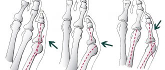
Stages of hallux valgus deformity of the big toe
Traumatologists and orthopedists at the Komarova Clinic identify several stages of deformation:
- First stage: displacement no more than 20 degrees. There are no unpleasant sensations, only aesthetic inconvenience.
- Second stage: displacement of 20-30 degrees. When walking for a long time, pain appears.
- Third stage: displacement of 30-50 degrees. The bunion prevents you from walking, it hurts, and it becomes difficult to find comfortable shoes.
- Stage four: displacement greater than 50 degrees. Constant pain and ongoing inflammation bring suffering even at rest. The deformity begins to affect other phalanges of the fingers.
Diagnosis of the stage of the disease
There are 3 degrees of the disease, which can be distinguished by the manifestations of symptoms and the appearance of the foot.
- Initial (moderate). The patient complains of discomfort when wearing shoes, which manifests itself in skin friction and the formation of calluses on the outer edge of the first metatarsophalangeal joint. A lump appears with redness and swelling of nearby tissues. Regular aching pain is often observed in the foot area. The radiograph shows a valgus angle of less than 20°.
- Average. Arthritis (inflammation of the joint) with characteristic redness of the skin is observed. The pain intensifies. External examination shows the displacement of the second finger to the first. Corns form in areas under the second and third fingers. The angle of valgus deviation increases to 20–40°. The x-ray shows subluxation of the 1st metatarsophalangeal joint.
- Launched. There is pain throughout the entire foot, and movement while wearing shoes becomes constrained. In many cases, patients become unable to work. The foot is significantly deformed. There is a strong displacement of the first finger, which ends up above or below the second. A large bony protrusion forms at the base of the first toe. The skin tissue under the second and third fingers becomes rough, and corns and calluses form. The radiograph shows that the valgus angle exceeds 40 ͦ. Subluxation or dislocation of the 1st metatarsophalangeal joint is clearly visible.
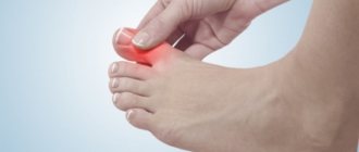
Consultation with a specialist is required for the following changes:
- swelling has appeared at the base of the thumb;
- the protruding bone is red and painful;
- the finger deviates outward;
- legs get very tired when walking;
- swelling is observed;
- Painful calluses and corns form on the soles.
If you have problems with foot deformities, you should consult an orthopedic doctor. During the initial examination, he will find out what the deviation of the thumb is, what condition the blood vessels are in, whether there are calluses, corns, and whether pain is felt in the joint. In addition, the doctor will evaluate the degree of mobility of the foot. To make a diagnosis, one examination is not enough; additional examination is necessary: x-ray in 3 projections, MRI, computer plantography.
The disease can be treated non-surgically only in the initial stages (1–2) with a deviation angle of no more than 20 ͦ. A variety of braces, orthopedic shoes, physical therapy exercises, and medications help well. The surgical method is indicated if the angle of deviation of the fingers exceeds 20 ͦ, the pain is constant, there is a disturbance in gait, and the protruding joint is motionless.
Conservative treatment of hallux valgus is contraindicated in the following cases:
- The presence of varicose veins, thrombophlebitis.
- Open, unhealed wounds on the feet.
- The patient experiences intolerance to fixing materials.
- Allergic reactions to medications were detected.
Caution should be exercised when prescribing treatment for patients with diabetes mellitus, obliterating endarteritis (the feet are vulnerable and require special care).
Complications manifest themselves in increased pain, decreased mobility of the feet, deformation of the fingers, pathology of the knee and hip joints, and gait disturbances.
If the patient does not adhere to the orthopedist's recommendations, this may provoke progression of the disease. Then surgical intervention is inevitable.
Treatment of a bunion on the big toe in Vladivostok
Baths won't help! No matter what adherents of traditional medicine write on the Internet, lotions, baths, compresses and iodine nets will not get rid of a bunion on the foot. All these measures can only temporarily alleviate suffering by relieving pain and heaviness in the legs. If the deformity is minor, it can be corrected using various orthopedic means.
For example, special orthopedic insoles are very effective - which can be individually made for each patient by a traumatologist-orthopedist at the Komarov Clinic.
At the initial stage of the disease, wearing a night abduction bandage, which fixes the big toe in the correct position, can alleviate the situation. The design does not allow you to walk with it, but it does not interfere with sleep. It turns out to be a kind of therapeutic sleep.
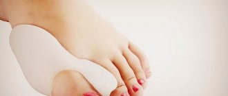
For daily wear, there are special corrective pads that protect the bones from pressure from shoes and form the correct position of the big toe, as well as various interdigital partitions.
Orthopedic insoles Sursil Orto in Vladivostok
At any stage of development of the disease, it is imperative to wear orthopedic insoles.
They will not only help to correctly distribute the load on the foot, relieving the problem area, but will also relieve tension from the joints of the legs and spine. The key to successful treatment of a bunion in its initial stage of development is an insole made individually for your foot, taking into account the characteristics of the foot, arch and size. Modern insoles are very thin (2-3 millimeters), easily fit into any shoes, that is, you can wear them with your regular shoes. And they are done very quickly, 20-30 minutes - and the insoles are ready.
And, of course, the traumatologist-orthopedist of the Clinic on Komarova will select an individual set of therapeutic procedures: massage, physiotherapy, physical therapy. All these methods will help stop the development of the disease and prevent further deformation of the foot.
Methods of surgical correction of thumb deformity in Vladivostok
However, today the most effective treatment for bunions on the leg is surgical correction.
There are many surgical techniques to return the finger to its normal position. “Reconstruction” of a hallux valgus in our clinic is carried out using the most modern methods. These techniques allow patients to return to their normal lives as quickly as possible. For the first time after surgery, it is necessary to wear special shoes that do not place stress on the forefoot. Then you can switch to orthopedic shoes, or move with the help of crutches or a stick. It is important to understand that the operation eliminates only a cosmetic defect (that is, the “bump” itself), but does not restore the transverse arch of the foot. To prevent the bunion from appearing again, you will have to wear only the right shoes with arch support and custom orthopedic insoles for the rest of your life.
Recovery period after surgery
The rehabilitation period varies and depends on the intervention technique. During this period, the patient takes painkillers and undergoes physiotherapeutic procedures. Over time, massage is added.
After surgery to remove cones on the big toe, it is recommended to wear special orthopedic shoes and insoles. Bandages and various fixatives are divided into night and day ones. The doctor will tell you what type of shoes or bandages to buy, how and when to wear them.
Preventing the appearance of a bunion on the big toe
However, no matter what innovative technologies and methods are, the best treatment is prevention. In this case, the right shoes, wide in diameter, with a round toe, made of soft materials and with a heel no higher than 4 cm. If you cannot refuse stilettos, then be sure to insert a special insole for high heels with a transverse corrector to support the foot.
By the way, running or jumping sports can also provoke the formation of a bunion on the big toe, since the front arch of the foot is overloaded during training. The solution to the problem is to wear special sports shoes for a specific type of physical activity.
And one more tip - there is nothing better for preventing “bumps” on your feet than walking barefoot on sand or small pebbles. This massage perfectly strengthens the ligaments of the foot and helps to avoid the development of flat feet and other joint diseases.
You can make an appointment with the clinic’s traumatologists-orthopedists for a consultation on the diagnosis, treatment and prevention of foot deformities by calling the outpatient department or 240 27 27.