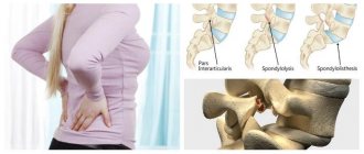
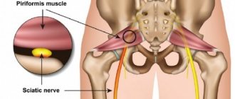
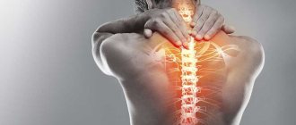
Clinical symptoms of intervertebral hernias cause excruciating pain, impaired sensitivity in the extremities, decreased range of motion in the back, and a noticeable decrease in a person’s physical capabilities. The ill-fated disease not only significantly depresses the quality of life, but can also lead to more dire consequences, including damage to the spinal cord or brain, as well as paralysis of the lower or upper extremities.
Unfortunately, vertebral hernias cannot be completely cured by conservative methods; this disease is accompanied by an irreversible pathogenesis leading to disc degeneration. The progression of degenerative-dystrophic processes can be prevented by using effective therapeutic measures in the early stages of the pathology, making it quite possible to achieve stable remission of the disease and live a normal life without experiencing pain and motor dysfunction.
But if the disease is left to chance, then with a high degree of probability surgery cannot be avoided. Surgery is the only salvation for patients with a progressive course of the disease, accompanied by persistent neurological symptoms, critical compression of the spinal cord or dysfunction of internal organs.
Today, the fundamental tactic for the need to operate on a hernia is microdiscectomy - a microsurgical procedure that can significantly minimize surgical trauma, reduce the risk of complications to an extreme minimum and achieve a relatively quick recovery for the patient. It is to this surgical intervention, which is a vital measure for many people, that we will devote our material.
What is microdiscectomy
At the moment, microdiscectomy is recognized as the most promising surgical method for this diagnosis. In Russia, the price for the procedure ranges from 60 thousand rubles or more. Microdiscectomy is a low-traumatic session for removing an intervertebral hernia, which involves excision of a pathological formation with maximum preservation of the disc; it is completely removed only in isolated cases. Resection manipulations are carried out under the careful supervision of a super-powerful surgical microscope using miniature microsurgery instruments. Anesthesia is usually carried out by administering general anesthesia, but local anesthesia is also possible.
Scar after 2 months.
This operation, in contrast to classical discectomy, is performed through a small incision, which is approximately 3 cm. With a conventional open discectomy, an outdated method, the incision reaches 10 cm. By the way, classical discectomy has not been practiced for a long time in countries with highly developed neurosurgery due to the high level of surgical aggression, serious impairment of the supporting functions of the spine, long rehabilitation and often developing serious intra- and postoperative consequences. But let's return to our considered technique.
About 50% of all such interventions for spinal hernias are performed on the lumbar region. It is worth noting that l5 s1 is the most common level, most often subject to disc correction, in 40% of cases. But the average age of patients, so to speak, of a surgical profile, according to clinical observations, is 40-45 years. Of course, a person at an earlier age (20-40 years old) or over 45 years old may have a rather unfavorable pathogenesis, which may need to be eliminated through surgery.
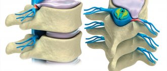
Hernia with complete stenosis.
The most important thing is that at the moment it has become possible for such patients to undergo highly effective microdiscectomy, influencing the vertebral and paravertebral structures as correctly as possible, without disturbing their anatomical and functional links. It is easily tolerated, and after it a person does not experience unimaginable pain. The effectiveness of the operation, we posted a video about it, has been proven by many years of clinical observations and is at least 90%. However, in 5%-10% of patients, a relapse of the disease is possible after some time.
In case of relapse, a repeat operation is indicated, which safely eliminates the newly formed hernia. It is worth saying that the secondary development of protrusion of the nucleus pulposus after microdiscectomy most often occurs during the first 3 months. Postoperative relapse is not a common phenomenon, but requires attention, because its occurrence is often influenced by poorly performed manipulations or poor rehabilitation after the intervention. Therefore, make sure that you undergo the treatment process in medical institutions with an impeccable reputation and extensive practical experience in this area.
The first in Russia to practice microdiscectomy was in Ramenskoye, in the spinal surgery department of the Central District Hospital of this district. By the way, many people from different cities try to come here, since this medical structure, as they say, has enormous experience in the field of surgical treatment of hernias.
How is a lumbar microdiscectomy performed?
Surgical intervention is ideal for the treatment of lumbar hernias of the spine and is considered the so-called “gold standard” for the pathogenesis of lumbosacral localization. As reviews show, if patients undergo such an operation no later than 3 months after its appointment, it mostly gives excellent results. Therefore, do not delay its completion, since late intervention reduces the chances of achieving a high effect. Additionally, we note that in Moscow the price for microdiscectomy of the lumbar segment is slightly lower than for removal of a hernia in the neck area.
Now let's move on to the actual characteristics of the operational process that you have to transfer.
- After administering anesthesia, an incision is made in the area of the diseased segment on the back under X-ray control; its length is usually 3-4 cm. Next, the doctor very carefully moves the muscles and fixes them in the desired position with a special device. Muscle tissue is not injured.
- After opening access to the problem disc, the surgeon, using a microscope with 8x magnification, carefully moves the nerve root so that it is not damaged during surgery. The ligamentum flavum is usually preserved.
- Then the specialist, carefully monitoring his actions through a microscope, removes the hernial mass with miniature microsurgical instruments. If sequestered cartilage fragments are found, they are removed.
- With this method, the intervertebral disc is generally not completely resected, it is preserved as much as possible, and it will continue to perform its functions in the future. To activate the regeneration of the fibrous ring, at the end its fibers are treated with a laser.
- At the end of the procedure, high-quality sanitation and disinfection of the surgical field is performed with further suturing of the cut skin layers.
Position of the patient on the operating table.
The duration of the surgical session ranges from 45 minutes to 1 hour. The patient is discharged after approximately 7 days, and along with the discharge he receives detailed instructions regarding his rehabilitation.
If we consider all the popular countries of medical tourism, the Czech Republic is the leader in all aspects. Czech specialists in microdiscectomy, however, as in any other areas of neurosurgery and orthopedics, enjoy high prestige throughout the world, since their professionalism does not raise the slightest doubt. And the prices here are the most affordable compared to other advanced countries.
Advantages of treatment at the Yauza Clinical Hospital
Doctors - scientists and practitioners
Neurosurgeons and vertebrologists of the hospital are scientists with scientific degrees, constantly adopting the experience of the best specialists in the world, practicing together with them in foreign and Russian clinics. All department specialists have the highest qualification category.
Expert equipment
The hospital's diagnostic and treatment equipment meets the latest requirements of medical science. Philips advanced digital tomographs (magnetic resonance and computer) are expert-level equipment that allows for highly accurate diagnostics.
Less traumatic
The department’s surgeons have thoroughly mastered the gentle technologies of minimally invasive surgical intervention in the treatment of the spine, which prevents bleeding and reduces the risk of other postoperative complications, shortening the rehabilitation period.
Individually for each patient
An individual treatment plan for each patient based on the results of high-precision diagnostics is developed with maximum compliance with the requirements of medical protocols and necessarily takes into account all the patient’s characteristics.
Innovations in techniques and materials
During surgical treatment of the spine, the most modern technologies and materials are used: minimally invasive instruments, endoscopic surgery, support of the surgical process with X-ray imaging and navigation tools.
Microdiscectomy of the cervical spine
The cervical region is considered the most vulnerable place on the spine, because it is densely supplied with a neurovascular network; moreover, in this area the spinal canal is more narrow, and the spinal cord passes into the brain in the upper part of the spine. Performing a microdiscectomy, as evidenced by professional reviews, will be more difficult in the cervical spine. But thanks to modern technologies, together with the excellent skill of the surgeon, there will be no problems.
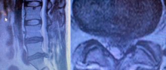
Cervical hernia on MRI
The surgical process proceeds, so to speak, in a vigilant and safe mode, which means that the arteries and nerves will not be damaged, the muscles will not be cut, they will only be carefully pulled apart. The main visualization tools are a microscope and an X-ray machine. Anesthesia is used mainly of a general type. Technically, the surgical session is somewhat different from the one performed in the lumbar area.
- The disc is accessed from the front rather than the back, meaning the incision is made on the front of the neck along a fold of skin rather than on the back. Its size also does not exceed 4 cm.
- A herniated intervertebral disc in the cervical area is usually completely removed, after which a stabilizer implant, or cage, is placed between the two vertebrae. This is a fairly simple system in design, but very reliable.
- The cage will contribute to the correct fusion of the two vertebral bodies together while maintaining the biological height of the ridge in this area, and will also not allow the functionality of the cervical area as a whole to be unbalanced.
- The session takes about 40-45 minutes, the patient is released from the hospital for 3-5 days, and then the patient needs to continue his recovery in a specialized medical facility.
Endoscopic type of microdiscectomy
This process of microdiscectomy is controlled not with a microscope, but with a light-conducting fiber-optic device called an endoscope. This is the most gentle tactic, which relates to a full ectomy, but it does not require general anesthesia, that is, the session is performed under local anesthesia.
An endoscope tube, at the end of which there is a miniature video camera, is inserted percutaneously into the spinal space, within the desired area, using a posterolateral approach. The incision is 2 times smaller than with a conventional microdiscectomy. It is approximately 1.5 cm.
The operating monitor receives video information in a multiply enlarged format, showing in real time the condition of the disc and adjacent vertebral tissues. Microsurgical instruments are inserted into the endoscopic probe, which is used to pinch off the protruding part of the cartilage. Having seen the free cartilaginous bodies exfoliated from the base of the disc, the surgeon also safely removes it. Pathological tissues are removed through the output section of the endoscope. Thus, the nerve root is freed from compression, and the pain syndrome disappears.
The price for endoscopic surgery is higher; in clinics in the Russian Federation, the price range starts from 100 thousand rubles, the maximum figure is about 300 thousand rubles. Percutaneous technology has similar goals and objectives as standard microdiscectomy. But the consequences and recovery time for endoscopy of a hernia at the lumbar level, as well as in other parts of the spinal axis, are significantly minimized. Moreover, reviews say that walking after such a minimally invasive operation is allowed within 2 hours, and if you feel well, you can leave the hospital the next day. It is worth noting that this method has been introduced into orthopedic practice quite recently, so it has not yet been mastered well enough in our country.
Rehabilitation and recovery after
In all cases, after microdiscectomy, whether it is done within the lumbar region or another area, a whole range of rehabilitation measures is always prescribed. Without a specific postoperative physical regimen, medication and physiotherapeutic treatment, surgical manipulations do not make sense, so you must undergo a rehabilitation program from beginning to end. Its development is strictly within the competence of a specialist in rehabilitation medicine and the attending surgeon.
Since the organization of restoration and control over the correctness of its implementation is carried out exclusively by the doctor, we do not have the right to recommend you a system of restorative measures. But we will try to form an idea of rehabilitation after this type of intervention.
- The patient is verticalized in the very initial period, usually within 24 hours after surgery.
- Walking and performing physical exercises are only recommended in a special corset, which will protect the operated area from unnecessary stress, unacceptable movements and injuries. The corset should be worn approximately 3-6 hours a day.
- Sitting is usually contraindicated for 2 weeks; a longer period (6 weeks) may be required.
- Lifting more than 3 kg of weight is strictly prohibited. Lifting weights of 5-7 kg and above is a taboo for life. Lifetime restrictions also apply to heavy physical labor and strenuous sports.
- From the first days, the patient receives broad-spectrum antibiotic therapy to counter the development of local infection. Painkillers and anti-inflammatory drugs from the NSAID group are also used. Vitamin-mineral complexes and vascular preparations are prescribed.
- The next day, therapeutic exercises begin, which are first performed in a lying position on the stomach and back. Basic exercises in the early period are based on dosed isometric contraction of the spinal and abdominal muscles, flexion and extension of the limbs, and high-quality breathing exercises.
- The complexity and duration of exercise therapy is increased gradually. The main goal of special exercises is to train and strengthen the muscular-ligamentous corset, develop good posture and practice correct movements, which will make it possible to normalize the spine and return a person to full physical capabilities.
- To activate blood flow, improve metabolism, stimulate the delivery of nutrition and oxygen to the vertebral and paravertebral structures, the specialist prescribes physical therapy procedures (magnetic therapy, laser therapy, electrophoresis, etc.).
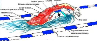
- Massages, acupuncture sessions, and swimming lessons are prescribed only by a doctor. Mostly, these measures are recommended for a certain period after half the rehabilitation period has already passed.
Magnetotherapy
It is not forbidden to follow the recommendations of Dr. Stupin; the famous doctor gives very valuable advice and guidance to patients who have undergone lumbar microdiscectomy. The popular doctor in his writings also describes a complex of exercise therapy after decompressive manipulations on the spinal column. However, remember that only your personal specialist should monitor your recovery after surgery and recommend all treatment and rehabilitation measures. Only he knows the whole state of affairs regarding your specific problem, the specifics of the operation performed, the general health status and a host of other individual nuances.
Based on feedback from patients, we conclude that the recovery period for people who flawlessly adhered to qualified medical recommendations lasts on average 5-6 weeks, maximum 2 months. However, an illiterate approach or lack of mandatory rehabilitation is a sure way to earn serious complications that can result in disability.
Working capacity is restored quickly and in most cases completely, but subject to adequate postoperative treatment, which is best done in specialized rehabilitation and health centers. Painful signs and paresthesias can decline gradually, and by the final point of rehabilitation they completely disappear.
Postoperative period
After surgery, the patient may experience pain and discomfort in the incision area. It must be borne in mind that the initial radicular pain may not completely regress immediately after surgery. To alleviate the condition in the postoperative period, painkillers and antibiotics are prescribed to prevent infectious complications. Under the supervision of a doctor, you will be instructed to stand on your feet immediately after you have fully recovered from the anesthesia. During the first four weeks after surgery, you should adhere to some minimal restrictions at home, such as not lifting more than 2.5 kg, and not doing sudden bending or stretching. In addition, you should not drive a car until your doctor allows it. It may take up to three months for the condition to improve. After discharge, you need to devote several months to recuperating. You should closely monitor your health and watch for symptoms that may indicate possible complications. Rehabilitation therapy influences successful treatment. It is prescribed for each patient individually, depending on the surgical operation, age, health, functional requirements, health problems and disability of the patient before the intervention.
Rehabilitation therapy has four main goals:
- Accelerating the time for symptoms to disappear, especially pain;
- Promoting faster functional recovery and
ability to work;
- Preventing symptoms from recurring;
- Prevention of complications and relapses.
Pain after microdiscectomy
A rational orthopedic regimen, physiotherapy and adequate drug care ensure a completely tolerable postoperative recovery. It is important to understand that it is normal to feel pain immediately after the procedure, as you have had a full surgery. As a rule, the pain syndrome is significantly reduced by 3-4 days, so be patient, soon you should not be bothered at all. But be sure to immediately notify your doctor if suddenly the pain does not decrease or intensifies, since it is possible that secondary negative reactions have occurred.
When, in addition to removing the hernia, lumbar fixation was performed.
Do not forget about the possibility of relapse, including in the long term. It also signals itself with painful signs, including lumbago, including impaired sensitivity in the legs and arms, which you have already encountered. To protect yourself as much as possible from re-development and exacerbation of the disease, you need to follow simple rules.
- This rule is addressed to women who want a child. Plan your pregnancy so that the birth occurs approximately 2 years after surgery. Doctors do not recommend that a woman after microdiscectomy become pregnant earlier than 12 months later.
- Do not sit exactly as long as the specialist told you. When you can sit, after 14 days or only after 1.5 months, the doctor will determine based on your progress in recovery. When this ban is lifted, do not stay in a sitting position for long.
- Do not ignore wearing a corset - you absolutely need it, strictly adhere to the standards for its use established by your doctor! Moreover, do not carry heavy weights and distribute the load evenly on each hand.
- As for sex, intimate life is not allowed for at least 7 days after spinal surgery. Throughout rehabilitation, intimacy should be unforced. In any case, do not hesitate to ask your attending surgeon about this issue; this is one of the temporary restrictions that also requires strict adherence.
- The next point is physical education. Let’s answer right away: is it possible to take physical training after microdiscectomy? No! Until your spine is completely restored, don’t even think about taking any sports standards. It is worth considering that intense running, jumping, leg swings, presses, twisting, pull-ups are not allowed after full recovery! People return to normal physical activity that does not involve increased stress after about 2-3 months.
- Do not drive a car during the entire recovery period. The same goes for traveling on public transport. Also, during rehabilitation, do not load the spine for more than 30 minutes, take rest breaks, taking a horizontal position. Try to maintain an optimized alternation of activity and rest after recovery.
- Every year, ideally 2 times a year, undergo sanatorium-resort treatment in a good sanatorium that specializes in the restoration of the spine and musculoskeletal system.
And the last thing is to regularly, throughout your life, do special exercises for the health of the spine, which qualified methodologists taught you back in the rehabilitation department. Your responsible attitude to what we have talked about will be rewarded with excellent results after surgical treatment, which will not take long to arrive. But most importantly, you will be able to protect yourself as much as possible from re-herniation both in the old place and in other intervertebral levels.
Results of the operation
- Fast patient recovery. Only 2-3 days of restricting the motor mode and 6-7 days of limiting the loads are necessary.
- Disappearance of pain within 1-2 days. Gradual relief of other symptoms of nerve compression.
- If the operation is carried out in the described volume, its effectiveness reaches 90%. In 10% of cases, relapse and reoperation are possible.
Microdiscectomy is a minimally invasive operation to remove a herniated intervertebral disc, performed by experienced surgeons at the Yauza Clinical Hospital, which will quickly relieve you of back pain, restore mobility, and improve your quality of life.
You can make an appointment by filling out the feedback form on the website or calling the specified phone number.
Reviews about microdiscectomy
After the operation, already at the time of discharge from the hospital, according to clinical observations, approximately 76%-80% of patients note that the pain in the neck or lower back has stopped completely. The positive dynamics are confirmed not only by the subjective feelings of the patients, but also by photographs taken during the control tomography. And only about 20% of people are discharged, saying that some pain remains in the back and/or limbs, but it is not as pronounced as it was before the operation.
As for sensory disorders, in approximately 60% of people upon discharge they decreased significantly, but not completely. Complete cessation of paresthesia in the limbs in the early period was determined by 20% of people. Exactly the same number (20%) noted that the nature of sensory disorders did not change at all, that is, it remained the same as it was at the preoperative level. It is worth noting that the absence of early postoperative changes towards improvement is almost always recorded in patients with an initially complex underlying disease, often aggravated by serious concomitant pathologies.
Residual or persistent pain and paresthesia for some time, localized in the arm, leg, back, spine, reviews of neurosurgeons should reassure you - this is not scary. The final normalization of once compressed nerve formations and weakened muscles will occur in the near future if you do not violate the basic medical requirements for rehabilitation. These signs should serve as a cause for alarm if they tend to intensify, have not completely disappeared after 2 months from the moment of manipulations on the spine, or arose in the long term against the background of a persistent absence of neurological symptoms.
3.When is the operation performed?
Microdiscectomy and discectomy are performed to relieve pain and restore spinal mobility. You may consider surgery in the following cases:
- You have very severe pain in your legs, weakness or sensitivity that prevents you from doing your usual activities;
- The listed leg symptoms do not improve after a month of non-surgical treatment;
- A medical examination showed that you have limited movement and abnormal sensations.
A microdiscectomy or discectomy is performed immediately if you have cauda equina syndrome:
- Urinary and fecal incontinence;
- Weakness and tingling in the buttocks, genital area and both legs.
About our clinic Chistye Prudy metro station Medintercom page!