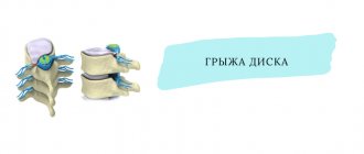
A herniated back, or intervertebral hernia, is a condition in which the intervertebral disc becomes deformed and protrudes beyond the vertebral bodies, compressing the nerve roots and spinal cord. This causes pain and other symptoms.
Most often, this disease occurs in older age, when degenerative processes develop in the spine. In approximately 75% of cases, surgical treatment is not necessary; the symptoms can be managed with conservative methods. When surgery is required, modern minimally invasive interventions are now used. They are performed by doctors at the international clinic Medica24.
Our expert in this field:
Abakirov Medetbek Dzhumabekovich
Traumatologist-orthopedist (vertebrologist)
Call the doctor Reviews about the doctor
A little about the structure of intervertebral discs and how a hernia develops. The intervertebral disc resembles the structure of a donut with filling. In the center there is a soft and elastic nucleus pulposus. It performs a spring function, softening vertical loads on the spine while walking, running, and jumping. Along the periphery there is a fibrous ring - it provides strength, due to which the shape of the intervertebral disc is maintained. Initially, degenerative changes develop in the intervertebral disc: it loses water, biochemical changes occur, due to which strength and elasticity decrease. If at this stage a person is bothered by symptoms, then most often neurologists diagnose “osteochondrosis”. Then the fibrous ring stretches, and a hernial protrusion forms on the disc. In the next stage, the annulus fibrosus ruptures and part of the nucleus pulposus comes out. Eventually, part of the nucleus pulposus breaks off—sequestration occurs. In such cases, the only option is surgery.
Under what conditions should the operation be performed?
Necessary conditions for determining indications for surgery:
- The presence of a substrate for the operation - hernia, instability, stenosis, which cause.
- Compression of nerve structures, blood vessels, which is the cause.
- Neurological deficit (weakness and numbness in the extremities, dysfunction of the pelvic organs) and severe pain.
Important! The operation eliminates compression of nerve structures and blood vessels, but does not eliminate the reasons that caused the compression.
When choosing surgery as a method of treating spinal hernia and its complications, the patient must understand the meaning of the proposed surgical treatment.
Diagnostic methods
The doctor’s task during the initial appointment is to identify symptoms in the patient that indicate damage to the spinal column. The doctor asks the patient about complaints, the nature of the pain, checks sensitivity and muscle strength. If signs are found that may indicate an intervertebral hernia, an examination is prescribed to confirm the diagnosis:
- Radiography. Intervertebral hernias are not visible on the pictures, but this study helps to exclude other causes of back pain, such as spinal curvature, fractures, and vertebral tumors.
- Computed tomography is a more accurate diagnostic method. During it, X-rays are also used, but the images are more detailed.
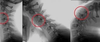
- Magnetic resonance imaging allows you to clearly visualize an intervertebral hernia, determine the level of its localization, and identify compression of the nerve roots.
- Myelography is an x-ray of the spine with the introduction of a contrast solution into the cerebrospinal fluid. The study reveals compression of the spinal cord and nerve roots.
Open operations
1. Discectomy. Removal of a “fallen out” intervertebral disc is a hernia (prolapse, extrusion, disc sequestration), which causes compression of nerve structures and blood vessels. The goal is to create decompression for compressed structures. The length of the skin incision on the back is up to 3 cm with the microsurgical method, with the endoscopic method - 1.5 cm. Area of application - lumbar spine.
2. Decompressive – stabilizing operations. Severe stenosis of the spinal and/or radicular canal with neurological deficit, where the cause is: hernia, arthrosis of the intervertebral joints, ossified (“petrified”) ligaments. Discectomy is combined with arcotomy, laminectomy (removal of the vertebral arch) and/or facetectomy (removal of the intervertebral joint), corpectomy (removal of the vertebral body). Simultaneous installation of various implants and stabilizing systems is necessary. Can be used in conjunction with navigation and endoscopic support. The skin incision is 3-15 cm depending on the number of operated vertebrae. The area is the entire spine, accessible from the back, front and side.
3. Stabilizing operations. Violation of the normal biomechanics and axial function of the spine is manifested by its instability, deformation, and pathological fracture. To prevent or reduce existing neurological deficits (paralysis), stabilization of the vertebrae with the help of special systems is necessary.
4. Palliative (alleviating), functional operations – rhizotomy and chondrotomy. Performed for spastic and persistent pain syndrome caused by the consequences of spinal cord damage. The purpose of the operation is to cut certain sections of the spinal cord and nerve trunks. As a result, the transmission of the pathological nerve impulse is blocked.
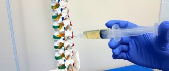
Percutaneous operations (by puncturing the skin) – “outpatient” neurosurgery
1. Radiofrequency denervation of the facet joints. It is used for facet syndrome (when the source of pain is altered intervertebral joints), special needles under image intensifier control (mobile X-ray unit) are installed in the area of the joints, through which radiofrequency effects are applied to the nerves of the affected joints. This eliminates pain.
2. Percutaneous puncture removal of disc herniation. The intervertebral disc is punctured with a special needle under image intensifier control, then its central part (nucleus pulposus) is removed using the following techniques:
- Percutaneous cold plasma nucleoplasty - evaporation under the influence of cold plasma.
- Hydrodisectomy – crushing and removal, high-speed fluid flow (900 km/h).
- Spiral nucleotomy is the introduction of a thin needle spiral, which makes rotational movements to remove part of the nucleus pulposus.
- Laser vaporization - removal occurs by exposure to a laser.
- Chemonnucleolysis - a special substance is introduced, leading to liquefaction of the nucleus pulposus and a decrease in its volume.
3. Vertebroplasty. Hemangioma, pathological fractures of the vertebral bodies impair the strength and stability of the spine, the geometry of the vertebra is disrupted, which can lead to damage to the spinal cord. The technique of execution is under image intensifier control, a thin tube with a guide, the vertebral body is punctured and filled with special cement.
3. Kyphoplasty. This is a similar procedure, but it allows not only to restore the strength of the vertebra, but also to compensate for its deformation, leading to a curvature of the spine that requires correction. Technique: under image intensifier control, special chambers are inserted into the body of the damaged vertebra through 2 hollow needles, which are then inflated. The pressure in the chambers is strictly controlled so that as a result the vertebra takes the required height without distortions (this is why two inflatable chambers are used). After the required position is restored, the chambers are removed and to fix the vertebra, the cavities in which the chambers were located are filled with cement.
4. Regeneration of intervertebral disc tissue. After removing the nucleus pulposus, part of the material is sent to a special laboratory, where normal cartilage cells are isolated and cultured (reproduction). Subsequently, the grown tissue is implanted into the intervertebral disc of interest using the percutaneous puncture method.
What consequences may occur after the operation?
Recovery after surgery is called an excellent result. The opposite situation is called bad consequences. The consequences are as follows:
- lack of effect from the operation;
- increased neurological status, pain syndrome;
- violation of the biomechanics of the spine - the appearance of instability or limitation of mobility in the spinal motion segment;
- intensification or appearance of a cicatricial adhesive process due to purulent-septic complications, bleeding, rough work of the neurosurgeon in the surgical wound;
- negative reaction to anesthesia (allergy, shock, exacerbation of chronic diseases), implants and stabilizing systems used;
- neurotic-depressive disorders;
- death. In planned spinal neurosurgery, this is a casuistry, where the cause is mainly acute pathological conditions during anesthesia (anaphylactic and other life-threatening types of shock, as well as rapid exacerbation of a previously undetected disease, for example, rupture of a cerebral aneurysm, acute infarction myocardium, etc.).
For an excellent result of the operation it is important:
- Correct determination of indications for surgery.
- Performing the operation at a high professional level.
- A well-planned and disciplined postoperative rehabilitation period.
Disturbances in the postoperative period are the main reason for the occurrence of poor consequences after surgery. In order to avoid them, proper rehabilitation is necessary.
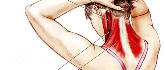
Development of a cervical hernia
The disease goes through exactly the same stages of development as hernias in other parts of the spine: tears or ruptures of the fibrous ring and the formation of protrusions, as a result of which the gel-like fluid of the nucleus pulposus leaks into the formed cracks. These disorders provoke the development of swelling, tissue integrity is destroyed, and pressure on the spinal canals, vascular tissues and nerve roots increases. Which are obliged to feed the brain, but due to such a violation they cannot perform their natural functionality.
Rehabilitation
The operation went well, the patient is confident that everything difficult is over. But for the patient, the most interesting period comes after the operation - the time of rehabilitation. The fullness of the patient’s life depends on rehabilitation and compliance with the doctor’s recommendations.
Principles of rehabilitation after surgery to remove a spinal hernia:
- Positive emotional attitude, despite the circumstances.
- The operation eliminates the consequences; only the patient himself removes the cause.
- Maintaining hygiene of movements (lifting and carrying heavy objects, running, jumping, prolonged and uncomfortable sitting, sudden and excessive physical activity)
- Regular physical education (general physical training and special exercises for the spine) FOR LIFE.
- Periodic course treatment - manual massage, physiotherapy, acupuncture, hirudotherapy. You can prevent pathological changes in muscles, ligaments and joints with the help of physical education, but you will not be able to eliminate existing disorders without medical help.
- Choose an activity you like - fitness, yoga, aqua aerobics, etc. and have fun.
- The health of the spine after surgery is not compatible with professional sports, weightlifting, contact martial arts, auto/quad/motorsports.
- If you are overweight, be sure to lose weight.
- Many professions have restrictions after surgery, which are decided individually together with the attending physician.
- Do not rely on medications; it is possible to correct the pathological condition with medication, but there is no cure.
To make a decision about surgery, the patient solves many issues, most of which are described in our article. But the main issue that the patient needs to decide is the choice of a neurosurgeon. So, a specialist answers all questions! He determines whether you need surgery, he performs it, and he makes recommendations.
Principles of criteria for choosing a doctor:
- It is not the hospital that will operate on you, but the doctor. This is especially true for a budget clinic. In private medicine, the specialist himself is the clinic. There are enough neurosurgeons in the state clinic, but it is not a fact that all of them specialize in spinal surgery. Go specifically to the doctor who was recommended to you.
- Neurosurgery is an extremely narrow specialization within the specialty itself. It is not possible to operate at the same high level on everything in neurosurgery - brain tumors, trauma, strokes, spine, etc. When it comes to herniated discs, this is a spinal neurosurgeon with experience in such operations of at least 150 - 200 per year. Science is a must. When communicating with the doctor, ask how many similar operations he has performed over the past month, and also look for his name among the authors of specialized articles in specialized medical journals. The Internet can help you.
- When deciding to undergo surgery, there should be no topics that are unclear to you. Do not hesitate to ask the doctor a variety of questions about the upcoming operation. Not only the answer is important, but also how they answer you. Calm, confident, without pressure.
- A top-class spinal neurosurgeon is a “piece product” with a corresponding price. In a private clinic the price is known. In a public clinic, you must make certain efforts so that the specialist chooses you from the flow of patients.
Causes of the disease
Most often, it is difficult to name the exact cause of a back hernia. Undoubtedly, degenerative processes that occur with age play an important role. Therefore, the disease rarely occurs in children and young people. This is mainly the lot of middle-aged and older people.
Other reasons:
- Heredity. Some people are born with weaker intervertebral discs and are more prone to developing herniations. This predisposition is inherited. Your risks are increased if close relatives suffer from this disease, especially at a young age.
- Obesity and overweight. Every extra kilogram puts additional stress on the spine. The main weight falls on the lower back, so intervertebral hernias are most common here. In second place is the cervical region, because it has high mobility. Problems with thoracic intervertebral discs are less common.
- Excessive stress on the spine. The development of degenerative changes and hernias is facilitated by: heavy physical work, frequent lifting of loads, intense sports such as weightlifting.
- Regular long-term stay of the spine in a monotonous, forced position. Because of this, the loads are distributed incorrectly, and certain parts of the intervertebral discs are constantly under increased pressure. For example, this happens with office workers, if the workplace is not equipped correctly, you have to constantly sit with your neck stretched forward. The lower back suffers from frequent flexion, especially when lifting loads from the ground.
All these reasons lead to chronic changes in the spinal column. But a hernia can also occur acutely, during an injury or when a person lifts something heavy.