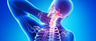
Chest pain ( thoracalgia ) is one of the most serious symptoms a person can experience. Sometimes even a doctor cannot immediately determine the cause of chest pain and find out whether this symptom is a sign of a life-threatening condition.
- Chest pain can be in any part and is caused by diseases of the heart, lungs, esophagus, muscles, bones, and skin.
- Due to the complex innervation of the body, chest pain may come from another part of the body.
- Chest pain may be caused by diseases of the stomach or other abdominal organs.
Symptoms of the disease
The symptoms of this disease are quite simple and it is with this variant of thoracic osteochondrosis that the diagnosis is usually quickly and accurately established. The clinical picture is represented by two main and a number of accompanying symptoms:
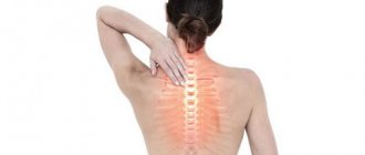
- Pain in the thoracic spine. The pain may intensify with movement, after static physical activity.
- Muscle tension in the thoracic spine.
- Crunching in the vertebrae when moving.
- Discomfort, crawling sensation in the interscapular area.
- An indirect symptom is an increase in the above complaints after hypothermia, prolonged exposure to a monotonous position (working at a computer).
How does vertebrogenic thoracalgia differ from thoracalgia due to ischemic heart disease?
*IHD - coronary heart disease.
Causes
Potentially life-threatening causes of chest pain include the following:
- An attack of angina or myocardial infarction. Chest pain in such cases is caused by impaired circulation in the coronary vessels, which can lead to myocardial ischemia. With angina pectoris, pain occurs during physical exertion, and with unstable angina pectoris even at rest. During myocardial infarction, the pain is usually intense and leads to the death of muscle tissue in a certain area of the myocardium.
- Aortic dissection (dissecting aortic aneurysm): The aorta is the main artery that supplies blood to vital organs of the body such as the brain, heart, kidneys, lungs and intestines. A dissection means a tear in the inner lining of the aorta. This can lead to massive internal bleeding and cut off blood flow to vital organs.
- Pulmonary embolism occurs when a blood clot enters one of the pulmonary arteries supplying blood to the lungs. This is a potentially life-threatening cause of chest pain that is not related to the heart.
- Spontaneous pneumothorax. Called a collapsed lung, this condition occurs when air enters the space between the chest wall and the lung tissue. Negative pressure in the chest cavity allows the lungs to expand. When spontaneous pneumothorax occurs, air enters the chest cavity, the pressure balance is disrupted and the lungs cannot expand. This in turn disrupts the oxygen supply to the blood.
- Perforation of internal organs: In a perforated organ in any area of the gastrointestinal tract, a hole or tear in the wall allows air to enter the abdominal cavity, which leads to irritation of the diaphragm, and can cause chest pain.
Other causes of chest pain that are not immediately life-threatening include the following:
- Acute pericarditis: This is inflammation of the pericardium (the membrane covering the heart)
- Heart defects such as mitral valve prolapse.
- Pneumonia: Chest pain occurs due to irritation of the pleura.
- Diseases of the esophagus can also present with pain similar to that of angina pectoris and are sometimes difficult to diagnose.
- Neoplasms (usually malignant) of the lungs can cause chest pain.
- Osteochondritis (Tietze syndrome): This is an inflammation of the cartilage tissue in the area where the ribs attach to the sternum. The pain is usually located in the middle of the chest, the pain can be dull or sharp, and can increase with deep breaths or movement.
- Shingles (herpes zoster) can cause quite severe chest pain, as the virus damages nerve fibers. The pain is usually located along the location of the herpetic rash.
Chest pain can also be caused by problems in the musculoskeletal structures.
- Rib injuries. A rib fracture can occur both during contact sports (for example, after a blow to the chest), and during falls and as a result of road traffic accidents. A rib fracture can sometimes be accompanied by lung damage and the development of pneumo or hemothrax. As a rule, diagnosing rib fractures does not cause any particular difficulties, since there is a clear connection between pain and injury.
- Vertebral fractures. Vertebral fractures may have a clear association with trauma (eg, a fall), but sometimes, especially in the presence of osteoporosis, the patient may not note a specific association with a specific incident of injury.
- Muscle injuries can occur as a result of overuse or poor technique during sports, which leads to muscle strain and pain in the area of those muscles. Muscle damage due to direct trauma is also possible.
- Joint damage. This is the most common cause of pain associated with the musculoskeletal system of the thoracic spine and chest. These disorders include damage to the intervertebral discs, the area where the ribs attach to the vertebrae, and the facet joints. The onset of pain can be gradual or sudden. Damage can occur as a result of a direct blow, sudden movement (sharp bending or twisting in the body, sharp extension), which leads to stretching of the ligamentous apparatus, joints, muscles, the development of an inflammatory process in the joint and muscle spasm. If such injuries are combined with poor posture, then the likelihood of developing degenerative changes in the joints is very high.
- In addition, the causes of pain may be dysfunction of the clavicular-sternal joints. Damage to these joints is usually associated with injuries from direct blows or ruptures of the ligamentous apparatus due to excessive loads. Pain may also be associated with injury.
- Intervertebral disc herniation. Disc herniations in the thoracic spine are quite rare and are associated with anatomical rigidity of the thoracic spine.
- Inflammatory diseases of the spine, such as ankylosing spondylitis (Bechterew's disease).
- Scheuermann-Mau disease. The pain syndrome is caused by severe hyperkyphosis and disorders of the biomechanics of the spine.
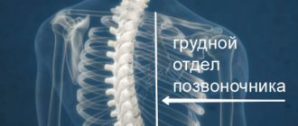
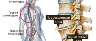
- Osteochondrosis of the thoracic spine. Changes in the intervertebral discs lead to compression of the nerve structures and the appearance of pain.
Treatment
Unfortunately, it is the localization of osteochondrosis in the thoracic region that suggests the least effectiveness and the speed of onset of the effect of therapy. Also, often, the treatment time for thoracic osteochondrosis increases to a month or more. Treatment of thoracalgia should be carried out by a neurologist. As with other localizations of the pathological process, therapy uses standard anti-inflammatory drugs (meloxicam, diclofenac, Celebrex, Airtal and others), muscle relaxants for existing muscle-tonic disorders (mydocalm, baclosan, sirdalud), neuroprotective drugs (B vitamins, thioctic acid etc.).
Chronic thoracalgia – pain syndrome of vertebrogenic origin
Chronic vertebrogenic thoracalgia is characterized by a recurrent long-term course. periods of exacerbation of pain are followed by remission, in which there are no unpleasant sensations and the patient considers himself completely healthy. Despite the feeling of general well-being, even during the period of remission, the syndrome of vertebrogenic thoracalgia is constantly developing. Further destruction of the nerve fiber occurs. The longer this syndrome lasts, the more difficult it is to restore the process of normal innervation of the soft tissues of the chest.
Thoracalgia as a pain syndrome can be present in a number of pathologies. For example, pain in the chest can be observed with pathologies of the pancreas, large intestine, gallbladder and liver. This syndrome may indicate general intoxication of the body and be observed with dysfunction of the endocrine system.
Mild thoracic thoracalgia syndrome may be a consequence of osteoporosis. This fact should be paid attention to by patients whose age has exceeded the threshold of 50 – 5 years. During this age period, secondary calcium deficiency may occur. It is important to monitor the condition of the large intestine, periodically check the condition of the thyroid gland, and take a blood test to determine the level of vitamin D.
In children, thoracalgia of vertebrogenic origin may be associated with rickets, a disruption of the process of formation of physiological curves of the spinal column. In adolescence, in almost 100% of cases, developing thoracalgia indicates poor posture. As a result of curvature of the spinal column, overstrain of the back muscles occurs. This causes sharp pain.
Exercise therapy exercises
Physical therapy exercises are very important, but they are ineffective in the acute stage. After all, the thoracic region has limited mobility and muscle spasms can sometimes only be relieved with medications. However, as a means of preventing the recurrence of thoracalgia, exercise therapy exercises come first. In case of illness, sets of exercises are indicated to strengthen the muscle corset, swimming, and cardio exercises in order to reduce excess body weight. It is undesirable to engage in martial arts (risk of injury), weightlifting (overload), basketball (frequent vertical loads).
Treatment of thoracalgia
To treat thoracalgia, it is important to make a correct diagnosis. Depending on the identified disease, its therapy is carried out, aimed at restoring the physiological structure of pathologically altered tissues.
During the first appointment, the vertebrologist examines the patient and recommends additional examinations as necessary. Then, based on the results of the examinations, the diagnosis is clarified and an individual course of rehabilitation therapy is developed.
Depending on the disease, the following manual therapy techniques can be used to treat thoracalgia:
- massage and osteopathy to improve microcirculation of blood and lymphatic fluid in the area of pathological tissue changes;
- traction traction of the spinal column - to eliminate compressive pressure from the radicular nerves and create conditions for straightening the compressed tissue of the fibrous ring of the intervertebral discs;
- reflexology – by influencing biologically active points located on the human body, it starts the process of regeneration (restoration) using the body’s hidden reserves;
- kinesiotherapy and therapeutic exercises improve muscle tone and speed up the healing process.
In some cases, laser treatment, electrical myostimulation and other types of physiotherapy are used in treatment.
You can sign up for a free initial consultation with a chiropractor or vertebrologist at our manual therapy clinic. During the appointment, the doctor will give you individual recommendations and tell you about all the treatment options for the identified disease that causes thoracalgia syndrome.
Treatment of thoracalgia in Samara includes methods that give the greatest effect:
- dosed traction and massage using the Ormed device
- manual therapy
- kinesio taping
- carboxytherapy
- ozone therapy (intravenous administration of ozonized saline solution and subcutaneous local administration of ozone)
- mesoinjection therapy with vascular drugs
- chronomagnetic complex "Multimag"
- physiotherapeutic techniques (high-top, ultrasound, electrophoresis)
- medical therapeutic blockades
Residents of Samara and Kazakhstan can undergo examination and treatment for thoracalgia at the FIRST NEUROLOGY clinic
Causes of vertebrogenic thoracalgia
What it is, thoracalgia syndrome, was described above in the article; you can find out about treatment below on the page. In the meantime, it's time to talk about the potential causes of pain.
Here is a list of pathologies in which vertebrogenic thoracalgia syndrome may occur:
- injuries of bone tissue (vertebral bodies, their processes, sternum, costal arches) - fractures and cracks;
- soft tissue injuries (intervertebral discs, ligaments and tendons, muscles) - sprains and ruptures, fiber separations;
- inflammatory processes (aseptic and infectious) - for example, herpes zoster;
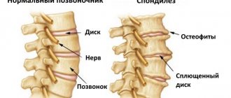
- degenerative dystrophic process of destruction of intervertebral discs (osteochondrosis and its complications, such as protrusion, extrusion and hernia;
- deforming arthrosis of the costovertebral joints;
- intercostal neuralgia;
- instability of the position of the vertebral bodies and their periodic displacement relative to each other and the central axis;
- stenosis of the spinal canal, including due to displacement of the vertebral bodies, prolapse of the dorsal hernia, cicatricial deformation of the dural sac, etc.;
- curvature of the spinal column (including scoliosis), poor posture;
- diseases of the internal organs of the chest (pneumonia, pleurisy, angina due to coronary heart disease, etc.);
- various tumor processes in the tissues of the spine, spinal cord and chest organs.
Thoracalgia is a pain syndrome and it can accompany any muscle fiber pathology. For example, if a person has a strong dry cough, then he will experience excessive muscle fiber tension. As a result, pain will occur. When carrying out primary differential diagnosis, this should be taken into account without fail.
It is also worth knowing that thoracic thoracalgia syndrome often occurs in people who are predisposed to it. It is expressed in the presence of risk factors such as:
- excess body weight - it is important to understand that every extra kilogram exponentially increases the shock-absorbing load exerted on the tissues of the spinal column, provoking their premature destruction;
- refusal of regular physical activity - the muscular frame of the back and paravertebral muscles do not work and do not provide adequate diffuse nutrition to the cartilaginous tissues of the spinal column;
- maintaining a sedentary lifestyle with predominantly sedentary work provokes stagnation of capillary blood and disruption of the trophism of all tissues;
- improper organization of sleeping and working spaces;
- violation of the principles of proper nutrition and insufficient drinking of clean water during the day;
- smoking and drinking alcohol provoke a decrease in the intensity of blood microcirculation in the lesions of the cartilage tissue of the spinal column;
- endocrine pathologies, for example, diabetes mellitus and diabetic angiopathy;
- poor posture, habit of slouching;
- incorrect choice of clothes and shoes for everyday wear and physical education;
- hard physical labor;
- rheumatic pathologies of cartilage tissue (rheumatism, systemic lupus erythematosus, multiple sclerosis, ankylosing spondylitis);
- constant stressful situations, including excessive psycho-emotional stress.
All these risk factors should be eliminated from your life if possible. During the initial examination, an experienced doctor interviews the patient to collect anamnesis and identify potential causes of tissue destruction in the spinal column. Therefore, you should not hide essential facts from him. This will help him more fully develop a course of treatment that will allow him to forget about the pain forever.