Almost all patients, after diagnosis, ask numerous questions about whether, with coxarthrosis, it is possible to lead their usual lifestyle and add some elements of sports to it.
Speaking in general terms, the doctor advises most patients not to abruptly, but radically change their usual lifestyle, since it is this that is the potential cause of the development of this pathology in 80% of cases. A sedentary lifestyle, excess calorie intake, refusal to play sports, incorrect posture and plantar feet are just a small part, the tip of the iceberg, of those health problems that ultimately led to the destruction of the hip joint.
The first thing to do after making a diagnosis is to find an experienced doctor, preferably an orthopedist or chiropractor. He will help you develop an individual therapy and rehabilitation plan. At the 1st and 2nd stages of coxarthrosis, complete restoration of the destroyed tissues of the hip joint is possible. But for this you will need to change your lifestyle, eliminate the effects of negative factors and carry out full restorative treatment using manual therapy methods.
If you need such help, then in Moscow you can make an appointment right now for a free appointment with a chiropractor or orthopedist in our clinic. The doctor will conduct an examination and review the results of the examinations. He will then create an individual treatment and rehabilitation plan for you.
To register, fill out the form at the bottom of the page or call the administrator at the specified phone number. He will agree with you on a convenient time to visit the doctor.
Next, we will answer the most common questions from patients about what can and cannot be done if coxarthrosis is detected (deforming osteoarthritis of the hip joint).
How to help yourself with arthrosis
Pain in the legs is a serious illness; the knee systems are the supporting mechanism of the body. The bones, tendons, and ligaments that make up the body are susceptible to damage and injury. Diseases associated with abnormal joint function, including arthrosis, do not allow patients to perform basic work normally. Many find it difficult to get out of bed, go downstairs, climb stairs, sit, or drive a vehicle.
It is important to tune in to serious, long-term treatment, including traditional methods, independent constant monitoring of your health. To monitor your weight and your joints in general, you shouldn’t give up physical therapy
When your legs begin to hurt, arthrosis is diagnosed, and measures to eliminate them must be discussed with your doctor. Then start training
To monitor your weight and joints in general, you should not give up physical therapy. When your legs begin to hurt, arthrosis is diagnosed, and measures to eliminate them must be discussed with your doctor. Then start training.
One of the best sports activities is swimming, water aerobics
It is important that exercises are carried out in water with a temperature of no more than 36°C
What physical exercises can you do at home?
For joint health and restoration of their function, it is recommended to engage in physical therapy. Such activities do not require much effort and time, and they are also not financially expensive. Depending on the stage of the disease, the doctor will recommend certain exercises.
Walking with arthrosis
Most orthopedists are almost unanimous: walking is beneficial for osteoarthritis of the knee joints. It not only has a preventive effect, preventing the onset of the disease, but is also a good remedy. While walking, a number of positive changes occur in the body:
- Improved blood circulation due to the functioning of the respiratory and cardiovascular systems - and, therefore, an increase in the delivery of nutrients to cartilage and bone tissue.
- Increased tone of working muscles.
- Strengthening the ligaments of the joint.
- Disappearance of stiffness.
- Expanding range of motion.
But even such a common activity as walking has its limitations. It should be noted that with grade 2-3 osteoarthritis of the knee, long treks, tourism, even a regular trip with bags from the store can become difficult. Although the load on the knee joint is significantly less than when running, you should not neglect safety measures by overloading the sore joint. Short walks are the best option for osteoarthritis of the knee joint.
Running and arthrosis of the knee joint
It is known that when running, the load on the knee increases more than five times compared to the normal state. Therefore, even against the backdrop of positive aspects (increased blood circulation, acceleration of general and local metabolic processes), overload of the articular apparatus has a significantly more negative effect. This can lead to accelerated cartilage destruction and disease progression.
You should not engage in the following active sports, which include running:
- Football.
- Tennis.
- Hockey.
- Basketball.
- Volleyball.
- Badminton.
- Skiing.
Doctors unequivocally state that running is contraindicated in the second degree of arthrosis of the knee joint. Even with initial changes in the joints, the benefits of running are not supported by everyone. Only light short runs can be recommended. But in any case, you should definitely consult with your doctor to assess the benefit-risk ratio. Whether to run or not with arthrosis of the knee joint will be decided only by a qualified specialist, having considered the pros and cons of each specific case.
Is it possible to squat with arthrosis of the knee joint?
You should also be careful with squats. Many doctors do not recommend performing them at all for osteoarthritis of the knee, while others recommend limiting them as much as possible, trying to ensure that the following conditions are met:
- Perform squats in such a way that you do not bend your knees completely. In this case, the load on the joint is less.
- If squatting is not yet recommended, then you can perform the following exercise: lying on your back and without lifting it from the floor, bend your knee, trying to pull your heel towards your buttock.
- It is good to perform squats for knee arthrosis in water, as this reduces the load on the joint.
It must be remembered that improper squats, even in healthy people, can lead to further development of arthrosis of the knee joint. Therefore, you should perform exercises according to medical recommendations.
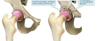
How does deforming osteoarthritis of the hip joint occur?
The hip (h/b) joint is the most powerful in the body, bearing the main load in human movement. It allows for greater mobility while maintaining stability under the pressure of the entire body weight. Such pressure is a serious test for articular cartilage, bone articulation - the acetabulum of the ilium and the articular head of the femur, which over the years leads to microdamage and degenerative changes in the joint.
Some diseases also play a role. Sometimes, for various reasons, the joint fluid changes its properties and no longer protects the cartilage well enough from friction. It becomes thinner, damaged and no longer protects the bones of the joint, which leads to their deformation. Pain appears.
This is how coxarthrosis develops. This is a severe progressive degenerative-dystrophic disease of the hip joints, often leading to loss of ability to work and disability.
How to swim for treatment and prevention
For joint pain, freestyle is recommended. During the period of remission, you can swim breaststroke. The water should not be cold - this can provoke an exacerbation. The optimal temperature is 35-36 degrees. It is recommended to exercise no longer than 30-40 minutes, gradually increasing the duration of water procedures. You should learn to breathe correctly in water by doing a short warm-up. If a person is a poor swimmer, it is recommended to use a life jacket. After exercise, you can do light muscle stretching, unless prohibited by your doctor. In general, the lesson is conducted according to the following plan:
- Warm up.
- Basic complex.
- Final gymnastics.
Degrees of coxarthrosis and symptoms of the disease
Symptoms of coxarthrosis usually appear in people over 40 years of age. Among them:
- Joint pain, often radiating to the groin or knee
- Gait disturbance
- Limitation of range of motion in a joint
- Muscle atrophy on the affected side
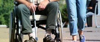
- In severe cases – disability
The severity of symptoms depends on the severity of changes in the joint.
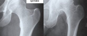
With coxarthrosis of the 1st degree, patients feel intermittent minor pain in the joint area, occurring at the beginning of physical activity, quickly passing after a short rest. The process of going up and down the stairs is disrupted. The patient is able to work. The range of motion in the joint is fully preserved.
X-rays show minimal narrowing of the joint space, and minor bone growths are possible. The head and neck of the femur are unchanged.
Coxartosis 2nd degree . At the second stage, the pain intensifies, occurs with a slight load on the joint (when getting out of bed, standing, short walking), and radiates to the leg (hip, knee). The patient has a noticeable limp. The range of motion in the joint is noticeably limited, and the ability to work is impaired.
X-ray reveals a narrowing of the joint space by approximately 1/3, the head of the femur has an uneven contour, and the femoral neck thickens.
With grade 3 coxarthrosis, the joint hurts both during movement and at rest, sometimes preventing the patient from sleeping. Mobility is severely limited. The patient needs support when walking. In fact, he becomes disabled.
On the x-ray, the joint space is greatly narrowed, the joint is deformed - the head of the femur is irregularly shaped with an uneven contour, the neck is wide and short.
In the early stages, it is easy to miss the development of the disease. The symptoms are mild and are often taken without due seriousness. To check the condition of your joints and not miss the onset of the disease, make an appointment with your doctor and get examined at a time convenient for you.
Are manipulations in water effective for the knee space?
The debate that water is beneficial for diseases in the knee cartilage area has been going on for a long time. In 2007, the Medical Council conducted studies examining the effectiveness of water exercises, exercises on land, and complete rest on the knee. Patients who previously experienced serious pain when walking, but noted positive improvements after water procedures, began to feel better.
To get a long-lasting effect, you should be responsible in following the recommendations. Visit the pool regularly for a long time. It is better to carry out the exercises prescribed by your doctor and attend a group of patients created for the treatment of diseases of the knee joint.
The exercises are effective if the patient cannot walk independently, for whom Nordic walking cannot be used for treatment.
Dr. Gitt's gymnastics complex
The first exercise using the Gitta method allows you to increase blood supply to the painful area, improve intestinal function, and massage internal organs. The patient should lie on his stomach, move his hips, as if rolling from side to side.
The next Gitta gymnastics exercise can be performed lying on your back, in a semi-sitting position. Spread your legs slightly to the sides, turn them alternately inward and outward.
The next two Gitta exercises are performed while sitting. We place our feet shoulder-width apart, pressing our feet firmly to the floor. Alternately bring and spread your knees. If pain occurs, you can place your knees wider. The last exercise consists of lifting your heels off the floor one by one.
If discomfort or pain occurs, you should stop exercising.
httpv://www.youtube.com/watch?v=embed/kuLeQzWh3Zc
How does swimming help with arthrosis?
Does swimming help young and old people with joint pain? Water procedures are intended for all age categories. Osteoarthritis of the knee joint can be obtained as a work injury or occurs as an age-related phenomenon. Visiting the pool and doing water aerobics helps normalize the performance of the knee and restore the mobility of the affected joints. How exactly does swimming help:
- The muscle structure relaxes, spasms and cramps are eliminated.
- Pain and discomfort are eliminated.
- During hydrotherapy, the knee can be extended or flexed with minimal resistance, which helps determine the presence of arthrosis.
- In clinical cases, when even painkillers cannot eliminate the pain, visiting the pool is the only way out. While the lower part of the body is in the water and remains afloat, a person does not feel 40% of his weight, therefore, pain subsides.
- All physiotherapy procedures cause severe pain and discomfort in the knee.
Is it possible to swim in the pool with arthrosis and severe pain? This situation is acceptable, but the duration of the therapeutic course will double.
Treatment of coxarthrosis
Conservative therapy
In the initial stages, conservative treatment of osteoarthritis of the hip joint can be effective, individually selected depending on the specific situation: the severity of the process, age, and the presence of other diseases.
To relieve pain at any stage of DOA of the hip joint, non-steroidal anti-inflammatory drugs are prescribed. Unfortunately, they have side irritating effects on the gastrointestinal tract, and their effectiveness weakens over time.
Muscle relaxants can be used to relieve muscle tension and improve blood circulation in the joint area.
The restoration of cartilage tissue is facilitated by the use of chondroprotectors.
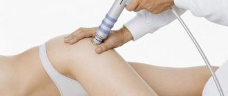
Hardware traction, physical therapy, physiotherapeutic treatment, manual therapy, post-isometric relaxation (the patient tenses and relaxes certain muscles, and the doctor stretches them during relaxation), osteopathy have a good effect.
Early diagnosis of the disease makes it possible to stop the development of coxarthrosis without resorting to surgical intervention. If you are worried about discomfort in your joints (even minor ones), immediately make an appointment with a specialist. This will allow you to detect the problem in a timely manner and select the most gentle treatment method.
To make an appointment with a doctor
Surgery
Endoprosthesis replacement of hip joint
If conservative therapy is ineffective, surgical treatment of osteoarthritis of the hip joint is indicated.
The Clinical Hospital on Yauza performs the most common and effective operation today for severe coxarthrosis - hip replacement (replacing it with a modern prosthesis from the world's best manufacturers DePuy, Zimmer, Smith & Nephew, Biomet).
Other methods of surgical treatment of coxarthrosis (DOA of the hip joint):
Arthroplasty – restoration of altered bone and cartilage structures
Osteotomy - The bones that form the joint are cut and re-aligned in an anatomically correct position. This operation sometimes allows you to preserve the joint and mobility in it.
Arthrodesis is a rigid (fixed) fixation of bones in the joint area with metal staples and screws. Allows you to keep the leg as a support, but makes it motionless in the joint.
Types of physical therapy
Exercise therapy for coxarthrosis of the hip joint includes the following components:
- swimming;
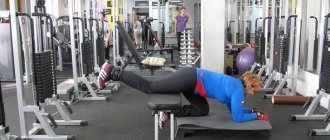
- medical simulators;
- physical training.
Swimming harmoniously affects all muscle groups, gently developing joints. In the pool, holding on to the side, you can perform the following exercises: fly-ups, leg pull-ups, “bicycle”. The resistance of the water will create the necessary load and ensure smooth execution. It is swimming and exercise in water that is prescribed primarily for joint diseases.
Many foreign and Russian doctors, including the famous Dr. Bubnovsky, categorically do not recommend an exercise bike or a bicycle due to compression on the joints. Only after endoprosthetics can you exercise in doses to strengthen your muscles. Walking on a treadmill is also not advisable.
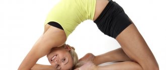
Yoga for coxarthrosis of degrees 1 and 2 strengthens and stretches muscles and ligaments, improves blood circulation. The main thing is to choose the appropriate exercises. For example:
- sitting on the floor, spread your legs apart (without tension), and try to reach your foot with your toes. But without jerks, but gently and gradually;
- sitting on the floor, one leg is extended forward, the other is taken back, bent at the knee and the foot is pulled up to the thigh of the straight leg. Painful symptoms are unacceptable.
Physical therapy for coxarthrosis currently includes many different techniques. The most popular programs are Dr. Gitt, Evdokimenko and Bubnovsky.
Is it possible to do sports and yoga with coxarthrosis?
The first question: is it possible to play sports with coxarthrosis? The answer to this is no, you can’t. Moreover, destruction of large joints of the lower extremities is an occupational disease of almost all athletes. It is important to distinguish sport from physical education. Sport is hard work to achieve record results. Physical education is an activity aimed at maintaining good health and sufficient body tone.
Feel the difference. If you go to the gym every day and lift a 60 kg barbell 30 times in order to build muscle, then this is a sport. It will lead you to destruction of the hip and knee joints, displacement of the vertebral bodies and disability. If you squat 10–15 times with dumbbells weighing 3–5 kg, then this is physical education. It will give you the opportunity to activate your muscles and improve your body tone.
You can do physical exercise with coxarthrosis, but sports are strictly contraindicated.
Second question: is it possible to do yoga with coxarthrosis? The answer is similar: yoga comes in different forms. There are complexes of simple health-improving gymnastics. And there are serious exercises that require fantastic body flexibility and endurance. You can do the first type. But it’s not worth pretending to be an experienced yogi who easily sits in the lotus position and throws his legs behind his head. For a person with coxarthrosis, this may cost the ability to walk independently in the future.
Swimming for the treatment and prevention of osteochondrosis
Osteochondrosis is a chronic disease, the essence of which is degenerative-dystrophic changes in the spinal column. In most people, the spine consists of thirty-three vertebrae, each of which is connected by an intervertebral disc.
The disc consists of an annulus fibrosus and nucleus pulposus. Thanks to these formations, the spine has the ability to bend. In addition, the discs perform the function of shock absorption. Degenerative changes in the intervertebral discs ultimately lead to rupture of the annulus fibrosus and displacement of the nucleus. The disease manifests itself as a pronounced pain syndrome, the localization of which depends on the level of damage to the spine. The lumbar spine experiences the greatest static loads, therefore osteochondrosis with all its neurological manifestations is most pronounced in this section. Many experts consider osteochondrosis to be a manifestation of the natural aging process of the spine. This is confirmed by the fact that osteochondrosis after the age of forty occurs in eighty percent of respondents. At fifty years of age, it occurs in one hundred percent of the population.
The complex of measures for the treatment of osteochondrosis necessarily includes physical exercise. Swimming is especially beneficial. During swimming, the body is in a weightless state, and there are no static, gravitational loads on the intervertebral discs. Physical activity is evenly distributed among all muscle groups. Therefore, spasming muscles relax, pain decreases, and blood circulation improves in the affected areas of the spine. At the same time, the paravertebral muscular frame is strengthened, which takes on part of the load, thereby unloading the intervertebral discs.
Causes and treatment of coxarthrosis
Coxarthrosis is deforming arthrosis of the hip joint.
Among degenerative-dystrophic diseases of the musculoskeletal system, it occupies one of the leading places.
Coxarthrosis can be primary (coxarthrosis of unknown etiology) and secondary, occurring against the background of hip dysplasia or congenital hip dislocation, aseptic necrosis of the femoral head, Perthes disease, trauma (bruise, fracture, dislocation, microtrauma), inflammatory process (coxitis). One or both hip joints may be affected. In primary cases, other joints (usually the knees) and the spine are often simultaneously affected. Most scientists believe that the trigger is a violation of blood circulation in the joint due to both deterioration of venous outflow and disruption of arterial inflow. One cannot but take into account the mechanical factors that cause overload of the joint, the incongruence of articular damage, leading to a redistribution of the load per unit area of the articular surface of the cartilage, as well as biochemical changes in the cartilage itself.
The main complaint in patients with coxarthrosis is pain, the nature, intensity, duration and location of which depend on the severity of the dystrophic process, i.e. from the stage of the disease.
There are three stages of coxarthrosis.
In stage 1, periodically after physical activity (long walking, running), pain occurs in the hip joint, less often in the hip or knee joint. As a rule, the pain goes away after rest. The range of motion in the joint is not limited, muscle strength is not changed, and gait is not impaired. Radiographs show minor bone growths that do not extend beyond the labrum. They are usually located around the outer or inner edge of the articular surface of the acetabulum. The head and neck of the femur are practically unchanged. The joint gap is unevenly slightly narrowed.
In stage 2, the pain is more intense, radiates to the thigh, groin area, and occurs at rest. After a long walk, lameness appears. The function of the joint is impaired. First of all, internal rotation and abduction of the hip are limited, i.e. flexion and adduction contracture is formed. The strength of the muscles that abduct and extend the hip decreases, and their hypotonia and hypotrophy are determined. The radiograph shows significant bone growths along the outer and inner edges of the acetabulum, extending beyond the cartilaginous lip. Deformation of the femoral head, its enlargement and uneven contour are noted. Cysts can form in the most loaded part of the head and acetabulum. The neck of the femur is thickened and widened. The joint space is unevenly narrowed (up to 1/3-1/4 of the original height). There is a tendency towards upward displacement of the femoral head.
In stage 3, pain is constant and occurs even at night. When walking, patients are forced to use a cane. There is a sharp limitation of all movements in the joint (flexion-adduction contracture) and hypotrophy of the gluteal muscles, as well as the muscles of the thigh and lower leg. A positive Trendelenburg sign may be detected. Flexion-adduction contracture causes an increase in pelvic tilt and an increase in lumbar lordosis. Pelvic tilt in the frontal plane, associated with weakness of the hip abductors, leads to functional shortening of the limb on the affected side. The patient is forced to step on his toes to reach the floor and tilt his torso to the affected side when walking to compensate for the tilt of the pelvis and shortening of the limb. This compensation mechanism leads to a shift in the center of gravity and overload of the joint. Radiographs reveal extensive bone growths on the side of the roof of the acetabulum and the head of the femur, and a sharp narrowing of the joint space. The neck of the femur is significantly expanded and shortened.
The diagnosis is based on clinical and radiological data. In addition to the stage of the disease, x-ray examination helps clarify the etiology of the process. In dysplastic coxarthrosis, flattening, bevelling of the acetabulum, changes in the size of the neck-diaphyseal angle, etc. are determined. In coxarthrosis, as a result of Perthes disease, or juvenile epiphysiolysis, the shape of the proximal end of the femur changes mainly. There is deformation of its head and neck (shortening, widening), a decrease in the neck-diaphyseal angle with the formation of the plow vara. The X-ray picture of post-traumatic coxarthrosis depends on the nature of the injury and the shape of the articular surfaces after fusion of the bones forming the hip joint. Differential diagnosis is carried out with osteochondrosis of the lumbar spine, coxitis and tumors of the pelvis and hip.
Treatment, due to the lack of a single pathogenetic mechanism for the development of the disease, is symptomatic in nature and is aimed at reducing pain and static-dynamic disorders of the musculoskeletal system. In this case, it is necessary to take into account the stage of the disease, the age of the patient, his general condition and the characteristics of clinical manifestations.
At stages 1 and 2 of coxarthrosis, treatment can be carried out in an outpatient setting. It is aimed at reducing pain, aseptic inflammation in periarticular tissues, improving tissue trophism and blood circulation in the limb, increasing joint stability and preventing other static disorders of the musculoskeletal system. During the period of exacerbation with severe pain, it is recommended to reduce the vertical load on the limb (avoid long periods of standing, carrying heavy objects, running). When walking for a long time, additional support on a cane is required. Analgesic and anti-inflammatory drugs are used (analgin, reopirin, amidopyrine, brufen, indomethacin, ortofen). To improve redox processes in cartilage tissue, vitamins, aloe, vitreous, rumalon and other drugs are prescribed. At home, you can use compresses with dimexide (10-15 procedures). In a clinic setting, electrophoresis of a solution of novocaine, dimexide, etc. is used, as well as ultrasound therapy, magnetic therapy, and laser therapy. After reducing the pain syndrome, manual massage of the lumbar region, hip joint, thigh and therapeutic exercises are performed, aimed at normalizing muscle tone, restoring mobility in the affected joint, followed by strengthening the surrounding muscles. Therapeutic gymnastics includes movements in the hip joint in positions of unloading of the joint (lying on the back, side, standing on the healthy leg, etc.). Use exercises aimed at strengthening the muscles that abduct and extend the hip. Standing on a healthy leg on a stand, holding onto a gymnastic wall, patients abduct and extend the thigh (freely, with a load, holding for 5-7 s, overcoming the resistance of a rubber bandage). It is better to perform hip extension while lying on your stomach with an amplitude of 10-20°; you can do this lying on the couch with your legs lowered to a horizontal level, or standing on all fours. Special exercises for the hip joint are carried out against the background of general developmental physical exercises, special exercises to strengthen the muscles of the back and abdominal wall in order to increase the stability of the lumbar spine. In a hospital setting, joint traction and hydrokinesitherapy are also prescribed. Joint traction is carried out on the patient's bed with a load of 5-7 kg for 3-5 hours a day, on a special traction table with a dosed load for 20-40 minutes, and also in water. During traction (10-15 procedures), patients are recommended to walk with crutches, unloading the leg. Joint traction is combined with manual massage of the thigh muscles, underwater jet massage with a water jet pressure of 0.5-1 atm for 5-8 minutes and physical exercises in water. These procedures are aimed at relaxing tense muscles, improving blood circulation in the limb and increasing diastasis between the articulating surfaces of the hip joint. In the future, the main attention is paid to therapeutic exercises aimed at increasing the stability of the hip joint by strengthening the periarticular muscles. Electrical stimulation of the gluteal muscles is performed (10-15 procedures). After treatment, patients are recommended to do therapeutic exercises at home, self-massage of the gluteal muscles and thighs, swimming, and skiing. Prolonged static loads on the legs, heavy physical labor, sports and rhythmic gymnastics, figure skating, aerobics, wrestling, and weightlifting are contraindicated.
In stage III coxarthrosis, conservative treatment, in addition to the indicated measures, includes intra-articular administration of Kenalog or Arthroporon. Treatment is carried out in a hospital, which provides the necessary regime for unloading the joint. Traction of the joint and physical exercises to increase mobility in the joint are contraindicated, because persistent (arthrogenic) contracture of the hip joint does not allow increasing mobility, and this attempt only causes additional microtrauma and increases pain. The main therapeutic measures are aimed at reducing pain, unloading the joint, and training compensatory and adaptive mechanisms. An orthopedic regimen and exercise therapy are used. Constant additional support is required when walking with a cane, and during an exacerbation - with crutches. Hydrokinesitherapy and therapeutic exercises should help improve joint stability, strength and endurance of the hip abductor and extension muscles, relaxation and stretching of the hip flexor and adductor muscles. Use isometric and dynamic exercises with range of motion in the joint within the limits of preserved mobility (until pain is felt). Manual and underwater massage is used with a water jet pressure of 1-2 atm for 10-15 minutes, electrical stimulation of the gluteal muscles. A well-trained muscular system helps to develop compensatory mechanisms even with gross changes in the joint. The period of formation and improvement of compensatory mechanisms is long and requires systematic training. Therefore, therapeutic exercises should be continued at home; strict adherence to the orthopedic regime for unloading the joint is recommended. Surgical treatment includes operations that preserve mobility in the joint (osteotomy, arthroplasty, endoprosthetics) and close the joint (arthrodesis). When determining the indications for choosing a method of surgical intervention, the stage of the dystrophic process, the general condition, age and profession of the patient, the condition of the other hip joint and the lumbar spine are taken into account. For stage I-II coxarthrosis and slight limitation of the function of the hip joint, various types of femoral osteotomies are most widely used. For stable fixation of bone fragments, the Trotsenko-Nuzhdin plate is used. With this type of fixation, additional external immobilization is not required in the postoperative period. To restore joint function, therapeutic exercises (general developmental breathing exercises, isometric muscle tension) are prescribed from the first days. From the 3rd-4th day they begin to facilitate movements in the knee and hip joints of the operated leg. From the 14th to 16th day, walking with the help of crutches without support on the limb is allowed. Up to 4 weeks, the exercises are light; after the sutures are removed, exercises in water can be used. In the future, manual and underwater massage and exercises to strengthen the muscles surrounding the hip joint are prescribed. Partial load on the limb is allowed 4 weeks after surgery, full - after 6 months. In dysplastic coxarthrosis, to increase the coverage of the head of the femur with the articular surface of the acetabulum, improve its centering, and reduce the load on the articular cartilage, various types of pelvic osteotomy are performed, which improves the biomechanical conditions in the joint. The most widely used is the Chiari pelvic osteotomy.
At stage III of the disease, it is impossible to stop the degenerative process, therefore the listed operations are considered as polliative. In this case, the most promising is hip replacement, which is carried out in the presence of a bilateral process with ankylosis in one of the joints, severe coxarthrosis and significant changes in the lumbar spine, with coxarthrosis and ankylosis in the knee joint on the same side. After surgery, a derotation boot is placed on the foot and lower leg for 3-4 weeks. From the 2-3rd day, easier movements are allowed in the operated joint in the sagittal plane, after 10-12 days - in the frontal plane. After 4 weeks, patients begin to walk with the help of crutches without support on the operated leg. Partial loading is allowed after 3-4 months, full - from the 5-6th month, provided the endoprosthesis is stable. After the start of walking, therapeutic exercises are prescribed to strengthen the muscles surrounding the endoprosthesis (in a lying position on the back, side, stomach). Classes continue in outpatient clinics or at home. At the same time, a manual massage is performed. Arthrodesis of the hip joint is performed for unilateral coxarthrosis of stage III in young people engaged in physical labor. After the operation, a plaster cast is applied for 5-6 months. During the period of immobilization, therapeutic exercises are prescribed (general developmental, breathing exercises, isometric muscle tension under a plaster cast and free movements in non-immobilized joints). When using the Umyarov plate for arthrodesis, plaster immobilization is not performed. In this case, from the 3-4th day, light movements are performed in the knee joint. You are allowed to get up after 3 weeks, walk with the help of crutches with partial load on the operated leg - after 4 weeks, with full load - no earlier than 4-5 months. when radiological signs of fusion of the femoral head with the pelvic bones appear. Sanatorium-resort treatment after surgical treatment is indicated after 6-8 months. The prognosis for life is favorable, the progression of the disease of unknown etiology is slow. With aseptic necrosis of the femoral head, the course of coxarthrosis is the most unfavorable.
Prevention. Specific prevention methods have not been developed. Primary prevention measures can be considered early detection and treatment of congenital hip dislocation and hip dysplasia, as well as clinical observation of adolescents with hip pathology. Secondary prevention consists of timely diagnosis of stage I coxarthrosis, systematic conservative treatment at intervals of 1-2 times a year, regardless of the severity of the pain syndrome, compliance with the orthopedic regime for unloading the joint, orientation towards “sedentary” professions, rational physical education, and control of body weight.
Bibliography: Krisyuk A.P. Deforming coxarthrosis in children and adolescents, Kyiv, 1982; Therapeutic physical culture, ed. V.A. Epifanova, p. 424, M., 1987; Abalmasova E.A. and Luzina E.V. Development of the hip joint after treatment of congenital subluxation and dislocation of the hip in children, Tashkent, 1983; Guryev V.N. Coxarthrosis and its surgical treatment, Tallinn, 1984; Korzh A.A. and others. Dysplastic coxarthrosis, M., 1986.
Swimming for the treatment and prevention of arthritis
Arthritis is an acute inflammation of one or more joints. Arthritis can be infectious, post-traumatic, metabolic. Infectious arthritis occurs due to the penetration of bacteria into the joint cavity. Post-traumatic develop after injury to the joint area. Metabolic arthritis is caused by disruption of biochemical processes in the body.
The main symptoms of arthritis are joint pain, limited movement, and swelling of the joint area.
Swimming is the only type of physical activity that is allowed during the acute phase of arthritis. Water cools the joint area, this in itself alleviates the patient’s condition. Smooth movements in the water massage the periarticular tissues, thereby improving the outflow of lymph and reducing swelling. Swimming also has a positive effect on metabolic processes in the body, due to which the root cause of articular syndrome disappears.
Is it possible to ride a bicycle with coxarthrosis?
Is it possible to use an exercise bike for coxarthrosis? Yes, it will be very useful, since there is practically no pronounced load on the articular surfaces, and the muscles work fully.
But it is impossible to say unequivocally whether it is possible to ride a bicycle with coxarthrosis. This type of physical activity is associated with a high risk of injury from a fall. An unsuccessful descent from a hill or sudden braking can lead to injury to the bone tissue inside the hip joint. This is a serious condition that can cause disability.
Therefore, take this issue seriously. Even if you really want to ride a bike, weigh all the possible risks. If you already have the 2nd stage of coxarthrosis, then it is better to refuse such entertainment until the condition of the cartilage tissue inside the joint is normalized.
Evdokimenko method
Exercises for coxarthrosis according to the system of rheumatologist Pavel Evdokimenko are based on strengthening and stretching the muscles without putting stress on the joint.
Combined exercises according to the Evdokimenko method are performed on each leg, first in a static version - a single lift and a long hold, and then in a dynamic version - lifting and holding for 1-2 seconds, slowly lowering. Dynamic options are repeated 10-12 times.
Daily combination complex:
- From a lying position, one leg is slowly raised 15 cm and held for 15-20 seconds. Also the second one. Then dynamic repetitions.
- From a lying position, one leg, bent at a right angle, is raised 10-15 cm from the floor, held for 30-40 seconds and slowly lowered.
- Lying on your back, bend your knees and spread them slightly to the sides. Feet on the floor, arms extended along the body. Perform a pelvic lift, leaning on your shoulders. “Hover” for 30-40 seconds, slowly lower. Then the dynamic option. Dr. Evdokimenko recommends this exercise only for grade 1-2 coxarthrosis.
Dynamic complex:
- Lying on your stomach, arms along your body. Straight legs are raised 15 cm from the floor, smoothly spread to the sides and brought together, and lowered down. 8-10 repetitions are performed.
- Lying on your side, bend your supporting leg at the knee, lift the other leg up 45 degrees, hold for 30 seconds, and slowly lower. Repeat 2-3 times.
- Lying on your back, raise one straightened leg 30-40 degrees, with weight, turn it completely inward from the hip, then turn it outward. Make 10-15 turns and lower.
Before classes, Dr. Evdokimenko recommends rubbing the joints with warming ointments, such as Espol or Nicoflex.
Causes of DOA of the hip joint
The causes and factors contributing to the occurrence of coxarthrosis may be:
- Injuries and long-term excessive loads on the hip joint (fractures, dislocations, fractures of the joint, as well as chronic permanent microtraumas in athletes, dancers).
- Congenital anomalies of the hip joint, dysplasia (up to 90% of cases), chondropathy.
- Past inflammatory process (autoimmune - rheumatoid arthritis and other systemic diseases, infectious - tuberculosis, etc.)
- Metabolic disorders
- Aseptic osteonecrosis
Most experts associate the development of coxarthrosis with impaired blood microcirculation in the joint area, caused by various reasons - sedentary work, low mobility, endocrine diseases, etc. The situation is aggravated by excess weight and curvature of the spine, which creates additional stress on the joints.
Without proper treatment, coxarthrosis leads to disability. The unbearable pain that appears at stage 4 of the disease is difficult to control even with strong painkillers. Don't let the disease change your life for the worse. Make an appointment with your doctor to get adequate treatment and stop the destruction of the joint.
Characteristic features of the course of arthrosis
Pain is a constant symptom of arthrosis. It increases after prolonged physical activity and changes in weather conditions. This disease is characterized by initial pain that can decrease a few minutes after the start of movement. At night, the pain may intensify, interfering with proper sleep. To get rid of pain, experts recommend planning visits to the pool shortly before bed. This guarantees no discomfort until the morning.
The disease occurs with periodic exacerbations. Relapse can be triggered by joint injuries, alcohol abuse, and exposure to stress factors. Arthrosis is accompanied by joint deformation, which can be slowed down with the help of special physical exercises. Without constant and moderate loads, the joint will bend, the pain will intensify, which will lead to disability and the need to resort to complex surgical interventions.
What rules should you follow when visiting the pool with arthrosis?
The key factors in choosing exercises are the person’s age, physical fitness and stage of the disease, which needs to be discussed in some detail.
Different degrees of stress can have a positive or negative effect on the patient’s health. If you overdo it at the initial stage of arthrosis development (incubation period), you can only accelerate the progression of the disease.
During the period of remission, methodological exercises are very important. You need to do smooth, slow, but long-term exercises to strengthen the condition of your muscles and joints. Swimming and arthrosis are compatible concepts, if you recognize the fine line between recommended methods and undesirable exercises.
In acute form, it is highly not recommended to exercise more than 15–20 minutes a day. This is due to the fact that in an aggressive form of arthrosis of the knee joint, the affected area becomes very swollen and red, and the existing pain does not decrease even in the pool.
Note: The patient needs to take into account not only the condition of the knee space, but also his own well-being, since a comprehensive therapeutic course involves exercise on land. It is necessary to calculate the forces evenly.
Exercises performed in the pool
Doctors often recommend using water procedures and Nordic walking for treatment. It is difficult for many to move; such patients have to stand in the water for physical therapy.
Physical exercises in the water space include manipulations:
- Walking in water. Walking in the shallow part of the water, in the deep. Walk forward, backward, sideways, forward with your back, which will help strengthen the muscles of the body.
- Physical training. Before starting classes, you should consult your doctor. The doctor will determine whether water procedures will harm the knee. Exercises include swinging your legs, marching in the water, and bending your knees.
- Often the pool has the opportunity to offer clients a gym, which in tandem with therapeutic “swimming” will bring benefits.
Exercises in a vertical position
Starting position: standing, feet together, lean on the back of the chair with both hands.
- Slowly do a half squat and also slowly return to the starting position. Number of repetitions - 10.
- Slowly stand on your toes, stretching upward, then slowly return to the starting position. Number of repetitions - 10.
Starting position: standing, lean your hand on the side of your healthy leg on the back.
- Raise your affected leg, then make a few gentle swings back and forth. Number of repetitions - 10.
- Raise your sore leg, then make a few gentle circular movements. Number of repetitions - 10.
httpv://www.youtube.com/watch?v=embed/hjkPeM4QVbI
In addition, you should follow a special diet and, of course, adhere to the recommendations of your doctor. Only in this case is it possible to improve the condition.
Gitta gymnastics
Vitaly Gitt is a famous chiropractor. The complex he developed, based on micromovements, has been successfully used for more than 15 years to restore the mobility of affected joints, even with severely developed pathology.
The exercises that Dr. Gitt suggests are performed repeatedly, measuredly, for a long time, the amplitude of movement is only 2-3 cm. Some of them can be performed while reading a book or working at the computer. Vitaly Gitt explains this technique by saying that the joint needs very little fluid for proper nutrition, but it must be produced constantly. At larger amplitudes, production occurs more actively, but the substance does not have time to be absorbed by the joints. That is why the Gitta method exercises, although simple, require patience and time. Suitable for grades 1, 2 and 3 coxarthrosis, without aggravating pain symptoms. Many patients confirm that Dr. Gitt’s gymnastics are truly healing, and do not require additional equipment.
A set of exercises for the treatment of coxarthrosis by Dr. Gitt:
- Lying on your stomach, arms under your head or along your body, feet on a small cushion, body relaxed. Rolls are made from side to side with a small amplitude. You should not strain your muscles or raise your pelvis. Gitt recommends performing this exercise three times a day for 10 minutes. Moreover, it is suitable even for severe coxarthrosis.
- Lying on your back. Legs slightly apart, turn each (without bending) left and right by 1 cm.
- Sitting on a chair, knees slightly apart, feet on the floor. Movement of the knees apart and together with an amplitude of no more than 1 cm.
- The healthy leg stands on an elevation, the sick leg swings left and right, back and forth. Performed 2-3 times a day for 5 to 10 minutes.
Dr. Gitt advises exercising between 3 and 6 hours a day. The result will only come after hard training.
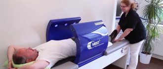
Vitaly Gitt has developed special simulators in the form of vibrating couches, which force all joints to work, making quick micro-movements, as a result of which the production of synovial fluid is activated, but without any load on the joint. Gitt uses similar devices for patients with serious injuries.
In addition to physical exercise, Dr. Gitt pays special attention to nutrition: the less protein in the diet, the healthier the joints. Because, in his opinion, an excess of protein products triggers autoimmune processes in the body
Painful symptoms are not allowed during training according to the Gitta system. You should either choose a comfortable position or reduce the amplitude.
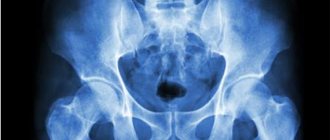
Diagnosis of the disease
A detailed survey and clinical examination by an orthopedic traumatologist helps to identify the first signs of coxarthrosis.
The basis of diagnosis is radiography (computed tomography) of the joint. Already at the first degree of coxarthrosis, a slight narrowing of the joint space and proliferation of bone tissue are detected. Subsequently, deforming osteoarthritis of the hip joint is manifested by deformation of the head of the femur, thickening of its neck, the appearance of osteophytes (bone spines), and the narrowing of the joint space becomes more pronounced.
Magnetic resonance imaging (MRI) of the joint can reveal changes not only in bone structures, but also in cartilage tissue, ligaments, and synovium.
Laboratory tests can exclude the presence of an inflammatory process in the body and identify signs of metabolic disorders.
If necessary, a diagnostic puncture of the joint is performed followed by examination of the synovial fluid.
Basic set of exercises
Having mastered the initial level, you can move on to the basic exercises using the Bubnovsky method.
These include exercises:
- "Jackknife". The straight leg is moved to the side. Try to grab your foot with your hands, fix the position, and go back. When performing, slight pain is allowed; over time, the muscles will get used to the load.
- The exercise can be performed while standing, alternately bending over one or the other leg. This exercise allows you to strengthen your joints and stretch your lower back muscles.
- Lying on your back, slowly raise your legs and try to touch the floor behind your head. This is a difficult exercise, but effective. You can start by simply raising your legs and trying to lower them behind your head. Do the exercise 20 times.
Is swimming good for joints and spine?
Details Category: How to cure osteochondrosis of the spine and joints at home
Today we will talk about swimming. Is swimming good for your joints and spine? The answer is clear – YES it’s good!
Swimming is recommended as a remedy for osteochondrosis, various curvatures of the spine, postural defects and many, many other diseases of the joints and spine.
If you have been to a doctor with problems with the spine or joints, then, in addition to physical therapy, I am sure that swimming was recommended to you as a therapeutic and preventative measure.
The healing properties of swimming for the spine and joints are based on Archimedes' law: any body immersed in a liquid loses as much weight as the weight of the liquid it displaces.
An average-sized person, weighing about 70 kg, weighs only about 3 kg in water. Those. in water you find yourself practically in a state of weightlessness.
So let's figure out why swimming is so beneficial for the spine, joints and other body systems.
Positive aspects of swimming for the spine and joints
1. When swimming, the joints and spine are unloaded, and the intervertebral discs are straightened.
So, in water a person weighs practically nothing, i.e. is practically in a state of weightlessness. That is why all loads compressing the vertebrae and intervertebral discs are completely removed.
If you are in the water (not even swimming, but just lying down), then the spine is completely unloaded, the intervertebral discs straighten and rest (within 40-45 minutes of being in the water, growth even increases due to the straightening of the discs).
2. When swimming, joints act with high amplitude.
Due to the fact that there is no compressive load during any type of swimming, all joints (including the joints of the spine) act with high amplitude in a variety of planes. Not only do you fully utilize your natural capabilities, but you can also expand them by increasing your range of motion.
And if joint mobility has decreased, then in water you can restore ease and range of motion much easier than on land.
3. Swimming is a great muscle workout.
Swimming is a great muscle workout. Moreover, with different methods of swimming, different muscle groups work.
In addition, when moving in water, we constantly have to maintain balance, for which the deep, internal muscles of the back and abs, which are also necessary to support the spine, are involved in the work.
4. When swimming, lung excursion increases.
As a person ages, the vital capacity of a person's lungs decreases. This occurs due to a decrease in the range of motion of the joints of the ribs and thoracic spine.
In older people, chest excursion (the distance by which the lower border of the lungs moves during breathing movements) is only 1-2 cm or even disappears completely.
When swimming, you need to master the technique of proper breathing: take a deep breath and exhale quickly. Greater depth and frequency of breathing movements will not only improve ventilation of the lungs, but also help to increase the range of motion of the thoracic spine and rib joints.
Swimming allows even in old age to use the joints of the ribs and spine for their intended purpose, to restore and maintain the necessary vital capacity of the lungs.
5. Swimming - calm and relaxation.
And the last point, but not the least important, is the positive emotional impact of swimming. No matter where you swim in the pool or in the lake, swimming is calming and relieves muscle and psycho-emotional tension.
This is how many pleasant and useful things you can get by regularly or periodically visiting the pool or swimming in open water.
And there are many styles you can swim with. Choose the swimming technique that suits you.
What style to swim for various diseases of the spine and joints
1. If you are stooped or have hyperkyphosis (increased thoracic curve), swim on your stomach, as this will extend the spine backwards and, accordingly, correct this type of posture.
2. For the same reasons, when flattening the thoracic curve of the spine, on the contrary, it is better not to use swimming styles on the stomach. In this case, swim more on your back.
3. For those who have not had physical activity for a long time, as well as for older people, it is better to swim breaststroke on the back or crawl (on the stomach or on the back) without extending the arms.
4. When swimming on your back, the spine is maximally unloaded, so if you have significant problems with the spine, it is better to lie down and swim on your back.
Is it possible to swim with coxarthrosis?
Many people ask whether it is possible to swim with coxarthrosis and how this activity can affect the condition of the hip joints.
Doctors strongly recommend swimming for any pathology of the musculoskeletal system. It is important that swimming becomes a regular activity. When in water, the human body does not experience the same high shock-absorbing load as on land. Therefore, you can not just swim, but also do water aerobics, develop the muscular frame of the body without the risk of damaging sore hip joints.
You can swim both in the pool and in open natural reservoirs. The only limitation is the low water temperature. If it is below 20 ° C, then the joints may be stressed. It is better to refrain from such water procedures.
How to choose a load level
Any physiotherapeutic procedures must comply with the standards of the lower pain threshold in the affected knee
Having considered all the benefits of swimming, it is important to remember that excess stress on the joints will have an adverse effect
Swimming for joints will be beneficial as long as a person reasonably calculates his strength. The level of load should be even on the upper and lower body. The key factor to consider here is the lack of sensation of water pressure on the sore knee area. As a rule, the load on the patient is selected by the attending physician on an individual basis, but when visiting the pool on your own, you need to remember several practical recommendations:
- Exercises for the upper and lower body are performed alternately. Thus, the blood circulation cycle is not disrupted.
- In advanced stages of arthrosis of the knee joint, the maximum duration of water procedures is 25 minutes, with a short interval of 2 minutes.
- The water in the pool should be warm. This way you can relax your muscles and have a longer session. The range of motion in the joints will expand significantly.
During the first lessons, it is very important to have an assistant or just a relative nearby. Joints that are unaccustomed to physical activity may not immediately get used to water procedures, which will seriously interfere with swimming.
Important to know: Doctors recommend that during your first sessions in the pool you only immerse your feet in warm water and do not perform any exercises. Thus, the knee space adapts to future loads and the first water aerobics classes will take place without serious pain.
Is it possible to walk, run and do squats?
The most accessible way for every person to increase their physical activity involves walking, running and squats. But is it possible to do all this with the destruction of the hip joints, which bear the maximum shock-absorbing load?
The answer to the question of whether it is possible to walk with coxarthrosis depends on your general condition and well-being. If you can do this and this process does not cause increased pain, then go. If walking without a cane is difficult, do not overcome the pain. This way you will only completely destroy the joint.
Is it possible to practice walking with coxarthrosis in order to significantly increase the load on the muscular system? Yes, this can be done, but only at the first stage of the disease. In case of 2nd degree coxarthrosis, it is better to wait while walking in a sports style and start restoring your physical shape with a complex of therapeutic exercises or kinesiotherapy under the supervision of a chiropractor.
Only after the muscular frame of the body is so prepared that it takes on most of the shock-absorbing load can it be possible to begin race walking. You can answer the following question in the same way: is it possible to run with coxarthrosis? Yes, it is possible at the 1st stage and only after consultation with an orthopedist - at the 2nd stage.
Whether it is possible to do squats with coxarthrosis is a very controversial issue. On the one hand, squats work those muscles that enhance diffuse nutrition of the hip joint. On the other hand, there is an additional load on worn-out cartilage tissue. It would be more correct to answer the question of whether it is possible to squat with coxarthrosis individually, objectively assessing the patient’s body weight and the condition of the muscular frame of the lower extremities.
If you have excess body weight and coxarthrosis develops, then you should exclude additional loads on the lower extremities until your body mass index approaches the physiological age norm.
Benefit of Aquatic Exercise
Water has been used for many years, even in ancient times, for therapeutic purposes. Will have a beneficial effect on muscles.
With the help of procedures, muscles can be relaxed and pain reduced. Hydrotherapy helps to stretch and bend the knee with the least resistance, which is important when diagnosing arthrosis.
You can visit a warm pool, which helps relax your muscles; cool water will relieve pain. With arthrosis, patients visit the pool and note changes in pain, which decrease when swimming. The body in water has good buoyancy
If a person is waist-deep in water, the body ceases to feel half of the existing weight. If the water is chest-deep, you can reduce the weight by another 25%. As a result, the knees are not affected by excess weight and are in a calm state. Exercising in water helps effectively strengthen the muscles in your knees. Activities prescribed by your doctor for treatment can cause serious pain. Such activities can be performed in water, the pain is unnoticeable.