Features of muscle pain
According to the latest statistics, in the European region, approximately 75% of the adult population regularly experience back pain. The numbers force us to think deeply about our own health and the disappointing prospects of facing myalgia.
To solve the problem (select treatment and prevention methods), it is important to know the typical factors that provoke the occurrence of pain. At the same time, you should realize that there are a huge number of muscles in the human body (over 600). In any of them, pain occurs under certain circumstances. Moreover, it can either signal minor “problems” in the body’s functioning, or be an indicator of the occurrence of a more serious problem.
The process of pain
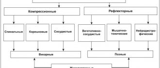
Pain during myalgia occurs both locally, in certain muscle groups, and spreads over large areas of the human body. Although there are several hundred different muscles in the human body, they can easily be divided into two groups based on the principle of their functioning.
The main task of skeletal muscles is to ensure human body movements. They also serve to communicate between the bone structures of the body. It would seem that it is in the skeletal muscles (due to their functional characteristics) that pain should most often occur.
In fact, smooth muscles are often susceptible to unpleasant sensations. This is especially true for people who lead a physically inactive lifestyle.
It is important to note! The walls of the hollow internal organs of the human body are the places in which the muscles of this group are located. The purpose of smooth muscles is to ensure the normal functioning of human organs and entire systems. They are found in blood vessels, heart, bladder, stomach and other organs.
Depending on which muscle the “failure” occurs in, the body signals through pain about a problem in a certain area of the body. Naturally, disruption of the normal functioning of the smooth muscle located in the stomach provokes discomfort in the abdominal area.
phlebolog.pro
Frequency and diagnostic criteria for restless legs syndrome (RLS)
The frequency of RLS, according to various sources, ranges from 2 to 10% among the adult population.
Diagnostic criteria for restless legs syndrome were proposed by Ekbom in the 60s, and a clear formulation of RLS as an independent disease was given in 1979, the last revision of the criteria was carried out in 2012. The diagnosis is established based on the patient's complaint, which must meet all 5 main criteria.
| Diagnostic criteria for restless legs syndrome (IRLSSG, 2012) |
| 1. An irresistible desire to move the legs, usually accompanied or caused by unpleasant sensations in the lower extremities. Sometimes the urge to move is not accompanied by unpleasant sensations; in addition to the legs, there may be a desire to move the arms or other parts of the body |
| 2. The desire to move the legs occurs or increases significantly in a resting position, sitting or lying down |
| 3. The urge to move and discomfort disappears during physical activity or stretching, and does not reappear while activity continues. |
| 4. The urge to move and discomfort increases in the evening or at night |
| 5. Symptoms should not be due to other medical conditions, seizures, or foot tapping. |
Additional criteria:
|
If your complaints do not meet the criteria for Restless Legs Syndrome, then you have a completely different disease and you should continue the diagnostic search. How to determine the cause of leg pain with a high degree of probability based on a complaint, read here>>
Differential diagnosis
RLS should be distinguished from other conditions with similar manifestations:
- night cramps
- positional discomfort of the lower extremities
- venous insufficiency
- arterial insufficiency
- akatasia
- polyneuropathy
- radiculopathy
- Parkinson's disease
Night cramps
more often they occur on one side, in the area of the foot or lower leg; they also have a circadian rhythm and appear at rest. In this case, a short-term muscle spasm occurs, which, unlike RLS, quickly disappears if you pull the sock toward you or do stretching.
Positional discomfort of the legs
characterized by localized pain in a specific small part of the leg or foot. The pain occurs after prolonged sitting and does not follow a circadian rhythm.
RLS differs from both of these conditions in that larger areas of the lower extremities are involved, the symptoms are persistent and long-lasting, and relief is provided only by walking or other activity.
For venous diseases (chronic venous insufficiency)
patients complain of discomfort in a standing position, which goes away when walking or lying with their legs raised up. Complaints may intensify in the evening or in the afternoon, while patients feel better in the morning. Taking dopaminergic drugs does not improve the condition of venous insufficiency.
Changes in the skin of the legs may be of vascular or neurotrophic origin. Trophic skin disorders are not typical for RLS.
For arterial lesions (atherosclerosis, endarteritis)
the pain appears when walking a certain distance, for example 200-300 meters, and quickly goes away at rest. This is called intermittent claudication. Unlike RLS, patients feel better at rest and have no desire to move.
Akathisia
is a condition that is most often a side effect of antipsychotic medications. Characterized by internal motor restlessness, the need to move, change position. It is difficult for the patient to sit still and remain in one position. However, there is no circadian rhythm, RLS mainly occurs at night and disrupts sleep. If the patient is taking antipsychotics, akathisia should be suspected first.
Polyneuropathy
manifests itself as a painful burning sensation in the arms or legs. Paresthesia (tingling, goosebumps), increased skin sensitivity, and painful touch are also characteristic. Neuropathy is not characterized by circadian rhythms and motor restlessness; movement does not bring relief. Dopaminergic treatment does not help.
Lumbosacral radiculopathy
differs from RLS by low back pain and asymmetric sensory paresthesia. It is necessary to keep in mind that radiculopathy can be combined with RLS.
Parkinson's disease (PD)
RLS may occur. Both conditions improve with dopaminergic drugs, although their pathogenesis is different. Parkinson's disease can mimic the symptoms of RLS, presenting with akatasia and restless legs. Restless legs is diagnosed in patients with Parkinson's disease who feel the urge to move their legs but do not meet the basic criteria for RLS.
RLS can be quite painful for the patient. However, this disease is treatable. It is recommended to base treatment on the severity of the disease and the presence of comorbidities.
According to the characteristics of the symptoms, three forms of the disease can be distinguished - intermittent RLS (periodic courses of treatment), chronic persistent RLS (daily therapy), refractory RLS (ineffectiveness of dopamine agonists / intolerance or side effects / enhancement effect in response to treatment).
Iron supplements
There is now convincing evidence that iron deficiency in the brain plays a role in the development of RLS. Treatment with iron supplements in patients with anemia resulted in significant improvement in several studies.
It is known that restless legs syndrome is detected in every fourth patient with iron deficiency anemia; the likelihood of developing RLS with anemia is 9 times higher compared to the general population.
It is recommended to examine serum iron levels
in all patients with RLS. When serum iron levels are below 75 ng/L, replacement therapy leads to significant improvement in most cases.
Conventional treatment begins with 325 mg of ferrous sulfate taken orally daily along with 100-200 mg of vitamin C to increase iron absorption.
Intravenous iron supplementation is known to rapidly replenish iron stores and require fewer administrations than oral therapy, but is less convenient. Intravenous administration of iron supplements is carried out when oral therapy is ineffective, intestinal absorption is impaired, and blood loss continues.
Non-drug therapy
Non-drug therapy is prescribed in all cases, but there is not much reliable data on effectiveness. Typically recommended:
- abstaining from alcohol and caffeinated drinks
- proper sleep hygiene
- pneumatic compression devices
- compression jersey (knee socks)
- gymnastics complex
- light aerobic exercise
Previously proposed vibration devices or massagers, when carefully evaluated, did not demonstrate benefits and their effect has not been proven.
Intermittent form (periodically occurring symptoms)
Symptoms occur no more than twice a week, but cause obvious discomfort. Non-drug treatment is started, and if there is no effect, medications are added.
Drug therapy includes carbidopa/levodopa, dopamine agonists, low-potency opioid analgesics (codeine or tramadol), or sedative hypnotics (temazepam or zolpidem).
When prescribing carbidopa/levodopa, one must remember the possibility of rebound syndrome, as well as amplification or “augmentation” syndrome. This complication of drug therapy is an increase in the manifestations of RLS and is characterized by an increase in the intensity of symptoms, their earlier appearance (during the daytime), a decrease in the asymptomatic period at rest, and the involvement of other parts of the body - arms, torso, face. Recent studies suggest that augmentation occurs more frequently with levodopa compared with dopamine agonists. The amplification phenomenon can cause worsening treatment outcomes, switching to the use of a drug of another class, or discontinuation of treatment.
Chronic persistent form of RLS
This form of RLS is characterized by a moderate to severe course, symptoms appear at least 2 times a week and are persistent. Before prescribing pharmaceutical therapy, it is necessary to explain to the patient that the goal of treatment is not to completely eliminate symptoms, but to reduce their intensity to a level where their quality of life is not affected.
Primary therapy usually includes either dopamine agonists or anticonvulsants (β2 calcium channel ligands) - Gabapentin, Pregabalin.
Recently, treatment is often started with anticonvulsants due to the difficulties that arise when taking dopamine agonists. Clinical studies have shown a decrease in the effectiveness of these drugs with long-term treatment, and some patients develop augmentation syndrome (increased complaints).
Other pharmacotherapy options include low- and intermediate-potency opioids.
Calcium channel ligands ?2? should be preferred in cases where the patient suffers from chronic pain, anxiety, insomnia, or impulse control disorders ICD (characterized by the inability to resist a strong impulse or urge to perform an act that is satisfying in the short term, despite long-term harm to both the subject and others people).
Conversely, dopamine agonists are prescribed when the patient has severe symptoms of depression, obesity/metabolic syndrome, risk of falls, or cognitive impairment, as these conditions may be aggravated by calcium channel 2 ligands.
table 2
Recommended doses of ligands ?2? calcium channels
| A drug | Initial dose | Effective dose |
| Gabapentin enacarbil | 600 mg | 600 – 1200 mg |
| Pregabalin | 75 mg | 150 – 450 mg |
| Gabapentin | 300 mg | 900 – 1200 mg |
Gabapentin is best prescribed when the patient's symptoms of RLS are accompanied by pain or there is polyneuropathy. Since gabapentin is known to cause peripheral edema, dizziness, and drowsiness, it is recommended to take 300 mg of the drug two to three hours before bedtime. Start with a low dose and increase weekly until symptoms improve or a maximum dose of 900–2000 mg is reached. Caution should be exercised when prescribing gabapentin to patients with renal failure.
Pregabalin is known to increase the risk of suicidal ideation, dizziness, fatigue, and weight gain. Despite these risks, pregabalin has been clinically demonstrated to improve treatment outcomes and has a significantly lower rate of augmentation syndrome compared with dopamine agonists.
Gabapentin enacarbil is a slow-release prodrug of gabapentin. Numerous RCTs have demonstrated its clinical effectiveness in reducing RLS symptoms. There are currently insufficient long-term studies examining its effectiveness or side effects beyond a 12-week period. Common side effects include dizziness and drowsiness, which appeared to be dose dependent.
Dopaminergic medications are among the most studied and widely used treatments for RLS. A meta-analysis of 29 RCTs using dopamine agonists found a greater than 50% reduction in symptoms (as measured by the International Anxiety Syndrome Scale) compared with placebo (61% versus 41%).
The FDA has approved three dopamine agonist drugs for the treatment of RLS: ropinirole, pramipexole, and rotigotine. It is generally recommended to start therapy at the lowest dose (Table 3) and instruct the patient to take the medication two hours before the onset of RLS symptoms to allow the medication time to work. For relief, the dose can be carefully increased by 1 mg at the end of the week, but never exceed the recommended dose. Common side effects include nausea, headache, dizziness and vomiting, which usually resolve within two weeks.
Table 3
Recommended dosages of dopamine agonists
| A drug | Initial dose | Maximum dose |
| Pramipexole | 0.125 mg/day | 0.75 mg/day |
| Ropinirole | 0.25 mg/day | 4 mg/day |
| Rotigotine | 1 mg/day | 3 mg/day |
Patients with RLS receiving dopamine agonists are at increased risk of developing augmented or impulse control disorders (ICD), such as pathological gambling, hypersexuality, compulsive buying, and binge eating disorder.
Initially, drug-induced augmentation was mainly associated with the use of levodopa. However, this phenomenon has also been reported in long-term trials of dopamine agonists. In fact, within a year of short-term use of dopamine agonists, augmentation rates ranged from 7-8%. Several retrospective studies have reported that the incidence of augmentation increases to 50% after 10 years of drug use. Since augmentation increases over time, and RLS is a chronic disease requiring ongoing treatment, β2 ligands are increasingly preferred. calcium channels compared to dopamine agonists.
Factors that increase the risk of augmentation:
- low serum ferritin levels
- use of certain medications that worsen RLS (such as recent cessation of opioids or antidepressants)
In case of severe augmentation symptoms, three options are offered. One is to switch treatment entirely to a long-acting dopamine agonist. The second option involves adding the ligand ?2? calcium channels and a gradual decrease in the original dopamine agonist. The goal is to withdraw dopaminergic drugs and replace them with β2 ligands. calcium channels, so patients should be warned that this approach may temporarily worsen their symptoms. If the attempt to withdraw dopamine fails, then the addition of a low dose dopamine agonist with a ?2 ligand can be continued. calcium channel. The third and final option involves tapering off the dopamine agonist followed by a 10-day washout period. This approach can lead to extremely severe symptoms of RLS, which may require low doses of dopamine agonists for relief.
Refractory RLS
When monotherapy fails due to tolerance, enhancement, or side effects, potential factors influencing therapy, such as the use of drugs that increase RLS, changes in sleep habits, or other sleep-disrupting pathologies, must first be considered. A common protocol involves combining primary therapy with another class of drugs (eg, calcium channel ligands for patients taking dopamine agonists, or vice versa).
Because both drugs provide therapeutic effects through different pharmacological mechanisms of action, combination therapy may provide better symptom control. In addition, patients' ferritin levels can also be retested and treated with iron supplements if deficiency is detected.
Long-term therapy
To date, most evidence-based treatment guidelines have been developed from studies that lasted no more than four months. This is a major limitation because RLS is a lifelong condition and the potential side effects of long-term drug therapy have been little studied. The effectiveness of the drug levodopa can last up to two years in 40% of patients, while the effectiveness of dopamine agonists such as pramipexole and rotigotine lasts for one and five years, respectively.
As for the ligands ?2? calcium channel blockers, pregabalin and gabapentin enacarbil have proven effective for treatment for one year. For other drugs there is not enough evidence to conclude their long-term effectiveness, such as gabapentin and many opioid drugs.
Due to the lack of evidence, it is clear that further research is needed to explore long-term pharmaceutical therapy for RLS. Some drugs, such as pergolide and cabergoline, have been completely discontinued in the treatment of RLS due to reports of fibrosis of the heart valves.
conclusions
Although significant advances have been made in the etiology, diagnosis, and treatment of RLS over the past decade, patients continually suffer from misdiagnosis and numerous unnecessary referrals. With improved diagnostic criteria, treatment strategies, and updated guidelines, primary care physicians have more resources than ever to diagnose and effectively treat RLS. Further study of the outcome of long-term drug therapy is clearly needed.
Literature:
- Differential Diagnosis and Treatment of Restless Legs Syndrome: A Literature Review Monitoring Editor: Alexander Muacevic and John R Adler Vishal Kwatra, corresponding author Muhammad Adnan Khan, Syed A Quadri, and Trevor S Cook
Muscle response
It is common for muscles to respond to a sequence of commands sent by the nervous system or brain. A reaction (reflex) to certain stimuli is also likely. When stimulated, that is, the appearance of a command or exposure to a stimulus, contraction (tension) of the muscle occurs.
After the stimulation stops, the reverse process begins, returning the muscle to its normal (relaxed) state. Such processes (tension-relaxation) can occur an infinite number of times every day.
During normal functioning of the body, cycles of muscle tension and relaxation remain practically unnoticeable to humans, much less cause pain. At most, you feel tired. Problems (feelings of pain) appear when the normal functioning of certain organs and (or) systems is disrupted.
Clinical case 2
Figure 3.
Clinical case 2. Patient T., 60 years old, with trochanteritis on the left. A - marking of anatomical landmarks and needle insertion points: 1 - projection of the iliac wing; 2 — projection of the ischial tuberosity; 3 - greater trochanter of the left femur; B — Performing a blockade of the trochanteric bursa under ultrasound control.
Patient T., 60 years old, complained of pain in the lower back with an intensity of up to 5 points according to VAS. The pain radiates along the posterolateral surface of the left thigh during physical activity; pain increases in the position of lying on the left side, as well as with physical activity up to 7 points. The patient has been experiencing pain for four months; the onset of pain is not associated with any traumatic factor. Notes the positive effect of taking NSAIDs. Concomitant diseases: arterial hypertension stage I, average risk, achieved normotension; diabetes mellitus type 2, compensated (target HbA1c level 7.5%).
When assessing the neurological status, no symptoms of damage to the central and peripheral nervous system were revealed. Lasègue's symptom is negative. When assessing the vertebral neurological status: tension and pain on palpation of the paravertebral muscles, the Schober test is negative, the phenomenon of advancing the left SIJ in the Piedal test, the Patrick test is positive, provoking pain in the area of the left SIJ, the Bonnet-Bobrovnikova test is weakly positive on the left. Palpation of the projection of the greater trochanter of the left femur is sharply painful (Fig. 3 A). A survey radiography of the pelvic bones and hip joints was performed, which revealed signs of stage I–II coxarthrosis on both sides. Based on the data obtained, a clinical diagnosis was formulated: “chronic lumbar ischialgia of musculoskeletal origin due to dysfunction of the left SIJ, left piriformis syndrome, left-sided trochanteritis.”
Therapy was prescribed with Ketoprofen 100 mg, one tablet twice a day, recommendations were given on maintaining habitual physical activity, limiting stress that provokes pain, and a complex of exercise therapy. At a follow-up appointment 10 days later, the patient notes a decrease in the severity of pain in the lower back to 2 points, confidently copes with a complex of therapeutic exercises, but the intensity of pain during exercise remains the same. Ultrasound of the left hip joint and periarticular tissues confirmed trochanteritis (bursitis of the trochanteric bursa of the gluteus medius muscle). Thus, in the genesis of nociplastic MPS in this patient, the leading ones are maladaptive myoplastic and chondroplastic mechanisms.
Considering the persistence of pain that significantly reduces the quality of life during exercise and the ineffectiveness of taking NSAIDs for more than two weeks, the patient underwent a therapeutic and diagnostic blockade under ultrasound control: 1.0 ml of 0.4% Dexamethasone and 2.0 ml of 0.5% Procaine in the area of the large trochanter of the femur (Fig. 3 B).
After the manipulation, 24-hour blood pressure monitoring was performed in the clinic. 30 minutes after the injection, the patient complained of a moderate headache and general weakness. A rise in blood pressure to the level of 175/90 mm Hg was noted. Art., treated with medication, increased glycemia to a level of 9 mmol/l. Taking into account risk factors, after stabilization of the condition, recommendations are given for monitoring glycemic levels (measurement of capillary blood glucose four times during the day) and blood pressure and their correction if necessary.
Figure 4.
Clinical case 2. Ultrasound navigation of the trochanteric bursa block of the gluteus medius muscle. A - ultrasound visualization of the manipulation. B — marking of landmarks during ultrasound navigation: 1 — shadow of the needle stroke; 2 — shadow of the needle tip and projection of the enthesis zone; 3 — distribution of the drug in the area of enthesis; 4 - periosteum of the femur; 5 - iliotibial tract.
When examined the next day, the patient noted a 50% decrease in the severity of pain when walking, however, fluctuations in blood pressure and glycemic levels during the day precluded consideration of the issue of continuing GCS therapy. When choosing a drug to continue local interventions, we took into account the lack of effect of the drugs Chondroreparant® Gialripayer® on the level of blood pressure and glycemia, and together with the patient a decision was made to continue injection therapy. Under ultrasound control, 2.0 ml of 0.8% Chondroreparant® Gialripayer®-10 was injected into the area of identified inflammatory changes; the procedure was well tolerated (Fig. 4 A, B). Then kinesiotherapy and oral NSAIDs were continued.
At the next visit seven days later, the patient reported regression of pain in the lumbar spine and a decrease in the severity of pain when walking to 3–4 points. During this period, she independently stopped taking NSAIDs. Upon examination, local pain is detected in the projection of the greater trochanter of the femur on the left, and therefore a decision was made to continue local injection therapy with Chondroreparant® Gialripayer®-10 at the same dose under ultrasound control; the blockade was tolerable without any problems. At the next visit seven days later, the patient noted complete regression of pain, but mild local pain persisted when palpating the projection of the greater trochanter on the left. Ultrasound revealed a small amount of free fluid in the trochanteric bursa on the left. To stimulate reparative processes, 2.0 ml of 0.8% Chondroreparant® Gialripayer®-02 was introduced. At subsequent visits, the patient had no complaints, pain on palpation regressed, and range of motion was restored.
Provoking diseases
Myalgia can be triggered by a variety of diseases (including viral and infectious). It may also be a consequence of a particular injury. There are not isolated cases when muscle pain begins to worry a person after taking certain medications.
The difficulty is that myalgia in certain cases is not limited to the area of a specific muscle. Pain may well have a negative impact on fascia, tendons, ligaments, and soft tissues, which are the “connecting links” between muscles, internal organs, and bones.
People do not always objectively perceive pain in the muscles, hoping that they will go away over time. Patients pay much less attention to myalgia compared to headaches or discomfort in the heart area. In fact, muscle pain can often indicate symptoms of dangerous diseases. If you do not seek medical help in a timely manner, serious consequences cannot be ruled out.
It is much safer to consult a doctor as soon as the first signs of muscle pain appear.
Treatment
Treatment for muscle pain depends on the cause of the symptom. Therefore, the most important factor determining treatment tactics is making an accurate diagnosis. For example, if muscle pain is caused by taking certain medications, then in such cases it may be enough to stop taking these medications or replace them with other medications. Drug treatment for muscle pain may include NSAIDs or analgesics, and even opiates.
Acute muscle pain
For acute muscle pain that occurs after an injury, it is necessary to provide rest and unloading, and in some cases, immobilization. In addition, local cooling with ice wrapped in a towel has a good effect in such cases, which helps reduce swelling, inflammation, and pain. In addition, it is necessary to stop the exercise that led to muscle pain. Treatment of muscle injuries requires quite a lot of time, since early restoration of normal loads can lead to chronic pain syndrome and excessive scarring of muscle tissue, and in severe cases to the development of myositis ossificans.
Chronic muscle pain
Treatment for chronic pain may include heat treatments as well as other treatments such as:
- Acupuncture and acupressure
- Electrotherapy (therapy through electricity)
- Electromyostimulation
- Physiotherapy
- Exercise therapy
- Manual therapy
Systematic exercises (physical therapy) are especially relevant when the cause of chronic pain is degenerative diseases of the spine, such as osteochondrosis, spondylosis, and disc herniation.
Surgical treatment methods are used for severe traumatic muscle injuries or when there is compression of the nerve roots.
Prevention of muscle pain consists of the following rules: maintaining a healthy lifestyle, sufficient physical activity, a balanced diet, proper ergonomics of the workplace, avoiding alcohol abuse and smoking.
Typical symptoms
Muscle pain often occurs along with other symptoms that are characteristic of certain problems in the body. An illustrative example is the consequences of a mechanical injury. In areas subjected to blows, abrasions, swelling, and local changes in the color of the skin surface (bruises) occur. Often such injuries are accompanied by pain in the muscles.
With myalgia, numerous additional symptoms are likely to occur. Among the most common:
- tendency to depression;
- loose stools;
- bouts of vomiting;
- muscle numbness;
- a sharp decrease in body weight;
- poor appetite;
- the appearance of swelling;
- insomnia;
- burning and tingling sensations in the muscles;
- difficulties while walking;
- convulsive muscle contraction;
- low concentration.
Symptoms typical of acute respiratory diseases may also occur. A person suffers from headaches. He cannot get rid of the feeling of fatigue. The feeling of chills adds to the problem. Fever occurs periodically. Pain in the throat is likely. A cough is also possible.
Bibliography
- Global Burden of Disease Study 2013 Collaborators. // Lancet. 2015 Aug 22; 386(9995):743–800.
- Hoy D, Brooks P, Blyth F, Buchbinder R. The Epidemiology of low back pain. // Best Pract Res Clin Rheumatol. 2010 Dec; 24 (6): 769–81.
- Pergolizzi J, et al. The chronic pain conundrum: should we CHANGE from relying on past history to assessing prognostic factors? //Curr. Med. Res. Opin. Feb 2012; 28 (2): 249–56.
- Alekseev V.V., Barinov A.N., Kukushkin M.L., Podchufarova E.V., Strokov I.A., Yakhno N.N. Pain: a guide for doctors and students, ed. N.N. Yakhno, Moscow "MedPress". - 2009. - 302 P.
- Nasonov E.L., Yakhno N.N., Karateev A.E. et al. General principles of treatment of musculoskeletal pain: interdisciplinary consensus. // Scientific and practical rheumatology 2021, No. 3 (54); pp. 247–265.
- Treede RD. The International Association for the Study of Pain definition of pain: as valid in 2021 as in 1979, but in need of regularly updated footnotes. // Pain Rep. 2021 Mar 5; 3(2):e643.
- Epifanov V. A., Epifanov A. V., Barinov A. N. Back pain. // MEDpress-inform - 2021. - 130 pp.
- Chou R, Deyo R, Friedly J., et al. Systemic Pharmacologic Therapies for Low Back Pain: A Systematic Review for an American College of Physicians Clinical Practice Guideline. // Ann Intern Med. 2021, 4; 166(7):480–492.
- Qaseem A, Wilt TJ, McLean RL, et al. Noninvasive Treatments for Acute, Subacute, and Chronic Low Back Pain: A Clinical Practice Guideline From the American College of Physicians. // Ann Intern Med. 2017; 166(7):514–530.
- Rozhkov D.O., Zinovieva O.E., Barinov A.N. et al. The state of skeletal muscles in chronic nonspecific pain in the lower back and approaches to therapy // Eff Pharm Neuro 2021, No. 11, pp. 24–35.
- Barinov A.N., Makhinov K.A., Rozhkov D.O. Some aspects of diagnosis and treatment of nonspecific back pain // Medical Council 2021, No. 10, pp. 122–130.
- Aydede M, Shriver A. Recently introduced definition of 'nociplastic pain' by the International Association for the Study of Pain needs better formulation. // Pain. 2021 Jun; 159(6):1176–1177.
When is it necessary to see a doctor?
Myalgia in certain situations may be one of the signals that serious (dangerous) processes are occurring in the body. Together with other symptoms, muscle pain may indicate the threat of meningitis. It is also not uncommon for pain to warn of the risk of a heart attack. They also occur before a heart attack.
In such cases, it is imperative to immediately consult a doctor. Below are characteristic symptoms, when they occur it is dangerous to hope that the pain will “go away on its own”:
- difficulty breathing, shortness of breath appears;
- sudden memory problems, frequent forgetting;
- seizures accompanied by convulsions;
- numbness of individual parts of the body;
- a sharp deterioration in vision, up to its loss;
- problems (rigidity) in the muscles of the back of the head, accompanied by elevated temperature;
- a constantly increasing feeling of weakness;
- immobilization of limbs;
- mental problems;
- disruption of normal perception of reality;
- inappropriate actions;
- frequent fainting without an obvious reason;
- pain in the chest, “radiating” to the jaw, neck or arms.
Such symptoms require immediate action. Delay (untimely medical care) can lead to irreparable consequences.
Causes of muscle pain
Pain in the skeletal muscle group is often a consequence of previous injuries. We are talking not only about direct mechanical effects on the human body that provoke corresponding consequences. Injuries and subsequent pain can occur if a muscle is torn. A similar situation occurs when they are stretched. The difference between both types of injury is the number of damaged muscle fibers. Tendon tears and ruptures also cause pain.
In both cases, the body is able to heal itself. Tendons and muscles are characterized by regeneration. However, severe tears require medical intervention. With such damage, the body's potential is insufficient. To return the structures to normal functioning, it is necessary to resort to prompt restoration.
Disturbances in the normal rhythm of the nervous system provoke the appearance of seizures. They can also occur as a result of excessive prolonged stress on the body. The result is excessive muscle contraction. The consequence is pain.
New perspective on musculoskeletal pain
MPS is classified according to its duration: if the duration of pain does not exceed 6 weeks, it is considered acute; if the pain lasts from 6 to 12 weeks, it is considered subacute; if it lasts for more than 12 weeks, it is considered chronic. It is assumed that the basis of acute pain syndrome is microtraumatization of the structures of the musculoskeletal system. A frequent factor in the development of acute or exacerbation of chronic pain is immediate excessive load against the background of prolonged static load, especially in a non-physiological position.
Prolonged static load itself contributes to overload and overstretching of the muscle, ligament or capsule of the facet joint. In this case, we can talk about the activation of pain receptors (nociceptors) in connection with tissue damage, that is, nociceptive pain. The average time for recovery and relief of nociceptive pain in this case does not exceed 2–3 weeks. However, in a number of patients, the duration of the pain syndrome extends beyond the period of physiological tissue restoration, and not only adjacent anatomical regions, but also distant anatomical structures begin to be involved in the pathological process of sensitization. To pathogenetically designate this process, the International Association for the Study of Pain (IASP) introduced the term “nociplastic pain” [6].
By definition, nociplastic pain occurs due to altered nociception in the absence of clear evidence of existing or potential tissue damage causing activation of peripheral nociceptors, as well as evidence of disease or damage to the somatosensory nervous system that could cause this pain. Altered nociception refers to false-adaptive (or maladaptive) plastic processes in the nervous (neuroplasticity), muscle (myoplasticity), connective tissue (chondroplasticity), bone (osteoplasticity) and other systems. The contribution of each of the above components in the formation of nociplastic pain is variable in different patients, which determines the response to the therapy.
Treatment of nociplastic MPS targeting the maladaptive neuroplastic component may include:
- muscle relaxants (channel modulators);
- antidepressants (stimulants of the activity of the antinociceptive system);
- brief psychological interventions (explanatory psychotherapy, cognitive behavioral psychotherapy [CBT], hypnotherapy, biofeedback, etc.).
To influence maladaptive myoplastic processes in MBS, the following should be used:
- kinesiotherapy;
- local injection therapy.
The basic therapy for maladaptive chondroplastic processes is considered to be:
- SYSADOA assignment;
- local injection therapy with hyaluronic acid preparations;
- kinesiotherapy.
The most difficult task is the correction of maladaptive osteoplastic processes due to their irreversibility. The range of therapeutic effects in this case varies in the following range:
- prescription of bisphosphonates;
- orthotics;
- surgical intervention [7].
The basis for successful personalized therapy for patients with nociplastic MPS is adequate diagnosis with identification of the predominant mechanisms of pain chronicity. The basis for diagnosing both acute and chronic pain is a clinical examination of the patient with a thorough history taking (to exclude danger symptoms - “red flags”), assessment of the neurological and vertebroneurological status. If the patient does not have any “red flags,” additional neuroimaging is not required.
Muscle pain caused by injuries
Almost any type of injury can cause pain in the muscles. In particular:
- stretching of muscle fibers;
- excessively repetitive movements;
- muscle fiber ruptures;
- excessive loads;
- blows with blunt objects.
In addition, pain occurs when the nerve is compressed. The phenomenon occurs when a person develops spinal stenosis. Compression can also occur due to a herniated disc.
Neuromuscular diseases and conditions that cause pain
Pain invariably accompanies certain diseases. The neuromuscular type includes:
- spinal cord damage;
- Charcot's disease, also called amyotrophic lateral sclerosis;
- stroke;
- brain damage;
- muscle infections (abscesses);
- dermatomyositis, accompanied by a rash on the skin and inflammatory processes in the muscles;
- Lyme disease caused by ticks;
- polymyositis, leading to muscle weakening and inflammation;
- Parkinson's disease, increasingly diagnosed in older people, affects the brain, accompanied by impaired coordination of movements;
- polymyalgia rheumatica, in which stiffness and pronounced muscle pain are observed;
- multiple sclerosis, also commonly seen in older citizens;
- muscle destruction, in particular rhabdomyolysis.
The list is impressive, and contains neuromuscular diseases that are a real problem for this century. Disappointing statistics indicate that the above diseases are increasingly being diagnosed by doctors. The number of sick citizens, of all ages, is constantly increasing.
Muscle atrophy in old age
Aging is a natural biological process, one of the components of the life cycle (birth, growth and development, flowering and decline of life). This inexorable law has existed for many millions of years and all living beings on Earth obey it - from the amoeba invisible to the naked eye to the “crown of creation” of nature - Homo sapiens. Currently, humanity has sufficient “baggage” of knowledge to refute the statement that old age is not a joy.
The weight of skeletal muscles in a physically strong person accounts for almost half of the total body weight (slightly more in men than in women). When you reach a certain age, muscles begin to decrease in volume. Scientists have calculated that after thirty years a person “loses” about one percent of muscle mass every year (and therefore becomes slightly weaker). This means that an elderly person over 70 years old is one and a half times weaker than a young person. The process of decreasing muscle mass and strength is called muscle atrophy in old age and is considered (as well as the appearance of the first facial wrinkles) the beginning of aging of the body.
Muscle atrophy in old age can lead to unpleasant consequences. For example, body weight increases. This occurs due to a decrease in muscle mass, because it is the muscles that most actively and efficiently burn excess calories coming from food.
Weight gain is the lesser of the evils that threaten muscle atrophy in old age. Because of it, the muscular frame, which serves as a reliable support for the spine, cannot cope with its “direct responsibilities” and the elderly person slouches. In addition, irritation and pinching of the spinal cord nerves may occur, accompanied by severe pain and disruption of the functioning of various organs.
Since muscle strength determines the quality of life of a person of any age, muscle atrophy dramatically complicates and worsens it. And this can continue for a long time - up to a complete refusal to move. At first, the older person begins to walk more slowly than usual. Then he leaves the apartment less and less often. Due to further muscle atrophy, it becomes increasingly difficult for him to maintain balance, he is afraid of falling and finally stops moving without anyone’s help. This behavior provokes the development of new, more dangerous diseases.
- Recommended articles to read:
- Social services for older people
- Diseases of old age
- Valuable tips on how to choose a boarding house
With a sedentary lifestyle, there is a risk of blood clots and blockage of the blood vessels of the heart and brain. The result is a heart attack or stroke—deadly diseases. There may also be congestion in the veins (most often in the legs). Their appearance is explained by the fact that the muscles “massage” the veins, helping venous blood, contrary to gravity, to move upward. It is for this reason that varicose veins and other vein diseases threaten people who are physically inactive. Physical inactivity also negatively affects the condition of the cardiovascular system, skin and musculoskeletal system.
It turns out to be a vicious circle - due to a decrease in physical activity, proteins break down, muscle contractility decreases and increases atrophy, and so on. Therefore, an elderly person must take immediate action to prevent muscle weakness from taking over. The question arises: what can he do, since age is making itself known and the process of muscle atrophy in old age is irreversible and inevitable?
If an elderly person unshakably believes that muscle atrophy as a sign of aging is irreversible and invincible, then, of course, “nothing can be done,” “sit and don’t rock the boat.” However, there are often known facts when people, regardless of age, continued to lead an active lifestyle and often surprised those around them with their liveliness of mind and incredible muscle strength. For example, masters of Chinese martial arts maintain excellent physical shape into old age and never get sick. The oldest bodybuilder Manohara Aicha (he is almost 100 years old) with his biceps causes the envy of thirty-year-old athletes.
To maintain muscle strength, you don't have to be a bodybuilding or martial arts "star". “Ordinary” physical activity (ideally recommended by a doctor) is enough, which will preserve and increase strength, tone and endurance. An active lifestyle, physical mobility and the desire for healthy longevity are the best prevention of muscle atrophy in old age.
Read the material on the topic: Seizures in the elderly
Other possible causes of muscle pain
Muscle pain can also occur as a result of a number of other diseases and special conditions of the body. At the same time, they pose no less a threat to human health, and in some cases can lead to fatal consequences. In particular, pain occurs when:
- pregnancy (at different times);
- acute lack of certain vitamins in the body (D, B12);
- frequent depression;
- fibromyalgia;
- red (systemic) lupus;
- renal failure;
- hypothyroidism;
- electrolytic disorders in the body, manifested by an imbalance of calcium or potassium in the blood;
- oncology.
Even with the flu and many other respiratory diseases, muscle pain is likely to occur. Another reason to think about the need for prevention as periods of infection approach.
Nutritional elements that help maintain muscle in old age
Old age involves a gradual loss of muscle mass (scientific name is sarcopenia), and only three key nutritional elements can avoid this biological process.
According to scientists, the most effective means of stopping sarcopenia is physical activity, which maintains muscle tone and improves the condition of the body's cardiovascular system.
And you simply cannot do without proper nutrition to solve this problem.
Scientists consider eating enough protein to be the first factor that helps maintain muscle in old age at the proper level.
This is a basic element, since protein is not only a “building” material that forms the muscles of the body, but also a substance that prevents the loss of muscle mass.
An elderly person should receive at least 1–1.2 grams of protein per kilogram of body weight from food every day. This consumption rate is calculated for people who do not suffer from metabolic disorders and kidney dysfunction.
The second element that helps maintain muscle strength in old age is the use of vitamin D.
According to scientific research, now in developed countries, approximately four out of five people who have reached old age have little vitamin D in their bodies. Therefore, doctors recommend taking vitamin supplements. This is all the more important because it is this vitamin that helps maintain a person’s mental abilities.
Scientists consider the third element to be the avoidance of destructive acids and inflammatory agents.
According to doctors, an elderly person should almost completely stop eating red meat and grains. He should get protein from dietary meat, and with plenty of vegetables (to improve metabolism).
Scientists say that older people who follow these tips lose muscle mass at the lowest rate.
Read material on the topic: Sanatorium for retirees
Test to find out the causes of muscle pain
You can determine the cause yourself, without going to doctors and without being a doctor by profession. To do this, it is enough to answer a few simple but extremely important questions:
1. Is the pain felt locally (in certain places) or throughout the body?
2. During what period (how long) do you experience pain?
3. What helps reduce pain or provokes its intensification?
4. Where exactly is the pain localized?
5. What medications are you currently taking or have you used until recently?
6. Are there other symptoms, attacks of fever, pain in the throat?
This kind of test determines to some extent the extent of the problem. However, it is unable to fully replace qualified medical advice.
Complications that arise as a consequence of muscle pain directly depend on the nature of the disease.
It is important to note! Modern medicine has an impressive arsenal of tools for effectively treating skeletal muscles, especially treating the spinal muscles.
Clinical case 1
Patient K., 56 years old, complained of pain in the lower back with an intensity of up to 6–7 VAS points, aggravated in an upright position and with static load (about 10 minutes). The patient has been experiencing pain for three years with periodic exacerbations (1–2 episodes per month), the average pain level is about 3 points, with exacerbations up to 6–7 points. A real exacerbation occurs within 10 days, when after a long stay on an uncomfortable chair and a sharp bend, the pain intensifies to 6-7 points at rest and to 7-8 points when moving. The pain is relieved by lying down and resting. On examination, he notices stiffness in the lumbar spine in the morning after sleep, regressing within 5–7 minutes. On the advice of relatives, she underwent an MRI of the lumbosacral spine, which revealed degenerative-dystrophic changes in the spine and a paramedian disc herniation LV-SI.
At the initial appointment, after collecting complaints and anamnesis, the neurological status was assessed; no symptoms of damage to the central and peripheral nervous system were identified. Lasègue's symptom is negative. When assessing the vertebroneurological status: left-sided thoracolumbar scoliosis, tension and pain on palpation of the paravertebral muscles, a sharply painful cord with a pain pattern recognizable to the patient is identified in the m erector spinae on the left. The Schober test is negative, but extension and bending to the right are limited due to pain, with elements of kinesiophobia, the phenomenon of advancing the left SIJ in the Piedal test, the Patrick test of the left SIJ is positive, the Bonnet-Bobrovnikova test is negative. With the patient lying on his stomach, sharp pain upon palpation in the projection of the left facet joint LV–SI, left SIJ. Based on the data obtained, a clinical diagnosis was formulated: “chronic lumbodynia of musculoskeletal origin due to dysfunction of the left SIJ, LV-SI facet syndrome on the left, MFPS of the erector spinae muscle.” Thus, in the genesis of nociplastic MPS in a patient, the leading role of maladaptive neuroplastic (chronic pain, kinesiophobia), myoplastic (MFPS) and chondroplastic (spondyloarthropathy) components should be considered.
Therapy was prescribed with Aceclofenac 100 mg, one tablet twice a day, Tolperisone 150 mg twice a day, recommendations were given on maintaining habitual physical activity, limiting pain-provoking loads, and a complex of exercise therapy. After 10 days, the patient notes a decrease in the severity of pain at rest to 5 points according to VAS, with movement - to 5-6 points, but cannot perform a set of therapeutic exercises due to pain. Considering the severe pain syndrome, which reduces the quality of life, its chronic course, as well as the low effectiveness of outpatient treatment, the patient was hospitalized at the Clinic of Nervous Diseases of the University Clinical Hospital No. 3 of the First Moscow State Medical University named after I.M. Sechenov.
Picture 1.
Clinical case 1. Patient K., 56 years old, with lumbodynia and osteoarthritis of the SIJ. Marking of anatomical landmarks and needle insertion points: 1 - projection of the spinous processes LIII–LIV; 2 - projection of the sacrum; 3 — projection of the iliac wing; 4 — needle insertion point for blockade of the LV–SI facet joint on the left.
The patient was offered to participate in a scientific study, the purpose of which is to diagnose and optimize the treatment of patients with MFPS. Considering the absence of specific changes in the spine according to MRI, to confirm the diagnosis and verify the leading pathogenetic nociceptive trigger, 2.0 ml of 0.5% Procaine was injected into the left SIJ under X-ray control. The patient noted a 50% decrease in pain intensity within 30 minutes after the injection. To differentiate the possible placebo effect of the invasive procedure, a repeat procedure was performed: 2.0 ml of 0.75% Ropivacaine was injected into the left SIJ. The pain decreased by 50% within six hours, against the background of which the patient began to cope more successfully with the exercise therapy complex. Considering the positive effect of diagnostic blockades with local anesthetic, a therapeutic blockade of the left SIJ was performed with a long-acting GCS solution (1.0 ml of betamethasone dipropionate and betamethasone sodium phosphate) and 2.0 ml of 0.5% Procaine (Fig. 1). During therapy, pain in the lower back decreased to 3–4 points at rest and to 4 points on VAS during exercise.
Taking into account the persisting pain syndrome, to verify the MFPS, an additional ultrasound control was performed: a power Doppler mode superimposed on the B-mode to determine the silent zone of absence of echo signals in the trigger zone when vibrations propagate from a source of external vibration. Ultrasound data revealed a myofascial trigger. However, taking into account the clinical data of involvement in the pathogenesis of pain syndrome of the facet joint LV-SI, to reduce the radiation dose under ultrasound control, a double diagnostic blockade of the joint was performed: 1.0 ml of 0.5% Procaine initially and 1.0 ml of 0.75% Ropivacaine again. A 50% reduction in the severity of pain corresponded to the duration of the anesthetic effect, which significantly reduces the likelihood of a placebo effect.
Figure 2.
Clinical case 1. Ultrasound navigation of a facet joint block. A - ultrasound visualization of manipulation; B — marking of landmarks during ultrasound navigation: 1 — shadow of the needle stroke; 2 — shadow of the needle tip and projection of the periarticular zone of the facet joint; 3 - periarticular distribution of the drug; 4 - dorsal surface of the sacrum; 5 - myofascial trigger.
When planning further therapeutic interventions, we took into account previous therapy with long-acting corticosteroids, therefore, in order to reduce the potentiation of side effects, we offered the patient blockade of the facet joint area with the drug Chondroreparant® Gialripayer®-10 (Fig. 1). The choice of drug was determined by the clinical picture of the pain syndrome: intensity, signs of a nonspecific inflammatory reaction of the facet joint against the background of degenerative changes. Periarticular injection of 2.0 ml of 0.8% Chondroreparant® Gialripayer®-10 was carried out under ultrasound control (Fig. 2).
Additionally, in order to study the state of contractile proteins of the skeletal muscle in order to optimize the kinesiotherapy regimen for MFPS, taking into account the structural and functional characteristics of the skeletal muscle, after signing the informed consent, we performed a biopsy of the myofascial trigger, verified using ultrasound. When performing SDS gel electrophoresis of proteins from a mini-sample of the patient's skeletal muscle, a decrease in the content of titin and nebulin was revealed (from 20 to 40%). Interpretation of the data obtained and assessment of the clinical significance of maladaptive myoplasticity will be possible after completion of the cohort recruitment and statistical processing of the material from our study [10].
During therapy, the patient notes a decrease in the severity of pain and an increase in the range of active movements. The CBT sessions allowed her to overcome kinesiophobia, her adherence to therapeutic exercises increased, and her fear of the progression of spinal changes detected on MRI and possible disability went away. At a follow-up examination 10 days after discharge, the patient noted regression of pain at rest. With prolonged static physical activity, pain occurs only at a level of 1–2 points, the patient engages in kinesiotherapy daily.
The peculiarity of this clinical case is the combination of three pathogenetic maladaptive mechanisms (neuroplastic, myoplastic and chondroplastic) of the formation of chronic nociplastic MPS, the relief of which was achieved using an interdisciplinary approach. The treatment plan was based on kinesiotherapy, CPT, rational pain relief using oral forms of NSAIDs, local therapy with long-acting corticosteroids, local administration of Chondroreparant® Gialripayer®-10, a pathogenetically based chondroreparative and non-pharmacological anti-inflammatory drug that reduces the cumulative risks of corticosteroids.
Diagnostic methods
To determine the causes of muscle pain and prescribe the appropriate course of treatment, the doctor must take into account two factors. The doctor studies the history of the disease. He also pays attention to the symptoms of the disease. Correct identification of the causes and the optimal method of treatment are the key to successful relief from muscle pain.
In most cases, problems are caused by excessive muscle tension. It usually occurs when a person is insufficiently active (sedentary work, inactivity). It is possible that the pain is a consequence of the injury. In the first case, there is no need for radical measures (surgery). Often significant changes can be achieved with the help of a special set of physical exercises.
Muscle training in old age
For older people over sixty years of age, classical strength training is not the best training option. Speed-strength, which increases muscle power, is more suitable.
Classic strength training includes exercises that should be performed at a slow pace (for example, at intervals of 3-4 seconds), while speed-strength training involves movements at maximum speed.
Scientific research has proven that it is muscle power that allows older people to cope with daily “motor” tasks (walking, getting out of a chair and up stairs, walking in the park, etc.). Medical scientists consider muscle strength to be the main component of functionality.
Specialists from Switzerland conducted special studies in 2011 and showed that training at an accelerated pace brings older people more benefits in terms of functionality than classical strength training.
Movements at the fastest possible pace, which is dictated by age, help increase muscle power and strength. Do not confuse speed-strength training with “coaching” weightlifters. Since muscle power training is a regular workout in the gym with excellent technique, “speed” exercises with lifting small weights.
Most scientific research did not use free weights, but special machines. Exercises were used aimed at increasing the power of large muscle groups. The training was conducted 2-3 days per week at an intensity of 70 percent of the maximum weight available to the elderly subjects, typically eight to ten repetitions.
The scientific studies involved elderly people who did not have problems with the joints, bone and cardiovascular systems.
An older person who does not have outstanding physical abilities should choose a lower-intensity workout option. For example, perform exercises not for eight to ten, but for 15–20 repetitions, thereby protecting himself from possible undesirable consequences.
- An older person who is around 60 years of age or older should exercise with light weights. He will only harm himself if he limits physical activity under the pretext “I’m not so young anymore and I need to take care of my old bones and heart.”
- Speed training allows you to maintain functionality and, therefore, guarantees a high level of quality of life until old age.
- It is recommended to train muscles three days a week in 2-3 sets of 8-10 or 15-20 repetitions.
- Each elderly person has individual abilities and capabilities, so the intensity of training must be selected for a specific case strictly individually.
Prevention
Muscles, like many organs of the body, have the ability to regenerate. The body is able to recover on its own. Naturally, if a person does not interfere with him with wrong actions, he will only aggravate the situation. A healthy lifestyle is the key to not only longevity, but also great well-being.
Several rules, strictly observed throughout life, allow you to maintain excellent health until the end of your days. This is also true for muscles, which can remain in excellent tone, despite any age-related changes.
- proper diet;
- active lifestyle;
- regular exposure to fresh air;
- optimal physical activity;
- proper organization of working time and place;
- cessation of smoking and alcohol abuse.
The rules are simple and accessible to every person, regardless of gender, specific professional activity, or age. Strict implementation will allow you to forget about pain in the muscles, prevent their occurrence in advance, and fully enjoy a healthy, fulfilling life. Naturally, even if you feel excellent, you should not forget about the need for regular medical examinations.
How to train muscles in old age
Upon reaching the age of sixty, every elderly person begins to feel the approach of old age - bones, muscles, cardiovascular, endocrine and other body systems are no longer as strong as in youth. Therefore, to maintain health, the importance of “strength” training for people over 60 is no less important than for twenty-year-olds.
The consequences of not doing strength training can be:
1. Loss of muscle mass.
When reaching the age of forty or fifty, the human body begins to “lose” muscle mass. This biological process is caused by a decrease in the production of anabolic hormones and a reduction in physical activity.
From anatomy we know that there are two types of muscle fibers: the first is “slow”, the second is “fast”. The second type of fiber produces forces that are 2-4 times greater than the “ability” of the first type. Therefore, it is the “fast” fibers that are responsible for muscle power and strength. Over time, an elderly person loses mainly type 2 fibers.
2. Loss of functionality.
One of the most important abilities that facilitates and improves the quality of life after sixty years, right up to old age, is functionality.
Old age involves a loss of muscle strength, and this process can lead to limited functionality and even disability, so you need to know how to strengthen muscles in old age.
Stage 1. Muscle pathology
Muscle pathology manifests itself in:
- loss of motor units;
- changes in muscle fibers;
- muscle death;
- decreased neuromuscular connection;
- slowing down the rate of muscle activation.
Stage 2. Impaired functionality
Characterized by a decrease in the speed of movement and recovery of strength.
Stage 3. Functional limitations
At this stage, the older person begins to move more slowly, for example, getting up from a chair or up stairs.
Stage 4. Onset of disability
The “saddest” stage is when an elderly person cannot leave the house without the help of a cane.
All these four stages “clearly” prove that giving up “strength” training slowly but surely leads to a deterioration in the quality of life of a person who has reached old age.
Read material on the topic: Nordic walking for older people