JSC "Medicine" (clinic of academician Roitberg) operates a Pain Treatment Center. More details about the Pain Treatment Center
Pain in the hip joint, as a rule, indicates the appearance of a pathological process in the anatomical structures of the joint itself or adjacent tissues and organs. Most often, pain is caused by mechanical damage, an inflammatory process, an infectious or endocrine disease. It is possible that there are other factors that can trigger the appearance of this symptom. Let's look at why the hip joint hurts and how to deal with this problem.
Causes
Among the factors that provoke the appearance of pelvic pain, we should first of all mention:
- infectious inflammatory process in the tissues of the joint capsule (arthritis);
- degenerative tissue changes, osteoarthritis;
- traumatic injury - dislocation or fracture of the femoral neck;
- aseptic necrosis affecting the head of the femur;
- inflammatory process in the tissues forming the joint capsule (bursitis);
- non-infectious inflammation caused by an autoimmune disease;
- tuberculosis process;
- pregnancy.
Pain in the pelvic bones is much less common in young patients than in older people. In the older age category (over 60 years), it is periodically or constantly present in 60-60% of patients, while children under 18 years of age suffer from it in barely 10% of cases.
Definition
Chronic pelvic pain syndrome is a condition characterized by:
- nonspecific pelvic pain of uncertain onset, present for at least 6 months
- absence of changes in organs and tissues that can explain the severity of the pain syndrome
- significant decrease in quality of life [4].
About 15–30% of all medical consultations are carried out for pain symptoms that are not medically explained [2, 3]. Often, thorough studies conducted by doctors of related specialties: gynecologist, urologist, proctologist, neurologist, show the absolute norm.
Pelvic pain in women
The joint connecting the pelvic and femur bones bears the heaviest load in a woman’s body, so its wear and tear is also extremely high. A common cause of its damage is a fracture of the femoral neck - an injury that most often affects women over 60 years of age. At this age, many of them develop osteoporosis after menopause. Bones become fragile, and any fall from height can lead to a severe fracture. In old age, its treatment usually requires surgery.
Another disease that often affects women is coxarthrosis, which is characterized by the gradual destruction of interarticular cartilage tissue. One of the typical manifestations is pain in the hip joint when walking, which subsides with rest. In women, the disease begins during menopause and progresses steadily, sometimes leading to complete inability to move independently.
MRI of the pelvic bones: preparation
The duration of the scan is 25-30 minutes; maximum immobility is required during the procedure.
No special measures are required to study this anatomical area: you must leave all objects containing metal outside the diagnostic room and choose comfortable, loose clothing without zippers, rivets, jewelry, etc.
Pain in the pelvis can be caused by a pathological process of internal structures (irradiation to the bones or joints). Therefore, to be able to evaluate parts of the reproductive, urinary and intestinal tract in the area of interest, 2-3 days before the scan, gas-forming foods and drinks should be excluded from the diet: peas, beans, cabbage, black bread, milk, kvass, beer, etc. For examination You should come after a 4-6 hour fast.
To prevent vegetative reactions to the administration of the drug, it is recommended to have a light snack 20-30 minutes before diagnosis with contrast. Eating a small amount before the procedure will not interfere with visualization.
MRI of the pelvic bones and vital organs of a given anatomical region is technically carried out in different ways: to study a specific structure, they use variable sequences and modes, wait a certain period of time to achieve contrast, etc. The doctor, with a preliminary assessment of the images and a clear suspicion of a disease of any organ may change the diagnostic algorithm. Proper preparation for an MRI will allow you to get better images. All identified changes will be described in the conclusion.
Take with you the results of previous diagnostics: X-ray and ultrasonography, CT, MRI, PET, hospital discharge. To conduct the study, you need a passport, a referral, and a Compulsory Medical Insurance/VHI policy (if the MRI is paid for by an insurance company).
Pelvic pain in men
The only pathology of the pelvic joint that affects only men is Perthes disease. This is a hereditary disease that consists of deformation of the head of the femur: it becomes flat rather than round. When walking, the patient has pain in the hip joint on the left side, if the pathology is localized on the left, or on the right side, if the head is deformed on the right.
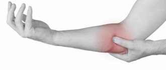
A disease that is more common in men is tendonitis, or an inflammatory process that affects the tendons. This is a disease of athletes and people forced to perform heavy physical work for a long time - loaders, masons, heavy weights, etc. Pain appears during heavy exertion, during active movements. With moderate and light loads, as a rule, there is no pain.
The next typically “male” disease is necrosis of the femoral head, and about a third of patients become ill at a young age - up to 30 years. The disease goes through several stages, and already at the very beginning the patient has pain in the hip joint on the right side (or on the left), and the pain is felt in the groin area, less often it radiates to the hip, knee or lower back. If left untreated, lameness appears over time and disability develops.
Stages of the disease
The basis for staging bone cancer is the international TNM system. It includes determining the primary tumor site, local spread of the tumor, and the presence of distant metastases.
Thus, there are 4 stages of bone tumors:
- Stage I. The malignant tumor does not extend beyond the bone or involves the cortical layer, and has a high or moderate level of differentiation. There is no damage to lymph nodes or distant metastases.
- Stage II. The tumor does not extend beyond the bone or beyond the cortex, but has a low degree of differentiation or is undifferentiated.
- Stage III. A neoplasm of any degree of differentiation without boundaries in the affected bone.
- Stage IV. Tumor of any size and any differentiation with metastases.
This cancer staging system is the basis for assessing a patient's survival prognosis. It also helps in selecting treatment tactics and the type of surgical intervention.
Pelvic pain in pregnant women
There are several reasons why hip joints may hurt in women:
- in recent weeks - lack of calcium or vitamin D;
- increased production of the hormone relaxin, which promotes sprains;
- exacerbation of latent hip dysplasia or osteoarthritis;
- increased stress on the joint due to weight gain;
- old injury.
Women who have pelvic pain during pregnancy should be more attentive to their health, because this condition can lead to an exacerbation of even minor diseases.
Ovarian cyst
The ovaries contain follicles - sacs in which eggs mature. A mature follicle ruptures during ovulation and releases an egg. If this does not happen, it turns into a cyst. Such ovarian cysts are usually harmless and can go away on their own, but they can cause pain and an enlarged abdomen. When the cyst is torsioned, a threatening condition occurs - severe acute pain should force you to immediately consult a doctor. Ovarian cysts are detected during a gynecological examination and ultrasound.
Chronic pelvic pain
Chronic is any pain in the pelvic area that continues with varying intensity for several months. Aching pain in the hip joint and surrounding area often occurs in women, but men also suffer from diseases that cause pain in the pelvic area. The causes of pain are varied, from osteochondrosis to urological and gynecological diseases. In each case, a thorough examination is necessary to identify the disease and prescribe adequate treatment.
Clinical picture of BOTP
The clinical presentation varies from patient to patient and may also change during pregnancy.
You can read about chronic pelvic pain here.
Pain
- Pain may begin around the 18th week of pregnancy and peak in intensity between the 24th and 36th weeks.
- As a rule, the pain goes away by the 3rd month after birth.
- Pain is localized between the posterosuperior iliac spine (PSI) and the gluteal fold, especially in the area of the SIJ and/or pubic symphysis.
- The pain may be local or radiating.
- Fortin's area is a rectangular area that extends from both VEPOs 3 cm laterally and 10 cm caudally. The person will often use one finger and point to the painful area, usually within this rectangular area.
- It was initially thought that pain below the knee could not be related to SIJ dysfunction, but Fortin et al. (2003) showed that pain from the SIJ can extend below the knee and be associated with SIJ dysfunction. Visser et al. (2013) also reported an association between SIJ dysfunction and intervertebral disc-related radicular pain.
- The pain may radiate down the back of the thigh and may occur in combination with (or alone) pain in the pubic symphysis.
- The pain may be described as stabbing, dull, shooting or burning.
- The intensity of pain on the VAS is on average about 5-6 points.
- It is useful to use a patient's pain distribution chart to differentiate between BOTP and BPBP. BOTP is localized under the PVPO in the buttocks, posterior thighs and groin (in particular, above the pubic symphysis).
- BSP is concentrated in the lumbar region, above the sacrum.
Functional complaints
Problems with movements such as
- Getting out of the car.
- Getting up from a chair.
- Limitation of mobility.
- Climbing stairs or walking.
- Standing for 30 minutes or longer.
- Standing on one leg or transferring body weight from one leg to the other.
- Rolling over in bed.
- Decreased ability to do housework.
- Pain/discomfort when lifting heavy objects.
Diagnostics
To determine the disease that causes constant pain in the hip joint, it is necessary to undergo a fairly extensive examination, which includes:
- X-ray of the hip joint, which allows you to detect or exclude traumatic damage, identify bone defects, growths and other pathologies;
- Ultrasound of the hip area, which reveals inflammatory processes and degeneration of soft tissues, calcified areas and other pathologies;
- CT scan of the hip joint, which is prescribed to clarify the diagnosis or in unclear cases;
- MRI to clarify the condition of the soft tissues of the joint capsule;
- puncture of joint fluid to remove effusion and study the composition of the infiltrate in order to detect the causative agent of infection;
- joint arthroscopy - endoscopic examination of the joint capsule using a probe inserted through a small incision.
In addition, the orthopedist, surgeon or rheumatologist prescribes laboratory tests for the patient in accordance with the presenting symptoms:
- general urine and blood tests;
- immunological blood test;
- biochemical blood test for rheumatic markers, etc.
MRI of the pelvis with contrast
MRI of the pelvic bones shows multiple foci, which indicates metastatic lesions
Contrast-enhanced magnetic resonance imaging improves imaging capabilities. An enhanced study is carried out to diagnose the tumor process, including detection of metastases in the pelvic bones, early detection of tumor recurrence after treatment, clarification of the severity of the inflammatory process, destruction, etc. Gadolinium-based contrast has a high safety profile and does not require a preliminary assessment of renal function. Side effects from the introduction of a paramagnetic agent are recorded extremely rarely. In 99.9% of cases, the study proceeds without complications. Contraindications to contrast:
- history of allergic reaction to gadolinium (during a previous MRI scan);
- receiving replacement cleansing therapy for end-stage renal failure;
- period of bearing a baby;
- early childhood.
General contraindications to MRI:
- implanted cardiac and neurostimulators, hearing aids, injectors for drug delivery, prosthetic joints, vascular clips, etc.;
- the volume of the hips is greater than the diameter of the drum;
- acute condition requiring resuscitation;
- mental and neurological diseases with uncontrolled motor activity.
Treatment
Clinic JSC "Medicine" offers the services of the "Pain Treatment Center". Experienced specialists will help solve the problem with any type of pain.
Since there are quite a lot of diseases that cause pelvic pain, the methods of treating them depend on the symptoms and disorders detected during diagnosis. For pathologies of bone and joint tissue, the following are usually prescribed:
- chondroprotectors, complexes of minerals and vitamins to strengthen bones, cartilage tissue and ligaments;
- muscle relaxants to reduce muscle spasms and relax tense muscles;
- non-steroidal anti-inflammatory drugs, in case of severe symptoms - steroids;
- drugs that improve capillary circulation to reduce swelling and manifestations of hypoxia;
- diuretics to stimulate fluid exchange and reduce swelling.
For inflammatory diseases, physiotherapeutic sessions have a good effect. Patients are prescribed electrophoresis, laser therapy, medicinal phonophoresis and other procedures. They are carried out at the end of the acute phase of inflammation to stimulate the recovery process.
If conservative methods are not effective enough, the patient may be prescribed:
- therapeutic blockade - the introduction of an anesthetic drug into the selected area to block the transmission of pain impulses along the nerve fiber, due to which spasmed muscles relax, the trophism of periarticular tissues improves, and blood flow normalizes;
- radiofrequency denervation - elimination of nerve fibers using a radiofrequency pulse through needles inserted into the joint tissue, which allows the patient to permanently relieve pain in the hip joint, radiating to the groin, knee or lower back;
- surgical opening and drainage - for purulent inflammation of the joint, followed by antibacterial therapy;
- removal of the tumor - in case of a malignant tumor process, after which chemical and radiation therapy is carried out to reduce the risk of relapse;
- hip replacement - in case of destruction, the head of the femur is removed and an implant is installed in its place, but in some cases not only part of the femur is replaced, but also the acetabulum of the pelvic bone.
The choice of method depends on the degree of damage to the joint and other tissues.
Complications
Malignant bone tumors, especially osteosarcoma, are characterized by hematogenous metastasis. The most common localization of metastases is lung and brain tissue.
It is also common for bone cancer to recur even after complete treatment. 95% of local relapses occur within 2 years after surgery. In this regard, radiography is recommended every 3 months during the first year after tumor removal, once every six months in the second year and subsequently - once a year.