O-shaped legs
The cause of unilateral O-shaped deformity in infancy is usually congenital hypoplasia caused by neurofibromatosis or fibrocystic dysplasia of the tibia. In contrast to physiological varus curvature, with this pathology, uneven deformation is observed (one lower leg is more curved than the other). The outcome of congenital hypoplasia of the leg bones can be pseudarthrosis.
Traditionally, rickets ranks high on the list of causes of O-shaped legs. And although this pathology is quite rare in pediatrics these days, it can occur, so it should always be excluded during differential diagnosis. It should be taken into account that rickets can develop in three periods of a child’s life: in utero (that is, congenital), at an early age and in adolescence. The cause of fetal rickets is vitamin D deficiency in the mother. Currently, this pathology is detected mainly in economically disadvantaged countries.
Infantile rickets occurs after stopping breastfeeding. At this stage of development, the child’s body requires a large amount of vitamin D. If the baby does not receive this vitamin in sufficient quantities, his bones become insufficiently strong and gradually bend when walking. An O-shaped curvature of both the legs and hips is possible. In some cases, an asymmetrical deformity is observed: varus curvature on one side is combined with valgus curvature on the other side. Anterior bending can also form - the so-called saber shins. Moreover, in contrast to damage to the legs with syphilis, when the legs are bent only anteriorly, a combination of deformation in the lateral and anteroposterior directions is observed.
Another critical age at which the likelihood of developing rickets increases is the period of intensive growth in adolescents. The cause of the deformation is a lack of exposure to the sun, a deficiency of vitamin D in food, unfavorable living conditions and some diseases. There is persistent late rickets, which, unlike the usual form of the disease, does not respond to treatment with standard doses of vitamin D. Persistent rickets develops due to genetic predisposition, chronic kidney disease and steatorrhea.
Intestinal rickets (rickets with steatorrhea) can occur with any type of long-term intestinal disorder. Occurs due to impaired absorption of fats, vitamins, phosphates and calcium. Similar disorders can be detected in adults, but in the latter case it is not rickets, but osteomalacia. The cause of the development of renal rickets is chronic kidney diseases that interfere with the retention of phosphates and calcium in the blood serum. With renal rickets, valgus deformity is most often observed, but an O-shaped curvature is also possible.
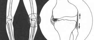
Another reason for the formation of O-shaped legs is Blount's disease (deforming osteochondrosis of the tibia). With this disease, there is not an arched curvature, as with rickets, but an angular curvature of the leg with the apex of the deformity at the level of the proximal epiphysis. At an early age (2-4 years), with Blount's disease, as a rule, bilateral curvature is detected; at an older age, only one lower leg may be curvature.
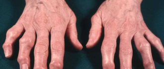
In adults, O-shaped legs can form due to Paget's disease (osteitis deformans), accompanied by damage to the femur and tibia. It is possible to damage both several and one bone, but more often several bones are involved in the process. The curvature is caused by excessive growth of bone tissue with insufficient destruction. In this case, the newly formed bone does not have sufficient strength due to incomplete calcification. It thickens and at the same time becomes soft, resulting in curvature and transverse fractures.
Plano-valgus deformity of the feet in children. Causes, symptoms, stages of development, treatment.
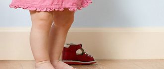
The musculoskeletal system of young children, like all organs and functions, is constantly developing. If you have ever observed babies, who often begin to walk at about 12 months, you have definitely noticed hesitant, waddling steps. The reason is that their feet do not have the natural arches of adults. Only by the age of 3 do the child’s legs acquire a normal structure.
At this age, it is necessary to pay great attention to the legs if, when walking, they bend inward - this is a planovalgus deformity.
stop, it is easy to see it on wet footprints when walking along the floor or, ask the child to join his knees together, while the feet will diverge. It is possible to correct the pathology, but if you start at the age of three, and if you start it, then in the future it threatens flat feet, arthrosis, leg pain, scoliosis, etc.
Why are children diagnosed with foot deformities?
The cause of the appearance of plano-valgus deformity can be congenital anomalies, but most often it appears due to weak muscles, or, on the contrary, very tense ones, as they sometimes say in tone. Large weight can also be a cause of incorrect children's shoes, where the child places the foot incorrectly, and she gets used to this position. Inactivity, past illnesses, lack of calcium, vitamins, etc. are also no exception.
How to determine?
The pathology is not difficult to determine, and if it is noticeable, you should not hope that everything will go away with age, it will not go away, and moreover, it may get worse.
If by the age of three the child:
- difficult to run;
- he often complains of pain in the knees and joints of the feet, even to the point of cramps;
- when standing at attention, his feet do not close together;
- when the knees are closed, the legs are X-shaped;
- When walking, the feet “fall” inward.
If at least one of the cases is noticed, you should immediately contact a specialist - a traumatologist, orthopedist. In frequent cases, the doctor only needs a visual examination of the patient to determine the diagnosis; for a more detailed study, for example, the cause, an ultrasound scan, computer diagnostics of the joints and a detailed measurement of the level of curvature are prescribed.
Stages of development
At the time of diagnosis, the doctor is supervised by the data that should be present for the normal formation of the foot and determines the height of the child’s arch (the norm is 4 cm) with an angle of no more than 125 degrees. The deviation of the heel from the vertical axis is acceptable within 5-7 degrees.
Based on the parameters, doctors distinguish 3 degrees of development of pathology:
- Easy. With a heel deviation of 10 degrees, the arch of the foot lowered to 1.5-2 cm, and an increase in the angle to 140 degrees. It can be easily corrected with correct and timely intervention.
- Average. There is a flattening of the arch up to 160 degrees with its height of 1 cm. The toes and heels are not comparable with the vertical axis by 15 degrees. Treatment is more complex and lengthy and requires professional intervention.
- Heavy. Here there is a complete flattening of the arches with a heel deviation of more than 20 degrees. Conservative treatment is useless; the help of a surgeon will be required.
What are the consequences of neglected deformation?
For boys, as for girls, plano-valgus curvature of the feet can turn into flat feet
. For the former, it is impossible to play football; for the latter, it is a problem to wear shoes with even the slightest heel. Regardless of the sex of the child, the spine is formed incorrectly, scoliosis, uneven posture, pain in the back and chest, and even disability appear.
Treatment
In order not to have to treat plano-valgus deformity in your child in the future, it is necessary to prevent this pathology. If earlier we grew up on the street, ran barefoot, we had more normal and high-quality shoes, but now, children spend their childhood in apartments on flat floors and often we buy them under the name of a good brand, for example, beautiful, but not comfortable shoes. Even such little things harm our kids, and we have no idea how much.
It is important that if the deformity is nevertheless determined by the parents, or even more by the doctor, there is no need to let everything take its course, it is necessary to begin immediate treatment. First of all, the cause is determined, for example, weak muscles, dystonia, lack of calcium, etc. And solving the problem of muscle dystonia - by doing a massage, and if there is a lack of calcium - taking vitamins, methods for correcting the deformity are simultaneously used. That is, the problem is solved comprehensively.
In the case of a congenital defect, a cast can even be applied to level it at the time of active growth with a further rehabilitation program ranging from therapeutic exercises to electrical muscle stimulation.
Gymnastics and massage
This technique helps strengthen muscles and ligaments. Exercises are prescribed by a doctor after determining the degree of deformity. They are very simple and can be done at home with a child of any age.
For older people, they will need to be performed on command: squatting, walking on toes, heels, on a turn and on the inside of the feet. Walking barefoot on various surfaces increases irritation of the nerve endings of the feet; there are special mats for this. In this way, the area of the foot is massaged, an impulse is sent to the brain that the leg is uncomfortable or slightly painful, after which the muscle “shortens” and the leg takes the desired position for this. The muscles are trained in the same way.
Massage is also the main method in treatment.
It is done not only in the foot area, but also along the shin and above the knee. This increases blood circulation, as if forcing the muscles to work. The massage is carried out in a course and only by specialists. Author: K.M.N., Academician of the Russian Academy of Medical Sciences M.A. Bobyr
IDEAL LEGS SHAPE
There are many opinions regarding the correct shape of the legs, but all this is from the realm of theory. When determining curvature, specialists use A. A. Artemyev’s classification, which he proposed in 2001.
The doctor was the first to talk about the parameters of the correct form from a practical point of view. The beautiful legs have three spindle-like spaces between the inner sides of the legs. These gaps are limited by the perineum, closed knees, muscles of the upper leg and ankle (Fig. 1).
How to detect?
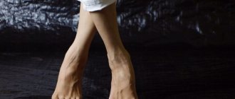
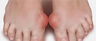
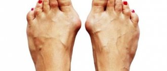
It is quite difficult for parents to independently notice the first signs of foot varus in their baby. Until he starts walking, they don't even know there's a problem. The disease develops slowly. Obvious signs of deviation do not appear immediately. A characteristic and obvious symptom of varus deformity in children is changes in the lower extremities, when in a standing position the legs are tightly pressed to each other and the knees do not close. The position of the legs resembles the letter "O".
Parents can suspect varus foot deformity in a child based on the following markers:
- erasing the soles of shoes from the outside, where the load and pressure are greater;
- outward deviation of the feet;
- awkward, uncertain gait;
- frequent falls when walking or running;
- leg pain;
- Difficulty straightening your knees.
What external changes in the legs look like with varus deformity of the feet in children can be seen in the photo:
HOW TO CORRECT CURVED LEGS BASED ON THE SHAPE OF LEGS
If you don’t want to hide imperfections, then you need to go to a surgeon. True curvature is corrected by orthopedic traumatologists during osteoplastic operations. This is not so much about eliminating a cosmetic defect as it is about preserving your health. Even a slight curvature of the bone changes the load on the joint, leading to severe diseases.
In case of false deformity, a plastic surgeon corrects the shape of the legs by injecting the patient’s own fat or installing implants. As a result, the inside of the legs becomes full, and the window between the knees and ankles disappears.