Infants' kneecaps are made from bone tissue and remain in the formative stage for a long time. At first, children have no kneecaps. At first they consist of cartilage tissue, and this has given rise to the myth that newborns do not have kneecaps at birth. Their formation ends by the age of 6. Lack of medical knowledge, or incorrectly presented information, leads to the formation of common misconceptions.
When children are born without kneecaps, this is a congenital pathology that can occur due to teratogenic effects on the fetus in the womb. In a child with physiological intrauterine development, sesamoid bones (including the kneecaps) appear at the 4th month of fetal development, but consist of cartilaginous bone, which is poorly visible on an x-ray.
Patella in children: when does it appear and can it be absent?
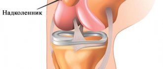
The kneecap plays a big role in the human body and body. It is worth noting that this is a large sesamoid bone. Starting from the age of three, this bone can be easily felt through the skin, and can be shifted left and right when bending or straightening the knee. The main function of this joint is to protect against displacement of parts of the femur and tibia, which form the knee joint. There is a myth that children are born without a kneecap. But we will find out whether this is true or not further.
Anatomical features
The patella (another name for the kneecap) is a type of sesamoid bone and is the largest of this entire group. The kneecap is shaped like a triangle, it is slightly convex on the outside and concave on the inside. The inner surface is covered with articular cartilage. It is the patella that helps us bend and straighten our legs, and also protects the knee joint from excessive displacement to the sides.
Cup formation
There is a myth that the kneecap in newborn babies is completely absent, but is formed closer to four months. But in reality everything is somewhat different. The calyx is formed in children already in the womb in the fourth month of pregnancy. But it is not always possible to detect it on an ultrasound examination, since it consists of cartilage tissue, while in adults it is made of bone tissue.
From two to six years of age, ossification nuclei form around this cartilage in a child. Gradually, all these zones merge with each other, forming the familiar kneecap. This happens around the seventh year of a child’s life. Children have kneecaps since prenatal development, and do not appear at a certain age.
Role and functions
The main and very important function of the patella is to protect the femur and tibia from displacement. When we bend and straighten the knee, the patella moves up and down, preventing the joint from moving to the sides.
In addition, on the sides of the knee joint there are cruciate ligaments, which further protect and enhance the strength of the joint. This is why children need to be careful with falls and impacts, because they do not yet have knee protection like adults.
Stages of formation of the patella and its functions
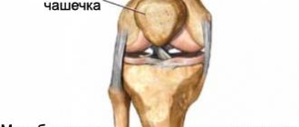
Sesamoid bones of the knee joint
The reason for the misconception can be that cartilage is less noticeable to the touch than bone tissue, or that the knees of an infant are characterized by some swelling that masks the presence of cartilage.
From the moment the sesamoid bones begin to form, and right up to birth, the kneecaps in infants undergo a certain development and actually acquire the usual shape of an adult patella, and the only difference is the structure that forms them. Therefore, the statement that children do not have kneecaps is not true.
In terms of the anatomical structure of the musculoskeletal system, a baby differs from an adult, but this also applies to the digestive and nervous systems. That is why he needs adult care and mother's milk. This is the only food that his digestive system can digest until about 4 months. It also has its own characteristics in the anatomical structure of the skeleton. An infant has more bones in its skeleton than an adult, but many of them are made of cartilage.
This also applies to the sesamoid bones, to which anatomy includes the patella. When the formation of the skeletal system occurs, the bones grow together and their number reaches the usual norm. The specific structure of the skeletal system and musculoskeletal system is provided by nature so that the body has the opportunity to grow. Once this process is completed, the child turns into an adult.
The main function that the kneecap performs in the body is to protect against displacement of the bones that make up the knee joint. While the baby is lying in the cradle, he is unable to move, and the knee joint is covered only by cartilage tissue. This is a sketch of the strong bone segment that will be needed to protect the femur and tibia.
We can assume that children’s kneecaps appear long before birth, but they develop in several stages until they acquire their natural appearance:
Bone formation begins in the womb at 4 months.
- in the womb, at 4 months, the formation of soft cartilage begins;
- at birth it is already present above the joint, but is difficult to palpate due to its soft structure;
- by the age of 2, when the ability to walk upright begins to appear and motor activity increases, the need arises to protect the joint, and growth zones are formed; from 2 to 6, growth zones (or ossification nuclei) lead to a gradual reduction of the sesamoid bones to more a state familiar to an adult;
- at 6-7 years of age, the once cartilaginous structure of the kneecap is completely transformed by growth zones into a bone structure located above the joint and protects it from displacement and injury.
A child of any age has its differences from an adult. Up to six months he has the ability to breathe and swallow at the same time, which he loses over time. In less than 3 days, a baby's excretory systems can excrete a volume equal to its weight. Newborns have excellent tenacity in the upper limbs, which they no longer have at one month of age. The peculiarity of the human race is that development occurs not only in the womb, but some segments and systems of the body begin to form after birth.
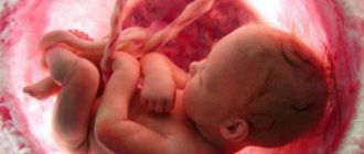
Newborn child
So the statement that all newborn children do not have kneecaps is incorrect and incompetent. They exist, but in the form of cartilage, not bone. Although a small percentage of babies born actually do not have them, this is an anomaly of intrauterine development, and not a characteristic feature of all infants.
Pathologies and absence of a cup
Is it possible to have a missing kneecap? Yes, but this already falls into the category of pathology. The problem occurs quite rarely. And it is more likely to occur in those children who have other disorders of the musculoskeletal system.
Doctors say that anomalies with the appearance and development of the patellas occur in children who have genetic failures or a negative impact on the mother during pregnancy. The main factors that can lead to improper development of the calyx or lead to its absence include:
- Radiation.
- Taking medications.
- Infections.
- Hormonal disorders.
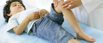
If any of these factors affects the mother in the first three months of pregnancy, the newborn will have an absent kneecap. If the expectant mother was exposed to negative influences in the subsequent months of pregnancy, then the child’s patella will be underdeveloped. If there is a suspicion that this bone is not developing properly, then after the baby is born, he is prescribed an X-ray examination and a full examination by an orthopedist.
Complete absence of the patella
This pathology is extremely rare. And if it is already observed in the baby, then only together with the pathological development of the tibia and femur. Children who were born without a patella often have congenital dislocation of the hip, tibia, and clubfoot.
This anomaly does not affect motor functions in any way. The child can jump, walk, run, and there is no pain or discomfort. The defect appears only as an aesthetic one. A slight lameness, weakness of the leg, and rapid fatigue are visible.
There is no treatment as such for this pathology. Although some resort to surgery.
Lobular patella
This pathology is detected in almost two percent of people who have had an X-ray of the knee joint. The problem is often discovered by chance and is more common in men than women. The lobular knee brace consists of two or three parts, but its size is normal and there is no pain or discomfort.
There is also no therapy. But you should always keep in mind that in children and adults with this defect, the risk of developing arthrosis is much higher. Therefore, it is necessary to prevent this disease in advance.
Also, children with this pathology are not recommended to engage in too active sports. And if you spend time on the sports field, be very careful about possible injuries and damage.
Children born to drug and alcohol addicts: possible developmental pathologies
Both ethanol and psychoactive substances are carried throughout the entire body through the bloodstream in almost a matter of minutes, easily penetrating the placental barrier. In addition, in pregnant women, due to the increased load on the liver and kidneys for physiological reasons, the metabolism of alcohol and drugs occurs more slowly, and accordingly, their toxic effect increases many times over.
When psychostimulants enter the fetal circulatory system, they cause vasospasm, increased blood pressure, and tachycardia. Hypoxia of the placenta develops, which is a common cause of premature birth and miscarriages. According to statistics, in 10–15% of addicts, pregnancy ends in stillbirth due to placental abruption and related complications.
To date, it has not been proven that children of parents of drug addicts and alcoholics develop various chromosomal mutations. In some of the author’s publications, based on the results of statistical studies, they indicate that in such “problem” families there is a high risk of having a child with Down syndrome, autism, cystic fibrosis, etc. Perhaps such data are due to the fact that often in such cases pregnancy is usually not planned, and, naturally, there is no question of examinations before conception, during gestation, or genetic consultations.
The most commonly diagnosed pathologies in infants
As a rule, the following developmental defects are noted in newborn children in families of alcoholics and drug addicts:
- craniofacial anomalies: narrow palpebral fissures, underdeveloped upper lip, elongated face shape;
- dysfunction of the central nervous system: increased excitability, convulsions, signs of cerebellar dysfunction;
- underdevelopment or abnormal development of joints, limb defects (short legs and/or arms, absence of fingers);
- heart defects;
- liver fibrosis, which is a serious risk factor and predictor of early death of a child;
- dysfunction of the kidneys and pancreas;
- genital defects;
- endocrine disorders.
If a woman systematically used drugs or alcohol during pregnancy, the fetus develops withdrawal syndrome, but due to congenital pathologies, dysfunction of the central nervous system, digestive and genitourinary systems, it is much more severe than in adults. Abstinence symptoms in newborns in families of drug addicts or alcoholics are characterized by:
- generalized convulsive syndrome;
- critical arrhythmia and hypertension;
- heart attack;
- cerebral hemorrhage;
- systemic severe hypoxia;
- temperature rise to febrile levels;
- renal and liver failure;
- encephalopathy.
How does the normal formation of the patella occur?
The common belief that children are born without kneecaps is incorrect. Children have patellas, but they are not as developed as in adults.
When do kneecaps appear in babies? This structural element is formed during the development of the fetus in utero, starting from the 4th month of pregnancy. It is a cartilaginous formation, not a bone one.
During development at the age of 2-6 years, this cartilage is surrounded by several growth zones - ossification nuclei. During growth, they gradually merge and form a single bone element.
Reference . Full patellas are formed by the age of 7 years.
As a result, we can say that the patellas do not appear at any particular age, but are formed during the growth and development of the child.
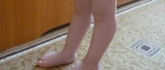
Babies' knees are very flexible and not as rigid as those of older children or adults. This leaves many parents wondering: Does their newborn even have kneecaps? When do knees become harder?
We reached out to two doctors—Dr. Chris Airey, Medical Director of Optimale, and Dr. Pierrette Mimi Poinsette, Medical Advisor for MomLovesBest—to find out the answer to this question and get more information about babies and children's kneecaps in general.
Do babies have kneecaps at birth?
According to experts, the newborn definitely has kneecaps. However, parents are often confused, and for good reason.
"It's kind of a common misconception that babies are born without kneecaps," Dr. Airey says. - But it is not so. Children are born with kneecaps made entirely of cartilage, which is why their knees are called "cartilaginous patellas."
It's not that babies don't have kneecaps. It's just that their kneecaps are made of a different material than the kneecaps of older children and adults.
Dr. Airey explains that cartilage is “a flexible, soft substance that protects joints from friction. Less hard bone means easier passage through the birth canal (for both baby and mom), and having soft, flexible knees is more conducive to our ability to crawl in early childhood.”
When do babies' kneecaps get harder?
However, at some point, we all develop stronger kneecaps. The process of turning the patella from cartilage to bone is called ossification and is usually complete by the time a child enters primary school.
“The kneecaps become bone between the ages of 2 and 6,” explains Dr. Poinsett. “The process is slow, starting from the center of the kneecaps and moving towards the edges.”
Because the knee joint is made flexible, some cartilage is still involved in its function. The point where the femur (leg bone) and the kneecap (knee bone) meet is provided with articular cartilage. This helps the knee and leg move smoothly together and protects the knees from injury.
Potential problems
Although your baby's kneecaps may seem more tender and sensitive than those of an older sibling, there is nothing to worry about. Dr. Poinsett assures that a child's kneecaps are no more susceptible to injury or health problems than an older child's kneecaps.
However, there may be some potential problems when transitioning from baby knees to more mature knees. In particular, a growing child may experience a condition called bilobed patella.
“Because the kneecap is formed from pieces that fuse together over time, the problem that can arise is a bilobed kneecap, where the pieces have not fused completely and result in a sort of split double kneecap,” says Dr. Airey. “Sometimes you can solve this on your own or manage it with physical therapy. It is important to monitor for pain and swelling in your child's knee area, as this may indicate a knee problem such as arthritis, or a tendon or ligament injury."
Common Knee Injuries
Children, especially those who are active in sports, are susceptible to injury from time to time. However, it is usually not the kneecaps that are the problem.
“Knee pain in children is most likely caused by a sprain or injury to the ligaments surrounding the knee,” explains Dr. Poinsett. “Injury to the kneecap occurs, but not often.”
However, knee injuries do happen and this is something parents should be aware of. The most common types of knee injuries occur due to overuse, sudden twisting, or direct impact to the knee and surrounding joints.
Some of the most common childhood knee-related injuries, according to Children's Healthcare of Atlanta:
- Fractures;
- Dislocations;
- Tendinitis;
- Anterior cruciate ligament rupture;
- Meniscus cartilage tears;
- Bursitis;
- Patellofemoral stress syndrome;
- Damage to articular cartilage.
Signs that your child may have a knee injury:
- Swelling in the knee area;
- Complaints of pain;
- Stiffness or inability to move your leg or bend your knee.
Pathologies
Having clarified the question of whether newborns have kneecaps, let’s move on to considering possible deviations in the development of this bone element.
Reference . Improper formation of the patellas is a pathology that is usually combined with other disorders of the musculoskeletal system.
This anomaly can be caused by various genetic disorders or the negative influence of factors on a woman during pregnancy:
- radiation exposure;
- medicines;
- infections;
- hormonal imbalances.
If a woman was negatively affected in the first 3 months of pregnancy, the baby may have a complete absence of patellas, and the child’s bone structure may be underdeveloped.
If the doctor has doubts about the development of the bone, an examination by an orthopedist and an x-ray are prescribed.
Missing kneecaps
This deviation is extremely rare and is often combined with underdeveloped bones. May lead to dislocation of the lower leg, hip, clubfoot, etc.
Reference . If, apart from the absence of patellas, no other ailments are detected, then the motor function of the legs is not impaired.
Only a noticeable defect and rapid fatigue during physical exertion appear.
Absence of bone does not require any therapy. If there are other violations, then surgical intervention is used.
Literature:
- Vostrikov V.V., Selizarova N.O., Grigorieva O.Yu. The role of hereditary factors in the formation of opiate addiction. - Pediatrician, 2015. - Volume IV, No. 4.
- Dmitrieva T.B., Igonin A.L., Klimenko T.V. and others. Psychotic conditions caused by abuse of opioids, cannabinoids, sedatives and hypnotics. - Narcology, 2003. - No. 1.
- Egorov A.Yu. Early onset alcoholism: current state of the problem. - Questions of Narcology, 2002. - No. 5.
- Sutulina I.M., Chernykh A.A. Long-term consequences of intrauterine drug exposure on the fetus. - Mother and Child, 2003. - No. 1 (12).
Do children have kneecaps?
The kneecaps are formed in children during intrauterine development for a period of 16 weeks. From the 4th month of pregnancy, the fetus begins the process of forming cartilage, which is necessary for further fusion and strengthening of the elements of the musculoskeletal system. The transformation of soft tissue into bone occurs in a child from 2 to 6 years of age. If the process of ossification of cartilaginous structures is disrupted, pathological abnormalities may develop.
Do newborns have kneecaps?
There are unfounded opinions that a child does not have patellas at birth. During the process of growth, the human body changes and strengthens, but such an important anatomical part of the osteoarticular system as the sesamoid bone in the knee is formed in the womb. The patella appears between 2 and 6 years of age. At this time, the cartilage tissue simply transforms into bone, and the child already has a knee pad. X-ray images do not clearly show the soft tissue, because of this there is a feeling that the joint is unformed, which provokes erroneous judgments about the period of cup formation in babies.
The absence of kneecaps in infants is considered a pathology caused by infection, hormonal imbalance, exposure to synthetic drugs or radiation.
Every pregnant woman worries about the proper and healthy development of the baby. Therefore, there are a lot of forums that raise the issue of the formation of the musculoskeletal system in children. There is no need to believe myths. If the baby really does not have kneecaps, then medical help cannot be avoided. Untimely treatment can cause immobilization of the baby and disability. All questions about the stages of a child’s growth should be discussed only with a doctor, and not rely on the opinions of others.
Normal development
A baby's kneecaps form in the womb during the 16th week of pregnancy. During normal fetal development, they consist only of cartilage tissue and many bones, which grow together only after birth. Newborns have much more bones than adults. As we grow older, small bone structures fuse together. The cartilage of the knee itself is surrounded by ossification nuclei (growth zones), which form the patella. The fusion of such zones occurs gradually, bone tissue is formed from approximately 2 years. Each child has completely individual growth periods, so the complete formation of the kneecap can be completed by the age of 6.
When do children start to develop kneecaps?
Patellas appear during fetal development, but until 2 years of age, the formed kneecaps are not visible on the skeleton, since they consist of soft, not bone, tissue. During the growth period, bone gradually develops. At the age of 2 to 6 years, a child’s cartilage is completely overgrown with bone tissue, merges with each other and forms the largest sesamoid bone in the skeleton. The growth and maturation of a child are individual parameters that need to be monitored not only by parents, but also by the pediatrician. With underdevelopment of the tibia and femur by the age of 6, disturbances in the formation of the kneecaps occur, which leads to clubfoot, dislocations and other pathological abnormalities.
Content:
- Children born to drug and alcohol addicts: possible developmental pathologies: 1.1. The most commonly diagnosed pathologies in infants
- What kind of children do drug addicts and alcoholics have: long-term consequences of parental addiction
- Families of drug addicts and children of alcoholics: psychology
It has already been proven that one of the causes of alcoholism and drug addiction is hereditary predisposition. Experts attribute this phenomenon to a disruption in the production of endorphins, genetically predetermined tolerance to alcohol and psychoactive substances. But children of alcoholics and drug addicts do not always follow in the footsteps of their parents; there are cases where in such socially disadvantaged families a child achieved success both in school and in future employment. Naturally, the life of children of alcoholics and drug addicts is very far from ideal. And we are talking not only about the psychological aspects of upbringing and growing up, but also about the state of health.
Anatomical features
The kneecap is also called the patella. It is the largest bone of the sesamoid group. Outwardly, it resembles a triangle, which is slightly convex on the outside and concave on the inside. The inside of the bone is covered with cartilage tissue.
With the help of a cup, a person has the ability to bend and straighten the lower limbs. Also, the patella region acts as a protection for the joint from unnecessary displacement to the sides. The cruciate ligaments are located on the sides of the bone. Thanks to them, additional protection and increased strength of the knee joint are provided.
Normal development of the knee
The idea that infants do not have kneecaps is erroneous. Newborns have patellas, but they are not as developed as in adults. Their formation begins during intrauterine development of the fetus. This occurs at 4 months of pregnancy.
In a baby, the patellar part consists not of bone, but of cartilage tissue. Around it there are ossification nuclei that appear between 2 and 6 years. Then these zones undergo fusion, and a single bone is formed. The calyx is fully formed by the age of 7 years.
Pathology
In medical practice, developmental disorders of the knee in children are often encountered. Doctors believe that deviations appear due to genetic failures and the adverse effects of external factors on a woman during pregnancy.
The following can have a negative impact:
- Radiation exposure.
- Taking medications.
- Infectious diseases.
- Hormonal disorders.
The impact of these factors is especially dangerous in the first trimester of pregnancy.
In the early stages, the fetal body begins to form. As a result, the newborn's kneecap may be completely absent or abnormal.
If there are doubts about the development of a child’s knee, doctors will prescribe an examination by an orthopedist and an X-ray examination.
Complete absence of the kneecap
Sometimes there are cases when the baby does not have a patella. At the same time, underdevelopment of the tibia and femur is often observed. Pathologies entail dislocations of the lower leg, hip, and clubfoot.
If, apart from the absence of a calyx, there are no violations, then this does not affect the functioning of the lower extremities. But a cosmetic defect is created, and the child can quickly get tired during physical activity.
If the baby was born without a kneecap, then therapy is not required. If there are additional disorders, surgical intervention is used, since the activity of the legs fails.
Lobular patella
The disease is detected rarely and accidentally during an x-ray. Male patients suffer more from pathology. The lobular patella is a cup of the knee that is of normal size, but is not complete. The bone consists of several parts.
No treatment is required for this disorder. However, patients need to be more careful, since the lobular patella is often subject to injury and the development of arthrosis. Therefore, sick children should engage in gentle sports and avoid extreme hobbies.
Congenital dislocation of the patella
Doctors consider this disorder to be a hereditary pathology, which is observed mainly in boys. In the presence of congenital dislocation of the knee, the joint becomes unstable, which greatly interferes with walking. People who have a cup with a similar disorder are more likely to experience arthrosis.
If a dislocation occurs, the patient is given an x-ray to assess the degree of displacement and underdevelopment of the patella. The photographs show that the bone is irregularly shaped and reduced in size. Elimination of pathology is carried out exclusively by surgery.
Thus, a person is born with a kneecap. But the final formation of bone tissue occurs by the age of 7 years of a child’s life.
Families of drug addicts and children of alcoholics: psychology
We should not forget about the psychological aspect of raising a child. Children of an alcoholic father usually suffer from a lack of attention from their mother, since she is often busy making money, getting her husband into treatment, keeping him from another binge, and hiding the problem that exists in the family from others. The child is forbidden to invite friends home, and he himself practically does not go anywhere. No one is interested in or monitors his progress or mastery of educational material. Due to the inability to communicate with peers and isolation, he may be subject to bullying.
The child witnesses frequent conflicts between parents, and sometimes becomes a victim of physical violence. Over time, he develops a certain stereotype of behavior and thinking, a pathological, “toxic” algorithm for building relationships with the opposite sex. Loneliness and a specific social circle become a trigger for the use of alcohol and psychoactive substances, and the commission of offenses at an early age. Children of fathers of drug addicts face similar problems. The situation is much worse when both parents suffer from addiction, and the child is left to his own devices and does not receive basic care or proper nutrition.
But is it possible to break this vicious circle? Does addiction really put an end to any attempts to create a full-fledged family? Experts emphasize that during pregnancy, as well as preparation for conception, there are simply no safe doses of drugs or alcohol (and this applies to both men and women), so the only option is to recover from addiction. Children of former drug addicts and alcoholics can be quite healthy if the future parents took seriously their health, were examined by specialized specialists, followed the recommendations of doctors, and completed a full course of psychotherapy and rehabilitation.