| Appointment with a dermatologist at the clinic. Call a dermatologist at home. | Reception is strictly by appointment, make an appointment by phone: +7 | Prices for services | Reviews about the clinic |
A rash on the legs should not be ignored, since its origin and causes can be different, and the consequences can be very serious. Often such rashes turn out to be a reaction to certain irritants and in this case they can quickly disappear. At times, a rash turns out to be a clear sign of one or another dangerous disease.
The appearance of an itchy rash on the skin of the legs is a good reason to visit a medical specialist. A dermatologist will be able to determine the cause of the rash and prescribe the correct treatment to get rid of such troubles.
Make an appointment with a dermatologist by phone or by filling out the online form
| Select a clinic | Demodicosis | Rash on shoulders | Calling a dermatologist to your home |
Answers to frequently asked questions about skin rashes:
- Which doctor should you contact for a skin rash?
- Is the skin rash contagious?
- What diet is necessary for skin rashes?
- What diagnosis is needed for a skin rash?
- Why is a skin rash dangerous?
- Why is it necessary to get tested for a skin rash?
- What diseases does a skin rash indicate?
- What examination is necessary for a skin rash?
- Which skin rash is dangerous?
- How to distinguish an allergic rash from an infectious one
- How to get rid of skin rashes?
- How to get rid of itching skin rash?
- What organs are affected by a skin rash?
- How to prepare for an appointment with a dermatologist?
- How to get checked for skin diseases?
- What diseases does a dermatologist treat?
- What tests should be taken by a dermatologist?
- What diagnostics can a dermatologist perform in the clinic?
- Where to go with a skin disease?
Causes of rashes
In fact, there can be a large number of reasons. For example, sometimes rashes occur due to mechanical stress when tight or uncomfortable shoes are used. As a result, the legs become red and chafed. Sometimes a rash appears after depilation.
A rash may appear after an insect bite. As a result, the person feels itching and begins to scratch the wound vigorously. Thus, some kind of infection may occur. When ticks bite, a hemorrhagic rash may well appear.
Another common cause may be a fungus. For example, mycosis refers to a rash on the feet due to parasitic fungi. The onset of such a disease is associated with the appearance of a watery rash and some cracks that appear among the toes. Then the skin particles begin to peel off and even purulent ulcers appear where there were initially blisters.
If we are talking about scabies, the rash is accompanied by severe itching. This happens because the scabies mite moves under the skin. When there is lichen, blisters may also appear.
When a rash is caused by allergic reactions, it is often associated with the use of cosmetics or certain detergents that have an allergenic effect. Reactions can be caused by fur, for example. Itching occurs due to dust mites and bed bugs. Food allergies caused by citrus fruits, chocolate, dairy products and so on are common.
Finally, we should not forget about urticaria as a possible cause of rashes. In this case, the rash may appear and then appear. Subsequently, the rashes become blisters - almost the same as those that can occur if a person is burned by nettles.
Doctors pay considerable attention to the hereditary causes of the rash. It could be eczema, for example. It usually causes bubbles containing liquid inside. Then they begin to burst and are replaced with dry crusts.
During diabetes, there may be itchy skin along with a rash on the legs. If normal blood sugar levels are restored and stabilized, the rash usually disappears.
Common symptoms and manipulations in dermatology:
- Skin rashes
- Calling a dermatologist to your home
- Itching in the urethra
- Itchy skin
- Skin rash
- Prevention of casual sex
- Skin neoplasms
- Pyoderma
- Pityriasis rosea
- Streptoderma
- Scabies
- Peeling skin
- Fungal infections
- Skin infection
- Pus on the skin
- Blisters on the skin
- Papillomas on the foreskin
- Sexually transmitted diseases
- Skin structure
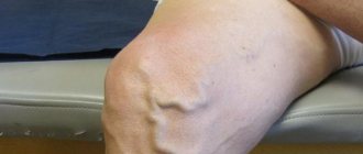
Skin rash in large joints
The terminal stage of HIV infection, which is characterized by severe immunodeficiency, i.e. the patient’s body becomes unable to independently resist infectious diseases. It can develop in different people after 2–15 years. The virus penetrates cells that have the CD4 receptor on their surface (T-helper cells, monocytes, macrophages, Langerhans cells, follicular cells of lymph nodes, microglia). Characteristic of HIV infection is chronic inflammation with damage to all organs and systems: autoimmune reactions, diseases of immune complexes and metabolic disorders lead to damage to the vascular endothelium and connective tissue with the development of cardiovascular, neurological, endocrine and osteoarticular pathologies. The source of infection is a person infected with HIV at any stage of the disease. The virus is transmitted through blood, semen, vaginal secretions, and breast milk. A case of AIDS is registered if a person with HIV infection is diagnosed with at least one of the following diseases (in the absence of other reasons for their development): 1) Bacterial infections (multiple or recurrent) in a child under the age of 13 years 2) Esophageal candidiasis 3 ) Candidiasis of the trachea, bronchi or lungs 4) Cervical cancer (invasive) 5) Coccidioidomycosis (disseminated or extrapulmonary) 6) Extrapulmonary cryptococcosis 7) Intestinal cryptosporidiosis with diarrhea for >1 month Cytomegalovirus infection (affecting organs other than the liver, spleen, lymphatics nodes) in a patient over one month of age 9) Cytomegalovirus retinitis with loss of vision 10) Encephalopathy caused by HIV 11) Infection caused by the herpes simplex virus: chronic ulcers lasting more than 1 month, or bronchitis, pneumonia, esophagitis in a patient in over one month of age 12) Disseminated or extrapulmonary histoplasmosis 13) Intestinal isosporosis (with diarrhea lasting more than 1 month) 14) Kaposi's sarcoma 15) Interstitial lymphoid pneumonia in a child under 13 years of age 16) Burkitt's lymphoma 17) Immunoblastic lymphoma 18) Lymphoma primary brain 19) Mycobacteriosis caused by M.kansasii, M. avium-intracellulare, disseminated or extrapulmonary 20) Pulmonary tuberculosis in an adult or adolescent over 13 years of age 21) Extrapulmonary tuberculosis 22) Other undifferentiated disseminated or extrapulmonary mycobacteriosis 23) Pneumocystis pneumonia 24) Pneumonia recurrent (two or more within 12 months) 25) Progressive multifocal leukoencephalopathy 26) Salmonella (non-typhoid) septicemia recurrent 27) Brain toxoplasmosis in a patient over one month of age 28) Wasting syndrome due to HIV
Correct treatment
Naturally, a rash on the legs needs timely and proper treatment. In case of allergic dermatitis, contact with the active allergen (which, first of all, is detected) must be excluded. It is possible to prescribe the correct diet and follow a normal regimen. For example, doctors do not recommend consuming foods containing nickel.
For dry skin on the feet, salicylic petroleum jelly is usually prescribed, which is applied to the feet three times a day. In case of swelling, you will need lotions, as well as special baths made with boric acid. Antihistamines, such as Telfast, Zyrtec, Suprastin, and so on, can also be used during therapeutic procedures. It should be noted that hormonal ointments that act locally are often used.
Publication date: 2018-02-07
Methods for diagnosing skin diseases:
- Diagnosis of skin diseases
- Diagnosis of skin diseases at home
- Diagnosis of allergic skin diseases
- Diagnosis of bacterial skin diseases
- Diagnosis of viral skin diseases
- Diagnosis of hair diseases
- Diagnosis of nail diseases
- Diagnosis of skin tumors
- Skin scraping
- Blisters on the skin
- Dermatoscopy
- Demodex tests
- Diagnosis of sexually transmitted infections
- Mushroom tests
- Skin scraping
YOU can call us: 8 (8452) 98-84-68 and +7-967-500-8468 or
Hemorrhagic vasculitis (synonyms: Henoch-Schönlein purpura, Henoch-Schönlein disease, rheumatic purpura, allergic purpura) is the most common disease from the group of systemic vasculitis. It is based on aseptic inflammation of the walls of microvessels, multiple microthrombotic formation, affecting the vessels of the skin and internal organs (most often the kidneys and intestines).
The main reason that causes this disease is the circulation of immune complexes and activated components of the complement system in the blood. In a healthy body, immune complexes are removed from the body by special cells - cells of the phagocytic system. Excessive accumulation of circulating immune complexes under conditions of predominance of antigens or insufficient formation of antibodies leads to their deposition on the endothelium of the microvasculature with secondary activation of complement system proteins along the classical pathway and secondary changes in the vascular wall.
As a result, microthrombovasculitis develops and changes occur in the hemostatic system: activation of platelets, circulation of spontaneous aggregates in the blood, pronounced hypercoagulation, decrease in plasma antithrombin III, thrombopenia, increased level of von Willebrand factor, depression of fibrinolysis.
In 1837, the famous German physician JL Schönlein described “anaphylactic purpura.” In 1874 his compatriot E.N. Henoch published a valuable work on the same disease.
The name “ hemorrhagic vasculitis ,” used only in Russia, was introduced in 1959 by the outstanding rheumatologist V. A. Nasonova. Henoch-Schönlein purpura still prevails .
Etiology
In the majority of patients (66-80%), the development of the disease is preceded by an upper respiratory tract infection.
The manifestation of the disease after typhus, paratyphoid A and B, measles, and yellow fever is described.
Other potential triggers for the disease may include:
- medications (penicillin, ampicillin, erythromycin, quinidine, enalapril, lisinopril, chlorpromazine)
- food allergy
- insect bites
- hypothermia
Sometimes hemorrhagic vasculitis complicates the development of pregnancy, periodic illness, diabetic nephropathy, liver cirrhosis, and malignant neoplasms.
Classification
By form
- cutaneous and skin-articular:
- simple
- necrotic
- with cold urticaria and swelling
- abdominal and cutaneous-abdominal
- renal and cutaneous-renal
- mixed
With the flow
- fulminant course (often develops in children under 5 years of age)
- acute course (resolved within 1 month)
- subacute (allowed up to three months)
- prolonged (allowed up to six months)
- chronic.
By degree of activity
- I degree of activity - the condition is satisfactory, body temperature is normal or subfebrile, skin rashes are not abundant, all other manifestations are absent, ESR is increased to 20 millimeters per hour.
- II degree of activity - a state of moderate severity, severe skin syndrome, body temperature rises above 38 degrees (fever), severe intoxication syndrome (headache, weakness, myalgia), severe articular syndrome, moderate abdominal and urinary syndrome. In the blood, the number of leukocytes, neutrophils, eosinophils is increased, ESR will be increased to 20-40 millimeters per hour, albumin content decreases, dysproteinemia.
- III degree of activity - the condition will be severe, symptoms of intoxication are pronounced (high fever, headache, weakness, myalgia). Skin syndrome, articular, abdominal (paroxysmal abdominal pain, vomiting, mixed with blood), severe nephritic syndrome will be expressed, and there may be damage to the central nervous system and peripheral nervous system. In the blood there is a pronounced increase in leukocytes, an increase in neutrophils, an increase in ESR above 40 millimeters per hour, there may be anemia, a decrease in platelets.
Clinical picture As a rule, hemorrhagic vasculitis is benign. Usually the disease ends in spontaneous remission or complete recovery within 2-3 weeks from the moment the first skin rash appears. In some cases, the disease becomes relapsing. Severe complications due to damage to the kidneys or intestines are possible.
There are several clinical forms of hemorrhagic vasculitis:
Simple (cutaneous) form; Articular (rheumatoid) form; Abdominal form; Kidney form; Lightning form; Combined lesion (mixed form). Clinically, the disease manifests itself with one or more symptoms:
Skin lesions are the most common symptom[7] and are one of the diagnostic criteria for the disease. A characteristic hemorrhagic rash is observed - the so-called palpable purpura, the elements of which rise slightly above the surface of the skin, which is invisible to the eye, but is easily identified by touch. Often individual elements merge and can form continuous fields of significant area. Sometimes individual elements become necrotic. At the onset of the disease, the rash may be petechial in nature.
At the onset of the disease, the rashes are always localized in the distal parts of the lower extremities. Then they gradually spread to the thighs and buttocks. Very rarely the upper limbs, abdomen and back are involved in the process.
After a few days, the purpura in most cases fades, acquires a brown color due to pigmentation and then gradually disappears. With a recurrent course, areas of pigmentation may persist. There are never scars (with the exception of isolated cases with necrotization of elements and the addition of a secondary infection).
Joint syndrome - often occurs together with skin syndrome, occurs in 59-100% of cases[5]. Joint damage develops more often in adults than in children.
The favorite localization is the large joints of the lower extremities; the elbow and wrist joints are less often involved.
Characterized by migrating pain in the joints, occurring simultaneously with the appearance of skin rashes. In about a quarter of cases (especially in children), joint pain or arthritis precedes skin lesions.
A combination of articular syndrome with myalgia (muscle pain) and swelling of the lower extremities is possible.
The duration of articular syndrome rarely exceeds one week.
Abdominal syndrome, caused by damage to the gastrointestinal tract, occurs in approximately 2/3 of all patients. It manifests itself as spastic abdominal pain, nausea, vomiting, gastrointestinal bleeding (moderate, non-dangerous bleeding occurs frequently - up to 50% of cases; severe - less often, life-threatening - in no more than 5% of cases). Severe complications such as intussusception, perforation, and peritonitis are possible. An endoscopic examination reveals hemorrhagic or erosive duodenitis, less often erosion in the stomach or intestines (any localization is possible, including the rectum).
Renal syndrome: the prevalence has not been precisely established; there is a significant range of data in the literature (from 10 to 60%). More often it develops after the appearance of other signs of the disease, sometimes one to three weeks after the onset of the disease, but in isolated cases it may be its first manifestation. The severity of renal pathology, as a rule, does not correlate with the severity of other symptoms. Clinical manifestations of kidney damage are varied. Usually isolated micro- or macroglobulinuria is detected, sometimes combined with moderate proteinuria. In most cases, these changes pass without a trace, but some patients may develop glomerulonephritis[7]. Nephrotic syndrome may develop.
Morphological changes in the kidneys range from minimal to severe nephritis with crescents. Electron microscopy reveals immune deposits in the mesangium, subendothelium, subepithelium, and glomeruli of the kidneys. They include IgA, mainly the 1st and less often the 2nd subclass, IgG, IgM, C3 and fibrin.
Lung damage: occurs in isolated cases. Patients with pulmonary hemorrhage and pulmonary hemorrhages are described. Damage to the nervous system: occurs in isolated cases. Patients with the development of encephalopathy, with minor changes in mental status, are described; there may be severe headaches, seizures, cortical hemorrhages, subdural hematomas and even cerebral infarction. The development of polyneuropathy has been described. Damage to the scrotum: occurs in children, no more than 35%, and boils down to swelling of the scrotum (which is associated with hemorrhages in its vessels). Lightning form. It is based on a hyperergic reaction, the development of acute necrotizing thrombusculitis. The disease most often develops in the first or second year of life, 1-4 weeks after a childhood infection (chickenpox, rubella, scarlet fever, etc.). Characterized by symmetrical extensive hemorrhages, necrosis, and the appearance of cyanotic areas of the skin (hands, feet, buttocks, face), which are confluent in nature. In the future, gangrene of the hands and feet, coma, and shock may develop.
Features of hemorrhagic vasculitis in children:
The severity of the exudative component; Tendency to generalize; Limited angioedema; Development of abdominal syndrome; Acute onset and course of the disease; Tendency to relapsing course.
Laboratory signs Nonspecific. An important sign to suspect the disease is an increase in the concentration of IgA in the blood serum.
RF is detected in 30% - 40% of patients. In children, an increase in ASL-O titer is observed in 30% of cases. Increases in ESR and CRP correlate with the degree of vasculitis activity.
Diagnostic criteria[edit | edit wiki text] There are classification criteria for hemorrhagic vasculitis recognized by the international community of rheumatologists, which have been successfully used in diagnosis for many years (since 1990)[8].
There are four of them, each given a clear definition.
Palpable purpura. Slightly raised hemorrhagic skin changes not associated with thrombocytopenia. Age less than 20 years. The age of onset of the disease is less than 20 years. Stomach ache. Diffuse abdominal pain, worse after eating. or intestinal ischemia (there may be intestinal bleeding). Detection of granulocytes during biopsy. Histological changes revealing granulocytes in the wall of arterioles and venules. The presence of 2 or more of any criteria in a patient allows a diagnosis to be made with a sensitivity of 87.1% and a specificity of 87.7%.
Other systems of classification and differential diagnostic criteria have also been proposed [9] [10].
Treatment First, a diet is necessary (allergenic foods are excluded). Secondly, strict bed rest. Thirdly, drug therapy (antiplatelet agents, anticoagulants, corticosteroids, immunosuppressants - azathioprine, as well as antithrombotic therapy). The following drugs are used:
disaggregants - chimes 2-4 milligrams/kilogram per day, trental intravenous drip. heparin in a dosage of 200-700 units per kilogram of body weight per day subcutaneously or intravenously 4 times a day, gradually withdrawn with a decrease in the single dose. activators of fibrinolysis - nicotinic acid. In severe cases, plasmapheresis or glucocorticosteroid therapy is prescribed. In exceptional cases, cytostatics such as Azathioprine or Cyclophosphamide are used. In general, the course of the disease is favorable, and immunosuppressive or cytostatic therapy is rarely used (for example, in the development of autoimmune nephritis).
Children must be registered at a dispensary. Conducted over 2 years. For the first 6 months, the patient visits the doctor monthly, then once every 3 months, then once every 6 months. Prevention is carried out by sanitation of foci of chronic infection. Regularly examine stool for helminth eggs. Such children are contraindicated in sports, various physical procedures and exposure to the sun.