Patients often turn to cardiologists with complaints of chest pain. Such unpleasant sensations are easily distinguished by various manifestations; they cannot be relieved with the help of drug therapy. Instrumental research methods do not allow us to identify pathologies associated with the functioning of the heart. When additional studies are ordered, osteochondrosis is diagnosed. The founder of the “Club of Former Hypertensive Patients,” the chief physician of the “Doctor Shishonin Clinic,” Alexander Yuryevich Shishonin, talks about the effect of cervical osteochondrosis on the heart.
How is the development of arrhythmia related to osteochondrosis?
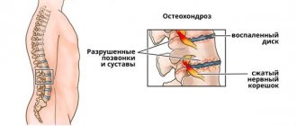
Osteochondrosis is one of the most common degenerative diseases of the spine.
Its effect on the body is overdetermined by the department it affects. For example, dysfunction of the autonomic nervous system, especially in individuals with thoracic and cervical osteochondrosis, can lead to hypotension, bradycardia and arrhythmia. In addition to this, deep venous thrombosis and coronary heart disease may develop. Most often, the progression of arrhythmia against the background of osteochondrosis is associated with compression of the vertebral artery, irritation of the nerve roots, hernia or protrusion of the disc.
Patients may complain of dizziness or even loss of consciousness, as well as nausea, weakness and blurred vision, and increased heart rate. Without treatment, symptoms progress and the condition worsens.
An arrhythmia is a malfunction of the heart that affects its speed or rhythm. Arrhythmia occurs when the electrical impulses that guide and regulate the heart's contractions do not function properly. They can be combined into several groups:
- acceleration of the rhythm (tachycardia);
- slowing down (bradycardia);
- premature contractions (extrasystole);
- chaotic or inconsistent formation of impulses (atrial fibrillation).
Depending on where the impulse originates, arrhythmias may or may not be sinus.
Arrhythmia during exacerbation of cervical osteochondrosis
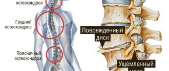
Cervical osteochondrosis not only makes the patient feel discomfort in the neck and limits movement, but also affects the functioning of internal organs, including the cardiovascular system. This leads to chest pain, rapid heartbeat, pulsation in the head, ischemia, cardiomyopathy, extrasystoles, and high blood pressure. This symptom complex is called “cervical angina”, “cervical angina”, collectively known as “neck syndrome”.
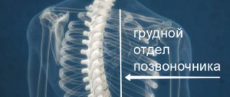
Lesion in the thoracic region and rhythm disturbances
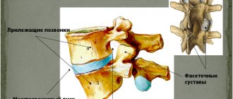
The thoracic region is the area of the spine below the neck connected to the ribs. It consists of 12 vertebral bodies with intervertebral discs. The disc acts as a cushion and also provides flexibility. The disc can wear out over time, due to aging or injury. Subsequently, the disc degenerates and the space between the vertebral bodies decreases.
Arrhythmia in osteochondrosis of the thoracic spine is manifested by pain in the upper or middle back and chest. If the disc is severely damaged, bone spurs can form and limit the mobility of the thoracic spine.
Spurs can cause a narrowing of the spinal canal. If the compression in the spinal cord increases, it can cause numbness, tingling and possible weakness in the limbs.
What happens in the vertebrae during osteochondrosis of the thoracic spine?
With osteochondrosis of this part of the spinal column, the degenerative process begins with the intervertebral disc. Changes occur in it at the biochemical level, due to this the disc swells for a while, then its height decreases. He ceases to cope with his function normally.
Then, tears and cracks appear in the peripheral part of the intervertebral disc - the fibrous ring. This predisposes to the subsequent development of intervertebral hernia.
The degenerative process also develops in the intervertebral and spinal-costal joints (spondyloarthrosis). This leads to narrowing of the intervertebral foramina through which the spinal cord roots exit, pinching, irritation and dysfunction of the latter.
Due to dysfunction of the intervertebral discs, the load on the vertebrae increases. A compensatory mechanism is activated - bone growths - osteophytes - appear along the edges of the vertebral bodies. This condition is called spondylosis.
If the first symptoms of the disease occur, visit the neurological center of the multidisciplinary international clinic Medica24. You will be examined by an experienced specialist and examined using modern diagnostic equipment.
Diagnosis and treatment
Determining the cause of the disease is the first step towards highly effective treatment. For this purpose, various methods and technological capabilities are used, but most often - radiography and MRI, electrocardiography and echocardiography of the heart.
Primary diagnosis is done using X-rays, and MRI can show us the extent of degeneration.
Arrhythmic heartbeat in osteochondrosis must begin to be treated by eliminating the cause. If structural changes in the heart have not yet occurred, the process can be reversed. That is, you need to focus on correcting spinal column disorders.
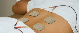
Non-steroidal anti-inflammatory drugs (for example, Ibuprofen) are used to relieve pain. In addition to this, there are a number of non-surgical treatment options that can help relieve pain:
- physiotherapy and exercise (McKenzie gymnastics, ultrasound therapy);
- extension (traction) of the spine;
- chiroplastic manipulations (to reduce joint dysfunction);
- osteopathic procedures;
- correction of motor mode;
- muscle relaxants;
- injections and blockades.
In some cases, surgical treatment is performed.
Effective treatment of symptoms of thoracic osteochondrosis
You can often cope with the symptoms of thoracic osteochondrosis at home. Many people “save” themselves, without doctors, with over-the-counter painkillers and painkillers, and by applying heat.
However, self-medication is only a temporary reduction in symptoms, and if done incorrectly, the problem can only worsen. During an exacerbation of osteochondrosis of the thoracic region, gymnastics and massage are contraindicated. On the contrary, at this time you should try to unload the spinal muscles as much as possible.
An experienced neurologist will correctly assess your condition and prescribe treatment that will help. The symptoms described in this article do not always indicate thoracic osteochondrosis; competent differential diagnosis is often necessary.
Visit a specialist. The neurological center of the multidisciplinary international clinic Medica24 employs experienced neurologists, uses modern diagnostic equipment, and treats diseases in accordance with the principles of evidence-based medicine. Clinic administrators are on duty at the telephone around the clock, call at any time.
We will call you back
Message sent!
expect a call, we will contact you shortly
The causes of thoracic osteochondrosis are not fully understood, and in general, this disease raises a large number of questions for many doctors. Some even argue that such a disease does not actually exist, and its manifestations are usually caused by completely different pathologies. By the way, in English-speaking medicine this term refers to completely different diseases.
conclusions
Osteochondrosis is a complex of degenerative disorders that affects the spinal column and can cause malfunction of internal organs, including the appearance of arrhythmias.
In most patients, osteochondrosis is treated without surgery. Physiotherapists recommend aerobic exercise (walking, cycling), strengthening the back muscles and skeletal traction. Surgery may be indicated if there are abnormal bone structures and neurological symptoms such as numbness, tingling and weakness in the legs.
In any case, treatment should be individual and comprehensive.
What reasons can lead to exacerbation of osteochondrosis of the thoracic spine?
The disease proceeds in waves - an exacerbation occurs, the symptoms of osteochondrosis become pronounced, then they subside - remission occurs. Factors that can provoke another exacerbation:
- In more than half of the cases, this occurs after sudden movements, prolonged stay in a monotonous position (static loads on the spine), intense physical activity, or heavy lifting.
- Hypothermia - both general and directly in the thoracic spine.
- Nervous tension, stress.
- Spinal injuries - acute (bruise), chronic (with prolonged and intense loads on the spinal column).
- Various infections.
Sometimes (in 15-20% of cases) exacerbation of thoracic osteochondrosis occurs for no apparent reason.
What is the relationship between depression and osteochondrosis?
The experience of neurologists shows that diseases of a physiological and psychological nature are often interrelated. The body cannot constantly work “like a clock,” especially if there is no regular “winding up.” An active lifestyle, physical exercise, proper nutrition, positive emotions - people deprive themselves of these pleasures in the pursuit of material well-being. Result: cartilage degeneration, circulatory disorders, and associated consequences: dizziness, inadequate reaction to psycho-emotional stimuli, increased anxiety, sometimes panic attacks.
What diagnostic methods are used for osteochondrosis of the thoracic spine?
For thoracic osteochondrosis, the doctor prescribes an examination, which may include the following diagnostic methods:
- Radiography. Usually photographs are taken in two projections: full face and profile. Based on the images in the photographs, you can evaluate the condition of the bodies and arches, articulations of the vertebrae, the height of the intervertebral discs, and the width of the spinal canal.
- X-ray contrast studies. Discography (images after the injection of a contrast solution into the intervertebral disc) and myelography (injection of a contrast solution into the space surrounding the spinal cord) help to detect at what level the spinal cord and its roots are compressed by the deformed intervertebral disc. Recently, these studies are practically not used, as more accurate diagnostic methods have appeared - CT and MRI.
- Computed tomography for thoracic osteochondrosis helps to detect the affected intervertebral disc, assess its size, the degree of “wear”, compression of the spinal cord and its roots.
- Magnetic resonance imaging allows you to examine the spinal cord and its roots, the degree of their damage, detect intervertebral hernia, and narrowing of the lumen of the spinal canal.
An important principle of the Medica24 international clinic is that we always have the interests of the patient as a priority. Our doctors never try to prescribe as many tests as possible. The scope of the diagnosis is exactly such that the neurologist can obtain all the necessary information, establish the correct diagnosis and prescribe the optimal treatment.
“Thyroid” causes of a “restless” heart
When the heart is “out of place,” we are usually talking about experiences of a psychological nature. But, if the heart is pounding with or without reason, the cause of the condition is no longer only in emotions, but first on the list of “heart provocateurs” are thyroid hormones.
Will make you worry not only the heart
Arrhythmia is a group of heart rhythm disorders that have a paroxysmal nature and can occur both after exercise and at rest.
During such an “attack,” the heart begins to beat faster or intermittently, and the minute volume of blood passing through the heart normally decreases significantly. Blood pressure “falls”. And patients at such moments may experience shortness of breath, headaches, dizziness and even lose consciousness.
The causes of this pathology may be hidden in defects in the conduction system of the heart itself, changes in the volume and rheological properties of the blood (for example, with dehydration or anemia) or an excess of thyroid hormones.
At the same time, T4 (or thyroxine) makes not only the heart “nervous,” because the hormone is a stimulator of absolutely all metabolic, energy and recovery processes in the body. And among the common symptoms of hyperthyroidism:
- general agitation and fussiness,
- unmotivated anxiety and irritability,
- insomnia,
- exhaustion due to normal nutrition,
- increase in body temperature,
- sweating, even at normal room temperature,
- increased thirst, frequent urination,
- increased pressure,
- redness of the skin and hair loss,
- Spastic digestive disorders (abdominal pain, diarrhea, constipation, etc.)
- and others.
Symptoms increase unnoticed, each time demonstrating greater resistance to standard treatment regimens.
As for the heart, then:
- with mild hyperthyroidism, the heart rate during an “attack” usually does not exceed 100 beats/minute;
- with moderate severity - on average, 120-140;
- and a severe form of pathology is accompanied by tachycardia above 140 beats.
Such arrhythmia, as already noted, is resistant to the action of conventional “heart” drugs. And, in the absence of correction of thyroid (“thyroid”) hormones, it can lead to the development of fibrillation and cardiac arrest.
When there is too much thyroxine
Hyperthyroidism, or excess thyroid hormones, can result from:
- excessive independent activity of the gland (diffuse toxic or nodular goiter, overdose of iodine-containing drugs)
- or an excess of the pituitary hormone TTT, which controls the activity of the gland.
In the first case, the cause of excess production of hormones may be either in the thyrocytes themselves (thyroid cells) or in stimulating antibodies. But the second - in most cases, in the pathology of the pituitary gland.
The exact cause of the disease can only be identified with the help of blood tests, ultrasound of the gland (with a biopsy in some cases), as well as CT or MRI, if necessary.
When is it time to see an endocrinologist?
The presence of arrhythmia along with several of the listed symptoms is a good reason to check the “health” of the thyroid gland. And in this case you will need:
- blood test for TSH,
- and free
- as well as antibodies to TSH receptors.
A decrease in TSH, even against the background of still normal or slightly elevated T4 and T3, indicates latent (subclinical hyperthyroidism); and if they significantly exceed the normative limits - about the clinical form (requiring immediate treatment) of the disease.
An increase in T4 and T3 simultaneously with TSH is a sign of pituitary pathology.
Detection of antibodies in the presence of symptoms is evidence in favor of Graves' disease.
Causes of depression in osteochondrosis
There are several reasons that cause depression when cartilage tissue is depleted. First of all, we are talking about impaired cerebral circulation associated with disorders in the cervical spine. A lack of blood flow, and therefore oxygen, leads to starvation of various parts of the brain responsible for certain important functions of the body. For example, vision, hearing, memory, speech, etc. Neurologists know that when faced with a shortage of resources, the brain “turns off” first of all the so-called “emotional background.” This is how nervous system disorders begin to develop: depression, aggression, panic attacks, insomnia, etc. In addition, a number of psycho-physiological reasons that develop in parallel in a person lead to a worsening of the condition:
- Constant suppression of symptoms. Acute processes are easier to overcome, and if the pathology is advanced, the body’s resources are depleted. As a result, a person is worried not only about pain in the back, neck, and lower back, but also any misunderstanding leads to stress.
- Waiting for an attack. Waiting for even a pleasant event is anxious, and when you are waiting for pain, a nervous state is guaranteed.
- Taking painkillers. By relieving pain in cartilage, a person suffering from osteochondrosis at the same time provokes the development of pathologies of the gastrointestinal tract, liver, and kidneys. This approach also leads to the emergence of phobias.
- Forced social and professional isolation. A person is not able to lead a normal lifestyle, it is difficult to do what he loves. Hence – psychological discomfort and depression.
- Deterioration of the functioning of the brain, which spends reserves on resisting the disease and is simply not able to “tune” other vital systems to normal functioning.
In patients who underwent treatment for osteochondrosis, symptoms of depression were observed 80% less often than in those who refused medical help. Conclusion: It is difficult to remain positive while waiting for the pain to come.