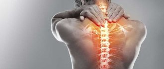
Pain relief for osteochondrosis can be achieved with the help of pharmacological drugs, traction procedures of the spinal column or through the use of osteopathy and pharmacopuncture. Everyone chooses exactly the technique that is more accessible to them at the moment. If you have nothing on hand other than ointment, then use it. Only at first
read the possible side effects and list of contraindications. In general, it is not recommended to take painkillers for osteochondrosis without a doctor’s prescription, as this can be dangerous to the patient’s health.
This article provides examples of which painkillers for osteochondrosis are prescribed by a doctor in a given case. This information is not a guide to action. Therefore, you should not consider it as advice on what painkillers you can take on your own for osteochondrosis without visiting a doctor.
The first thing worth remembering for all occasions is that taking any analgesics is possible only for 3 days. If the pain does not go away during this period, you should urgently seek medical help.
All painkillers for spinal osteochondrosis are divided into three groups: non-steroidal anti-inflammatory drugs, drugs that have a local irritant effect and muscle relaxants. Depending on the degree of destruction of the intervertebral discs and the stage of development of osteochondrosis, the doctor may prescribe one or another pharmacological agent. As a rule, in the early stages, the use of local irritant action is sufficient, due to which capillary blood flow increases and the process of primary regeneration of damaged tissue occurs. For protrusion of the intervertebral disc and compression of the radicular nerve, non-steroidal anti-inflammatory drugs are used. They relieve swelling of soft tissues and restore blood microcirculation. But at the same time they have a large list of contraindications and side effects. Therefore, it is not recommended to take them for a long period of time.
In advanced cases, with total damage to the cartilage tissue of the intervertebral disc, hormonal drugs are prescribed. They quickly eliminate the effect of inflammation, but quickly destroy the bone tissue of the vertebral bodies by leaching calcium salts.
Muscle relaxants make it possible to provide pharmacological assistance for static muscle tension in the area of the affected area of the spinal column. These drugs paralyze the ability of myocytes to tension. This has the opposite effect on the condition of the cartilage tissue of the intervertebral discs. Since the muscular frame of the back during protrusion and intervertebral hernia takes on most of the shock-absorbing load, thereby providing compensation for damaged tissue, the effect of muscle relaxants negates this effect. Neighboring vertebral bodies further compress the intervertebral disc. This can lead to its total destruction, the appearance of multiple hernial protrusions of the nucleus pulposus, etc.
How to properly relieve pain for osteochondrosis is described in this article. The most effective and safe methods of providing assistance for degenerative dystrophic lesions of the intervertebral disc are discussed here. If you require qualified help from a neurologist for this disease, then you can make a free appointment with a doctor at our manual therapy clinic in Moscow. Here you will be offered pain relief assistance without the use of potent pharmacological drugs.
Causes of osteochondrosis of the lumbar spine
Osteochondrosis is based on the destruction of the intervertebral disc - a flat cartilaginous formation that connects the vertebrae and ensures their mobility. The base of the disc is a soft nucleus pulposus surrounded by a fibrous (dense) ring. The core is 88% water and cushions the movement of the spine during vertical loading, such as lifting weights or walking. The function of the annulus fibrosus is to prevent the nucleus from moving away from the center of the vertebra. Also, the correct positioning of the core is facilitated by many ligaments and the muscular “corset” of the spine1,11.
At the age of 35-40 years, the blood supply to the disc core and its metabolism weakens, and the water content in it decreases to 60%1. But age is not the only and far from the main cause of lumbar osteochondrosis.
The following factors play a role in the development of the disease1,5,11:
- prolonged standing or sitting, especially in an uncomfortable position;
- overload of the spine associated with walking in high heels;
- poor posture, flat feet;
- nervous tension, chronic stress;
- overweight, metabolic disorders;
- poor physical development;
- congenital anomalies of the structure of the spine;
- heredity.
Most of these factors are related to the modern rhythm of life. It makes its own changes in a person’s habits, which turn out to be very harmful to the spine. The main problem is the imbalance between physical activity and the ability of the spine to bear a certain load. If the spinal muscles lose tone but are regularly overstrained, the load is transferred to the intervertebral discs and ligaments11. On the one hand, this situation creates conditions for microtrauma11, on the other hand, the risk of blood stagnation in the muscles increases, which prevents normal blood flow in them5.
In addition, the lower back bears the heaviest load11. And if the work involves heavy lifting, sudden movements, turns, flexion and extension, the pressure on the intervertebral discs increases many times over11. Therefore, wear of the intervertebral discs in the lumbar region is more often observed in people of certain professions - athletes, loaders and drivers5.
to come back to the beginning
Stages of lumbar osteochondrosis
When an intervertebral disc is damaged, its displacement is observed with the formation of an intervertebral hernia6, but all this happens gradually, so there are several stages in the development of the disease6,7:
- Stage 1 is characterized by unexpressed pain that occurs when lifting heavy objects. The patient can no longer tolerate prolonged physical activity, but at rest the pain, as a rule, does not bother him.
- In stage 2, the distance between the vertebrae decreases, which leads to pinched nerve endings. Sharp pain (lumbago) appears, radiating to the buttock, thigh and lower leg, forcing the patient to take a forced position with the torso tilted to the healthy side.
- At stage 3, the fibrous ring is destroyed and an intervertebral hernia is formed. The hernial protrusion compresses or irritates the roots of the spinal cord, the pain becomes severe, and is accompanied by disturbances in sensitivity (numbness of the limbs) and motor activity (weakness of the muscles of the leg or foot).
- By stage 4 , a pronounced deformity of the spine occurs, the patient moves with great difficulty, and the pain syndrome may be less disturbing.
What painkillers are available for cervical osteochondrosis?
Many patients do not know what pain medication is indicated for them for cervical osteochondrosis. The fact is that in most cases, pain syndrome in this localization of the degenerative dystrophic process is associated with excessive tension in the muscles of the collar zone. Therefore, standard non-steroidal anti-inflammatory drugs in such situations do not provide a visible positive effect.
What painkillers can be used for cervical osteochondrosis:
- muscle relaxant "Mydocalm" in the form of tablets and injections;
- “Capsicam” ointment for external use (the warming effect provokes muscle fiber relaxation);
- "Voltaren" in the form of a patch that gradually releases the active substance and effectively eliminates pain for 12 - 15 hours.
You can use any painkillers for cervical osteochondrosis only after consulting a doctor. This localization of dorsopathy is often associated with impaired cerebral blood supply. Therefore, not all drugs can be used.
For example, non-steroidal anti-inflammatory painkillers for osteochondrosis of the cervical spine can provoke the development of hemorrhagic stroke. These drugs can reduce the degree of platelet aggregation and increase possible bleeding. If the vascular wall is weak during a drop in blood pressure, bleeding may develop in the cerebral structures. In most cases, this results in permanent disability or death for the patient.
Muscle relaxants as painkillers for osteochondrosis of the cervical spine can be used only if it is possible, after the onset of their action, to lie down and relax the muscles of the collar zone. If you plan to continue working, this may lead to negative consequences. In particular, these painkillers for osteochondrosis can cause:
- increased compression pressure on the intervertebral disc;
- destruction of the fibrous ring;
- the formation of cracks and tears on its surface;
- the appearance of an intervertebral hernia;
- sequestration of intervertebral hernia;
- compression of the radicular nerve and the development of radiculitis;
- disturbance of innervation of the upper extremities;
- disturbance of heart rhythm due to compression of paired cranial nerves.
If you experience pain in the neck and collar area, we recommend that you seek medical help from a neurologist as soon as possible. The doctor will conduct an examination, rule out the possibility of displacement of the vertebral bodies, and perform a procedure for traction of the spinal column. In the future, a full course of manual therapy will be required to completely restore the damaged intervertebral disc.
Diagnostics
If you experience back pain, you should consult a neurologist. The history is followed by an examination of the patient, during which active and passive range of motion, muscle tone, limb strength, reflexes and sensation are assessed6.
The following instrumental diagnostic methods are used:6
- magnetic resonance imaging (MRI is a method for studying the layer-by-layer structure using magnetic fields);
- computed tomography (CT is a method for studying the layer-by-layer structure using x-rays);
- in rare cases, electromyography (a method for studying electrical potentials arising in muscles);
- functional spondylography (x-ray examination of any part of the spine).
You may additionally need to consult a urologist, gynecologist and proctologist1.
Advantages and disadvantages of cervical ultrasound
Each medical procedure has its own pros and cons. Fortunately, ultrasound examination of the cervical spine has more benefits. Let's look at some of them:
- 100% safety of the method. Clinical studies have shown that ultrasound does not harm the body. Even when compared with radiography or computed tomography, ultrasound has a significant advantage.
- Duration of the procedure. An ultrasound examination of the cervical spine is performed in less than half an hour.
- Study protocol. The necessary indicators are recorded, then deciphered by the ultrasound doctor and printed out. Immediately after the procedure, the patient receives information about the state of his health.
- Non-invasive. Ultrasound is absolutely painless; it does not cause a person physical or moral discomfort.
- No special training required. Before a spinal examination, the patient does not need to follow a diet or other preparatory measures.
- The only disadvantages that can be noted are the need for additional research if any abnormalities are detected in the patient.
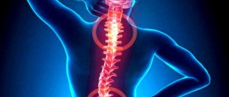
Methods for treating pain in lumbar osteochondrosis
The main manifestation of osteochondrosis of the lumbar spine is pain, which limits mobility, reduces the performance and quality of life of patients. Based on this, therapeutic measures in the treatment of this disease should primarily be aimed at pain relief4.
Pain with osteochondrosis of the lumbar spine can be 4:
- acute - lasts up to 6 weeks;
- subacute - from 6 to 12 weeks;
- chronic - more than 12 weeks.
In the treatment of osteochondrosis, conservative (non-surgical) and surgical methods are distinguished8. Therapy is selected individually, depending on the patient’s condition, his complaints and the data identified by the doctor during the examination4.
To relieve pain, complex drug therapy is used7.
Among the medications prescribed by the doctor may be4,7:
- muscle relaxants;
- antispasmodics;
- analgesics;
- non-steroidal anti-inflammatory drugs;
- tricyclic antidepressants;
- anticonvulsants;
- chondroitin sulfate and glucosamine.
Nonsteroidal anti-inflammatory drugs have been used successfully for the symptomatic treatment of back pain15. From this pharmacological group, the doctor may prescribe drugs containing naproxen15, for example, MOTRIN ®, which is intended for adult patients and adolescents over 15 years of age. One dose of MOTRIN ® is sufficient for up to 12 hours12. It should be borne in mind that the dosage and frequency of taking the drug is determined by the doctor.
Back in the 90s, patients with lower back pain were advised to limit active movements and observe strict and prolonged bed rest. This approach did not justify itself: it turned out that prolonged physical rest is harmful, since muscle function is impaired in this state. And this, in turn, leads to the pain becoming chronic and even often intensifying. According to research, acute low back pain is less of a concern for active patients. Therefore, by avoiding bed rest and following the recommendations of your doctor, you can recover faster13,14.
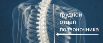
What painkillers should I take for osteochondrosis of the thoracic region?
The most effective and safe pain reliever for thoracic osteochondrosis is a traction procedure of the spine together with a session of osteopathy or pharmacopuncture. These methods allow not only to eliminate the symptom of pathology, but also to start the process of restoration of damaged tissue. Before taking painkillers for osteochondrosis of the thoracic region, it is important to understand that they do not have a therapeutic effect. All pharmacological drugs with an analgesic effect do not have a therapeutic effect. They relieve certain symptoms, for example, pain, spasm, stiffness, etc. At the same time, the disease continues to develop.
Do not look for an answer to the question of what painkillers to take for osteochondrosis, but rather be interested in effective and safe ways to restore damaged cartilage tissue of intervertebral discs.
Non-drug therapy
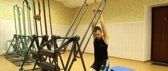
Physical therapy plays an important role in the treatment of lumbar osteochondrosis5,10,11. To prevent the development of the disease and alleviate its symptoms, the lower back muscles must be kept in good shape. Dosed physical activity and exercises help strengthen the muscle corset, increase range of motion and prevent complications5,11.
There are various complexes, which usually include exercises such as “bicycle”, “scissors”, lifting the body, performed in a lying position11. It is useful to do the “penknife” exercise: lying on your back, you need to press your hips to your chest and your head to your knees, “folding” like a penknife. It is recommended to do the exercise 10 times in total11. Therapeutic swimming also helps with osteochondrosis5.
It is better to carry out training under the supervision of a specialist in physical therapy, since many exercises are allowed only in the interictal period, and bending the torso forward at a certain angle can displace the disc even more5,11.
In addition to physical therapy, for lumbar osteochondrosis they use5,10:
- corsets;
- “stretching” the spine;
- manual therapy and massage;
- physiotherapy (magnetic therapy, laser therapy, ultrasound therapy);
- acupuncture.
Considering that osteochondrosis in advanced cases can lead to complications, it is important to pay close attention to the spine at any age. Prevention of disease and pain requires active action - normalizing weight, choosing comfortable shoes and regular physical activity. If the pain is severe, you should consult a doctor who will select the optimal treatment.
The information in this article is for reference only and does not replace professional advice from a doctor. To make a diagnosis and prescribe treatment, consult a qualified specialist.
to come back to the beginning
Literature:
- Biktimirov R.G., Kedrov A.V., Kiselev A.M., Kachkov I.A., Osteochondrosis of the spine - Almanac of Clinical Medicine - 2004. P.329,330,333,334, https://cyberleninka.ru/article/n/osteohondroz-pozvonochnika/viewer
- Human anatomy: textbook. -method. manual / M. V. Ulitko, I. M. Petrova, A. A. Yakimov; [under general ed. M. V. Ulitko]; Ministry of Science and Higher Education education Ros. Federation, Ural. federal n-t. - Ekaterinburg: Ural Publishing House. University, 2021. - 88 p. ISBN 978-5-7996-2447-7
- Oleynik A.D., Kovalev S.A. // Criteria for a comprehensive assessment of foci of lumbar osteochondrosis // Bulletin of the International Scientific Surgical Association - 2008 P. 84 https://cyberleninka.ru/article/n/kriterii-kompleksnoy-otsenki-ochagov-poyasnichnogo-osteohondroza/viewer
- Nikiforov A.S., Avakyan G.N., Mendel O.I., Modern approaches to the treatment of complications of spinal osteochondrosis - RMZh No. 26 from November 26, 2010 - P.1633 https://www.rmj.ru/articles/nevrologiya /Sovremennye_podhody_k_lecheniyu_osloghneniy_osteohondroza_pozvonochnika/
- Velichko T.I., Loskutov V.A., Loskutova I.V. // Exercise therapy and therapeutic swimming in orthopedics // 2014 -2.5 — Osteochondrosis https://monographies.ru/en/book/section?id=7270
- Gushcha A.O., Konovalov N.A., Dreval O.N., Grin A.A., Dzhindzhikhadze R.S., Arestov S.O., Dreval M.D., Kashcheev A.A., Vershinin A. V.V., Asyutin D.S., Korolishin V.S., Clinical guidelines for the diagnosis and treatment of herniated intervertebral discs of the lumbosacral spine. - 2014 - pp.4,7,8 https://www.mst.ru/information/manual/lumbar_disc_herniation.pdf
- Pilipovich A.A., Treatment and prevention of osteochondrosis - 2015. — S.1,17,18,19 https://cyberleninka.ru/article/n/lechenie-i-profilaktika-osteohondroza/viewer
- Rodichkin P.V., Shalamanov N.S. Pathophysiological rationale for complex therapy of intervertebral hernias -2012-P.32 https://www.vmeda.org/wp-content/uploads/2016/pdf/31-36.pdf
- Instructions for medical use of the drug Motrin ® tablets, Reg. number P N002874/01 ///o-motrin
- Buchakchiyskaya N.M., Maramukha V.I., Maramukha A.A., Maramukha I.V., Non-drug treatment of patients with neurological manifestations of osteochondrosis of the lumbar spine - Science of Life and Health - 2013. P.104 https://cyberleninka.ru/article/n/nemedicamentoznoe-lechenie-bolnyh-s-nevrologicheskimi-proyavleniyami-osteohondroza-poyasnichnogo-otdela-pozvonochnika-sovremennyy/viewer
- Therapeutic physical education for spinal osteochondrosis: textbook / O. V. Bezrukova, G. I. Bulnaeva; GBOU VPO IGMU of the Ministry of Health of Russia. – Irkutsk: IGMU, 2013. – 58 p.
- Frick et al. Efficacy and safety of naproxen sodium and ibuprofen for pain relief after oral surgery. Current Therapeutic Research. 1993;54(6):619-27.
- Clinical recommendations. Diagnosis and treatment of musculoskeletal (nonspecific) pain in the lower back. Russian Interregional Society for the Study of Pain. 2021
- Cochrane Database of Systematic Reviews. (2010.) “Advice to rest in bed versus advice to stay active for acute low-back pain and sciatica.” https://pubmed.ncbi.nlm.nih.gov/20556780/
- Svetoslav Nikolaev Stoev, Stanislav Radoslavov Gueоrguiev, Vasil Georgiev Madzharov, Hristina Viktorova Lebanova (2021). “Naproxen in Pain and Inflammation – A Review”, International Journal of Pharmaceutical and Phytopharmacological Research, 11(1), pp.142-148.