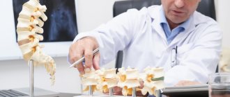
Physical activity for arthritis: 8 useful tips
Physical Activity for Arthritis: 8 Helpful Tips
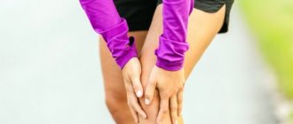
Pain makes people suffering from arthritis avoid physical activity, which is actually the best way to relieve it.
arthritis, physical activity, physical therapy
For 7% of Russians suffering from rheumatoid arthritis, joint pain causes them to refuse even light physical activity. However, numerous studies confirm that nothing relieves arthritis pain like exercise. What kind of physical activity is suitable for people with arthritis? What ways will make training more gentle?
Prohibited sports
Incorrectly selected physical activity leads to pain and exacerbations
When studying the topic of rheumatoid arthritis and sports, it is important to know that some physical exercises and activities can cause relapse and complications: renewed inflammation, increased pain and swelling. Sometimes associated injuries also occur. If you are diagnosed with knee arthritis, the following activities are strictly contraindicated:
- fast walking and running;
- long descents and ascents of stairs;
- intense strength training, as well as those that include sudden movements;
- jumping in place and jumping rope;
- snowboarding and ice skating;
- water aerobics and cold water swimming.
Is it possible to exercise if you have knee arthritis? With this disease, exercise therapy is the basis of therapy and effective prevention, but real results can only be expected with complex treatment. This means that you need to follow a diet and nutrition regimen, lead a healthy lifestyle, and take prescribed medications.
Separately, it should be said about the features of permitted sports. Not everything is so simple with such activities:
- Skis. There are two types of skiing: flat and mountain. The first is considered very useful for a sore knee, because it puts less stress than when walking. The main thing is to go straight, since a skating step (the ski is moved to the side) has a destructive effect on the joint. Mountain descents are strictly prohibited, as there is a significant risk of injury.
- Yoga and Pilates. In general, both of these activities are aimed at unloading joints, improving blood circulation, stretching and oxygenation. But both Pilates and yoga contain a large number of positions and asanas that can cause harm. These types of physical activity should be performed under the supervision of an instructor who understands the characteristics of the disease.
- Bicycle rides. If you have arthritis, you can only ride a bike on a flat surface. Strong shaking when driving over bumps causes destruction of diseased joints.
It should be remembered that each situation requires an individual approach. For example, if prohibited cardio training is really needed (when the cause of arthritis is excess weight), then running and jumping can be replaced with race walking.
The main priority is pain control.
The key to successful exercise for people with arthritis is pain control. Before starting training, be sure to visit a doctor who will determine the type of arthritis and prescribe the necessary anti-inflammatory drugs and procedures. Once the symptoms are relieved, the person can do any exercise that does not worsen the disease. The attending physician can also recommend exactly those exercises that will be useful and warn about the dangers of certain types of exercise.
Stretching for joint mobility
Regular stretching is very important to increase joint mobility. To relieve joint pain and stiffness, take a hot bath before stretching. Also, to reduce your risk of injury, warm up with light cardio: 10 minutes of walking in place or on an elliptical machine is ideal. Perform stretching exercises for 30 seconds without sudden movements or jerks.
Finger Stretch Make a fist with your hand. Then unclench and try to straighten your fingers as much as possible. Repeat this exercise twice a day, gradually increasing the number of repetitions to 20. You can also squeeze the resistance band to further strengthen the muscles.
Wrist muscle stretch This exercise should be performed while sitting at a table. The front part of the arm should be positioned on the table, and the hand should hang down. With your right hand, gently pull your left hand towards your shoulder. Then switch hands. Gradually increase the number of repetitions to 20, perform the exercise twice a day.
Elbow stretch Extend your arm forward so that it is parallel to the floor. The palm should face up. With your other hand, pull the palm of your outstretched arm so that your fingers point toward the floor. Hold it in this position for 30 seconds. Then do the same, only in the starting position lower the palm of your outstretched arm down.
Stretching the thigh muscles Sit or lie on your back with your legs slightly apart and straight at the knees. Turn your knees towards each other, your toes should touch. Stay in this position for 5 seconds. Then turn your knees the other way. Repeat this exercise twice a day, gradually increasing the number of repetitions to 5, 10, and then 20.
Ankle Stretch Stand facing a wall with your palms pressed against it. Place one foot forward, the other back. Slowly lean forward, making sure to keep your heels flat on the floor. Feel the tension in your calf and Achilles tendon. Hold this position for 30 seconds, repeat 3 times, then do the same with the other leg.
Don't forget about proper nutrition.
There is no magic diet that will relieve pain, but that doesn't mean you shouldn't eat healthy. You need to properly combine diet and exercise: for example, with a lot of strength exercises, the body needs protein for muscle growth (egg whites, almonds, chicken, salmon). With a strong cardio load, you need to provide the body with enough carbohydrates (fruits, whole grain bread, brown rice).
Expert: Galina Filippova, general practitioner, candidate of medical sciences Author: Olga Gorodetskaya
The material uses photographs belonging to shutterstock.com
Static exercises
If regular strength training is not possible due to severe joint pain, try static (isometric) exercises. With such loads, the muscles tense without visible movement.
1. Strengthening the chest muscles This exercise should be performed while standing. Place your hands at chest level and press your palms together as hard as you can for 5 seconds; then rest for 5 seconds. Repeat 5 times. Gradually increase the duration of the load to 10–15 seconds.
2. Strengthen your shoulder muscles To do this exercise, stand with your back to the wall and your arms down. Without bending your elbows, try to press your hands against the wall. Hold this position for 5 seconds and rest. Repeat 10 times.
3. Strengthening the thigh muscles Such exercises strengthen the thigh muscles, which are the main support for the knee joints. Sit on the floor or bed, straighten one leg and bend the other at the knee. Then tighten your thigh muscles on your straight leg as hard as you can, count to 6, and relax. Then do the same with the other leg. Repeat the exercise 5 times, gradually increasing the bar to 10–15 repetitions 2 times a day. Attention! If these exercises cause pain, ask your physical therapy instructor to find a different routine for you.
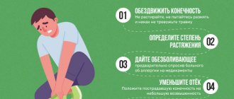
Rules
When any form of arthritis is detected, only the attending physician can select the optimal sports complex that the patient will perform for medicinal purposes. This is due to the need to apply dosed physical activity to the affected joint, since exceeding its permissible intensity leads to aggravation of pathological disorders. To do this, it is necessary to conduct a detailed examination of the patient using laboratory and instrumental diagnostic methods in order to establish the stage of development of the disease and the exact localization of the source of inflammation.
There are certain requirements for conducting sports activities:
- It is forbidden to carry out physical training during periods of exacerbation of the pathological condition, since the additional load on the damaged joint contributes to an increase in the intensity of the inflammatory reaction.
- After the manifestations of arthritis are stopped and the disease again goes into remission, sports training should be resumed gradually, starting with minimal stress on the joints.
- During the lesson, the patient should not perform exercises that would result in axial loads being applied to the damaged joints.
- During training, significant bending of the damaged joints should be avoided.
- When performing physical therapy exercises, you should not take analgesics to reduce the intensity of pain, as this can lead to additional injuries to the damaged joint.
- Before starting a set of exercises, the joints and muscles must be prepared - massage them for several minutes, making circular movements.
- The patient should not perform exercises at the limit of his strength. If the feeling of fatigue increases, you need to take a short break from training. If performing any exercise is accompanied by intense pain in damaged joints, it must be excluded from the sports complex.
To make it easier to perform physical exercises, after pre-warming the damaged joints, it is advisable to do a short warm-up - perform a series of movements with a small amplitude in the joints affected by arthritis.
Train at home
You can also train your muscles at home with or without equipment. Doing squats, wall push-ups, lunges and other exercises that use your body weight will strengthen your muscles and help protect your joints.
A physical therapist can show you exactly what exercises to do. An expander is an excellent, inexpensive option for home use.
On the subject: How to get rid of pain from rheumatoid arthritis?
Back to contents
Diagnostics
Diagnosing rheumatoid arthritis in the early stages can be difficult. Unfortunately, there is no pathognomic analysis that allows a confident diagnosis of rheumatoid arthritis. But nowadays, the diagnosis of rheumatoid arthritis is based on factors closely associated with this disease. There are a number of criteria for diagnosing rheumatoid arthritis:
- Morning stiffness in and around joints for at least one hour.
- Simultaneous swelling in three or more joints
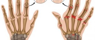
- The presence of swelling in the wrist or fingers.
- Symmetrical involvement of similar joints on both sides.
- Rheumatoid nodules are tumor-like formations in the skin (mainly in the elbow area).
- The presence of an increased level of rheumatic factor in the blood.
- X-ray signs of changes in the hands and wrists with damage to the bone tissue around the joints characteristic of rheumatoid arthritis.
The diagnosis of rheumatoid arthritis can be reliably made if 4 or more factors are present. The first four factors must be present for at least 6 weeks. The doctor needs to see the joints when the process is activated due to the following reasons:
- Patients cannot always describe their symptoms.
- Rheumatoid arthritis may present like other joint diseases
- Patients sometimes do not pay attention to mild pain and endure it until it is time to seek medical help.
- Some diseases that mask rheumatoid arthritis can make it difficult to diagnose: - gout, fibromyalgia, other autoimmune diseases (for example, SLE).
In view of the above, early diagnosis can be difficult. In fact, the average time from the onset of symptoms to diagnosis is almost 9 months.
Although diagnosing rheumatoid arthritis is not an easy task, it is extremely important to correctly identify the disease. A late diagnosis can be fraught with consequences due to the early involvement of internal organs in the process. Experts believe that timely treatment allows for good long-term results.
The doctor needs to carry out a number of measures in order to make a diagnosis:
- Medical history It is important to provide your doctor with information about the frequency, intensity, and time of day when symptoms occur.
- Physical examination The examination allows you to visually assess the presence of an inflammatory process in the joints.
- Laboratory tests These include both general tests and detection of rheumatic factor and high levels of sialic acid or the presence of autoantibodies, changes in the level of various enzymes, etc.
- Radiography allows you to diagnose any damage to bone tissue.
The place of physical rehabilitation in the treatment of juvenile arthritis
Rheumatology is one of the fastest growing specialties, and successes in treating patients, including those with such a serious disease as juvenile idiopathic arthritis (JIA), are undeniable. Timely prescribed adequate basic therapy, including genetic engineering therapy, significantly reduces the activity of rheumatic disease, as a result the patient’s condition improves, the range of motion in the joints expands, which allows the child to lead an age-appropriate active lifestyle [1–3]. Nevertheless, rehabilitation measures aimed at both improving the function of the affected joints and strengthening the child’s body as a whole and increasing his endurance are important for juvenile idiopathic arthritis. Therapeutic physical education (physical therapy) and other rehabilitation activities accustom the child to the need for constant exercise and help develop the correct stereotype of motor activity for life. However, at present there is not enough methodological literature devoted to the rehabilitation of children with rheumatic diseases; the available literature concerns mainly adult patients, so discussion of the problem is relevant and timely.
JIA is one of the most common and disabling rheumatic diseases in children. The incidence ranges from 2 to 16 per 100,000 children, and is more common in girls than boys. In the Russian Federation, the prevalence of JIA in children under 18 years of age reaches 62.3 [4, 5]. The classification and nomenclature of JIA includes the identification of seven variants of the course of the disease (according to the classification of the International League of Associations for Rheumatology (ILAR)):
1) systemic arthritis; 2) polyarthritis: negative for rheumatoid factor (RF); 3) polyarthritis: positive for the Russian Federation; 4) oligoarthritis: a) persistent and b) spreading; 5) enthesitic arthritis; 6) psoriatic arthritis; 7) other arthritis that: a) does not meet any of the categories or b) meets the criteria of more than one category.
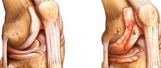
To date, the etiology of juvenile idiopathic arthritis remains unknown. The mechanism of development of the disease is based on the activation of cellular and humoral immunity, probably in response to the appearance of a foreign or altered self-antigen. As a result of complex interactions, activated T-lymphocytes, macrophages, fibroblasts, synoviocytes produce pro-inflammatory cytokines, causing a cascade of pathological changes with the development of progressive inflammation in the joint cavity. Uncontrolled reactions of the immune system lead to the development of acute immune inflammation with its transformation into chronic inflammation with the development of pannus and irreversible destruction of articular structures [4, 5].
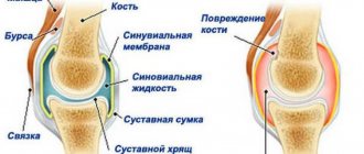
Articular syndrome is the leading symptom complex of all forms of idiopathic arthritis, and, from the point of view of rehabilitation, it should be assessed as a manifestation of maladaptation of the musculoskeletal system, in which the following typical pathomorphological processes are observed: inflammation, circulatory disorders, dystrophy and degeneration. As a result of these processes, pain, distortion, and deformation occur, leading to dysfunction of the joint. This pathological chain inevitably leads to a deterioration in the quality of life and disability of the child.
Rehabilitation, or restorative treatment of children, is a process that includes a set of measures. Rehabilitation measures are aimed at preserving the functionality of the affected joints and stabilizing the pathological process. In the rehabilitation of children, compliance with dietary recommendations is important; daily routine, physiotherapeutic methods of treatment; reflexology; Spa treatment.
Our publication focuses on kinesiotherapy methods, which occupy a central place in the physical rehabilitation of children with juvenile arthritis. By the term “kinesitherapy” we mean positional treatment (including orthotics), exercise therapy, massage, manual techniques, mechanical and occupational therapy.
Among the general recommendations, it should be noted that the child should sleep on a comfortable bed; an orthopedic mattress is preferable, not too soft, but not too hard. If the joints of the lower extremities and spine are affected, orthopedic shoes can be used, but, in any case, shoes with a hard back. Disability of our patients is often caused by damage to the joints of the hand. Therefore, it is necessary, with the help of doctors, exercise therapy methodologists, and parents, to form the correct stereotype of movements in the wrist joint to correct possible or existing ulnar deviation in it. So, you need to maintain a straight axis when performing all movements, including when performing physical therapy exercises (position on the edge of the palm), avoid positioning the hand towards the little finger. To prevent the formation of “swan neck” type deformities, it is recommended to reduce the load on the terminal phalanges - that is, a “cushion grip” is developed, thickened cone-shaped handles and pencils are used.
One of the key places in the rehabilitation of children with lesions of the musculoskeletal system is occupied by physical therapy. As a result of inflammation and painful sensations in the joints, a forced compensatory limitation of limb mobility occurs and, as a result, hypoxic and subsequently hypotrophic processes in the muscles [6, 7]. Exercises allow you to maintain and restore range of motion in affected joints, prevent the development of muscle wasting, and maintain muscle strength and endurance. Physical activity is a proven method of preventing osteoporosis, the risk of which is increased in patients with JIA [8]. Also, exercise therapy in childhood stimulates psychomotor development, is an excellent means of distraction from illness, and is an element of psychotherapy.
When carrying out exercise therapy, the level of load is dosed individually depending on the functional and age capabilities of the child. It is advisable to carry out a set of exercises 2-3 times a day (by a methodologist and trained parents). The complex necessarily includes breathing exercises, as well as exercises for developing correct posture, strengthening the muscle corset, and activating large and small muscles of the limbs.
Exercise therapy has virtually no contraindications, with the exception of the period of high activity of JIA, accompanied by fever, other systemic manifestations of the process, severe pain, and pronounced humoral changes. In this situation, for the purpose of early prevention of the formation of contractures, the so-called. passive gymnastics, when work with joints is carried out by a physical therapy instructor or a trained parent; the range of motion in the joints is determined within the pain-free corridor [6]. Passive gymnastics may also be necessary for young children if the child himself is not yet able to follow the instructions of the methodologist. The use of this method may be indicated in the case of active complicated uveitis in patients with JIA if joint development is necessary. According to individual indications, after performing a complex of passive gymnastics, treatment with a weighted position can be used.
Treatment with positioning using weights is also used in the presence of formed joint contractures. According to the method, the joint is brought to the “extreme” position in the direction of limited movement and a weight is fixed on it, the gravity vector of which coincides with the vector along which movement is limited. Depending on the age and condition of the patient, the lesson time ranges from 10 to 30 minutes 3–5 times a day. When performing this manipulation, a gradual slow passive stretching of the periarticular muscle-tendon apparatus occurs, which leads to an improvement in the motor function of the joint. However, according to the children’s department of the Research Institute of Rheumatology, the use of weights (including “cuff traction”) has worked well for damage to the knee joint, but in the acute period it only worsens the condition of the elbow joint [8].
Orthosis is an important method of rehabilitation treatment for patients with JIA, the main goal of which is the correction of pathological deviations of the joints and the formation of their correct functional alignment. The use of orthoses during physical therapy exercises is especially effective for maintaining and consolidating the achieved results during the development of joints.
The orthosis mode and its use are selected individually in each case. Practice shows that the most “in demand” are orthoses for the wrist, knee, and ankle joints. If necessary, some orthoses can be worn at night [4, 6].
It is recommended to wear a “Schanz collar” if the cervical spine is affected. The height of the treatment collar should be equal to the distance from the bottom to the top of the neck, that is, to the jaw in front and to the base of the skull at the back. It is recommended to wear the collar for a total of 1.5 to 3 hours a day, depending on the age and individual characteristics of the child [6].
The myofascial component has a certain significance in the formation of chronic pain syndrome and the development of pain contractures. Myofascial pain syndrome (MPS) is a myalgia manifested by local and/or referred pain, the source of which is the myofascial trigger point (MTP). MTT is a group of compacted, as if frozen muscle fibers, in which there is an area of intense pain [9]. MTT is characterized by the presence of a constantly or not always palpable compaction (“cord” or “nodule”) within the muscle, as well as a sensory disorder, which most often manifests itself as pain. The mechanism for the appearance of MTT is believed to be irritation of sensitive nerve endings in joints, muscles and ligaments, which leads to chronic excitation of mechanoreceptors involved in the formation of pain, and physical and metabolic muscle tension occurs. The structure of muscle contraction changes, satellite trigger points are formed in the muscles that perform a compensatory function. An important part of stress is hypoxic (ischemic) damage. Trigger points can contribute to the formation of tendinosis due to disruption of the dynamics of muscle contraction, local overload of the tendon and the occurrence of relative hypoxia in these areas [9, 10]. This mechanism is also implemented in patients with JIA: when local pain occurs, the “muscle protection” mechanism is activated, and the resulting spasm leads to a decrease in motor activity. In a state of physical inactivity, conditions are created for ischemia of the muscular system, which is one of the prerequisites for the development of MBS [9]. The pain syndrome can be “reflected” from spasmodic periarticular muscles and tendons: thus, according to our observations, when they are relaxed using manual techniques, pain in the joint area has a clear tendency to decrease. Manual influence on the area of the myofascial pain point in our patients leads to a pronounced softening of the dense cord and an increase in the pain threshold. It was also noted that with myofascial relaxation, in a large proportion of cases the severity of enthesopathies decreases. In order to correct the myofascial component of the pain syndrome, our patients use the post-isometric relaxation (PIR) method, as well as soft manual (osteopathic) techniques.
PIR is used in the presence of myalgic syndrome. By relaxing spasmed muscles, the ischemic component of the formation of MBS is eliminated, their blood supply is restored, and pain is reduced. In addition, PIR has proven itself well when working with joints in which restriction of movement has developed due to both severe pain and painless spasms of the periarticular muscles and tendons, as well as due to a decrease in the size of the joint space. The exercises are performed as follows: the patient moves towards the restriction to the “barrier”, then fixes the joint in this position for 1–2 minutes and relaxes the tension. The training regimen is selected individually and performed by a trained specialist.
In the case of multiple trigger points and the patient’s increased sensitivity to pain, it is possible to use osteopathic correction of myofascial tension. The actions are carried out with the lightest touches at the level of the muscle fascia, which makes it possible to achieve relaxation of the muscle-tendon system without causing pain. Typically, this technique is used 1–2 times, after which the patient is transferred to a combination of PIR + exercise therapy to establish positive feedback between one’s own work and the achieved result [6].
Osteopathic correction and PIR techniques are used both in the stage of remission and in the stage of clinical and laboratory exacerbation of the disease and can accelerate the reduction of pain, improve the well-being and motor function of the joints, and help prevent the formation of joint contractures. Unfortunately, these techniques are not always applicable to small joints of the hands and feet; they have proven themselves much better on large and medium-sized joints [6].
Manual massage is also included in the complex of rehabilitation measures for JIA. It prevents the increase in muscle hypotonia and malnutrition and is aimed at eliminating muscle imbalance in the periarticular muscles. Massage can be indicated at the stage of proliferative changes in the joints without exacerbation of the disease. Methods generally accepted in childhood are used. The joint area is not massaged. Massage is contraindicated for acute synovitis, febrile syndrome, serositis and visceritis, humoral activity above grade 1, and the presence of general contraindications (acute respiratory viral infections, skin diseases, etc.) is also taken into account.
Mechanotherapy is one of the methods of medical rehabilitation. It is based on the use of dosed movements performed by patients using special devices. The method is more suitable for older children. Contraindications for mechanotherapy are the presence of bone ankylosis, severe pain in the joints, severe muscle weakness, and impaired congruence of the articulating surfaces of bones [11].
Rehabilitation and treatment of our young patients is ultimately aimed at ensuring a high quality of their future life, adaptation to living conditions and future work. Occupational therapy, or “occupation therapy,” promotes integration into public life and socialization of children with juvenile arthritis. Occupational therapy is of great importance in cases of damage to the small joints of the fingers, allowing to slow down the progression of impairment of their motor functions. Modeling, beading, knitting, playing the piano, etc. are recommended. This method is also useful as a means of increasing general and mental tone, and allows the use of play techniques in younger children. Receiving the product of labor serves as an incentive for better performance of work and includes elements of competition and creativity. The correct stereotype of hand function developed in the process of occupational therapy is reinforced when performing everyday manipulations.
In conclusion, I would like to note that, despite all the successes of modern drug treatment of juvenile arthritis, physical methods of rehabilitation occupy an important place in the complex of patient management. The rehabilitation plan is drawn up individually, based on the problems of a particular patient, and not just on his diagnosis. Rehabilitation should be an activity not only for doctors and methodologists, but also for the patients themselves and their parents. The tactics of rehabilitation measures and the program should be drawn up together with them; with this approach, the patient becomes the central figure of rehabilitation treatment and an active participant in this process.
Literature
- Konopelko O. Yu., Zholobova E. S., Rozvadovskaya O. S., Elyashevich V. Ya., Nikolaeva M. N. Etanercept in real clinical practice in the treatment of patients with active juvenile idiopathic arthritis) // Scientific and Practical Rheumatology. 2013, 51(1): 44–47.
- Neroev V.V., Katargina L.A., Denisova E.V., Starikova A.V., Lyubimova N.V. Efficacy of genetically engineered biological drugs in the treatment of uveitis associated with rheumatic diseases in children // Scientific and Practical rheumatology. 2012, 53(4): 91–95.
- Michels H., Nikishina I.P., Fedorov E.S., Salugina S.O. Genetic engineering biological therapy of juvenile arthritis // Scientific and practical rheumatology. 2011, no. 1, 78–93.
- Zholobova E. S., Shakhbazyan I. E., Ulybina O. V. et al. Juvenile rheumatoid arthritis. Guide to pediatric rheumatology / Ed. N. A. Geppe, N. S. Podchernyaeva, G. A. Lyskina. M.: GEOTAR, 2011. pp. 162–245.
- Pediatric rheumatology: clinical recommendations for pediatricians / Ed. A. A. Baranova, E. I. Alekseeva. M.: Union of Pediatricians of Russia, 2011. 236 p.: ill.
- Geppe N. A., Meleshkina A. V., Makarova M. R., et al. Physical rehabilitation of children with juvenile arthritis: a textbook for students of medical universities: local electronic publication. M.: Publishing house GBOU VPO First Moscow State Medical University named after. I. M. Sechenova, 2014.
- Milyukova I. V., Evdokimova T. A. Therapeutic physical education: The latest reference book / Ed. ed. prof. T. A. Evdokimova. St. Petersburg: Sova; M.: Eksmo Publishing House, 2003. 862 p.
- Shelepina T. A. Therapeutic gymnastics in complex therapy in patients with juvenile chronic arthritis // Modern rheumatology. 2013, (3): 64–6.
- Ferguson LW, Gerwin R. Clinical Mastery in the Treatment of Myofascial Pain. Lippincott Williams & Wilkins, 2005.
- Naomi Lynn Gerber, Siddhartha Sikdar, Jen Hammond, Jay Shah. Brief Overview and Update of Myofascial Pain. Syndrome and Myofascial Trigger Points // Journal of The Spinal Research Foundation. Spring. 2011, vol. 6, no. 1.
- Physical rehabilitation. Textbook for students of higher educational institutions / Under the general editorship. prof. S. N. Popova. Ed. 3rd. Rostov-on-Don: Phoenix, 2005. 608 p.
A. V. Meleshkina1, Candidate of Medical Sciences A. V. Bunin N. A. Geppe, Doctor of Medical Sciences, Professor S. N. Chebysheva, Candidate of Medical Sciences
GBOU VPO First Moscow State Medical University named after. I. M. Sechenova Ministry of Health of the Russian Federation, Moscow
1 Contact information