A mobile dynamic system - the human spine, which allows you to maintain the shape that nature gave, carries out the functions of ensuring and preserving the spinal cord. Various diseases and injuries of the spine are a possible impetus for the development of degenerative-dystrophic processes, which result in a disease such as vertebral retrolisthesis.
Vertebral retrolisthesis
- posterior displacement of a vertebra, when the normal relative position of neighboring vertebrae is lost, which can occur at any age.
Upright posture determines the anatomical features and biomechanics of human movements, which explain the appearance of vertebral retrolisthesis in areas where there is a physiological deviation from the axis of the spine - in the cervical and lumbar region. The thoracic region is least susceptible to this pathology.
The most common pathology of retrolisthesis is retrolisthesis of the cervical spine, with the smallest size of the vertebrae susceptible to traumatic lesions. Less often, doctors are faced with the diagnosis of retrolisthesis of the L5 vertebra, which bears the maximum load during movements, due to the fact that the sacral region following it has minimal mobility.
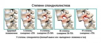
Retrolisthesis has the following classification, showing the degree of stability of the motion segments, which depends on the size of the displacements:
- I Art. — offset up to 25%;
- II Art. — offset from 25% to 50%;
- III Art. — offset from 50% to 75%;
- IV Art. — offset from 75% to 100%.
The long course of the disease with retrolisthesis, with an increase in the size and extent of vertebral displacements up to 10 millimeters, leads to an irreversible phase. As a result of the displacement of the vertebra - vertebral retrolisthesis, there is an increased load on the tissues surrounding it, and with a prolonged incorrect load ratio, the facet joints are overloaded, resulting in the development of spondyloarthrosis.
The developing pathological process affects:
- vertebral discs with fibrous rings;
- anterior longitudinal and capsular ligaments;
- end plates with their cartilages.
In this case, the stability and activity of joint activity is disrupted; asymmetry of muscle tone is observed; disturbances occur in the ligamentous apparatus and fascia.
The occurrence of retrolisthesis, with a posterior displacement of the vertebra, is usually the result of trauma or other damage to the spinal column:
- with long-standing damage to intervertebral discs;
- in a situation where the spinal segments are overloaded, having different directions during movement;
- compression fracture;
- ligament rupture.
The L 3 and L2 vertebrae are the least susceptible to retrolisthesis, which can only occur as a result of direct injury to the spinal column. Often a pathological process, vertebral retrolisthesis, develops when intervertebral discs soften with abrasion and rupture. When there is no support for the lower vertebra with any pathology, displacement of the upper vertebra occurs. Older people undergo vertebral retrolisthesis as a consequence of a disease such as arthritis, when disc tissue is depleted.
The following reasons can provoke the development of the process of vertebral retrolisthesis:
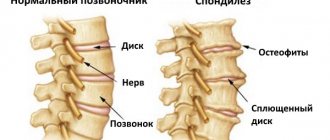
- diseases of the spine - spondylosis and osteochondrosis;
- the presence of congenital weakness of vertebral structures;
- hereditary factor;
- tumor processes in the spine and surrounding structures;
- the presence of age-related changes in the structures of the spinal column.
Among people predisposed to a disease with vertebral displacement, a large percentage is occupied by men, whose area of activity includes heavy physical labor, activities with short-term, intense loads on the spine in athletes - weightlifters, wrestlers, acrobats. The possibility of developing vertebral retrolisthesis in young children arises from injuries and participation in traumatic sports.
Sign up for a free consultation +7
Clinical manifestations of vertebral retrolisthesis
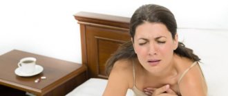
The pathology may not manifest itself for quite a long time, occurring in a latent form with pain that occurs in a person as a result of physical activity. There is a gradual increase in pain intensity with the addition of accompanying symptoms. The patient complains of pain of a certain localization. Their severity is determined by the pathological process, leading to disruption of the motor functions of the segment due to the fact that the soft tissues become unstable.
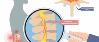
The pressure that a displaced vertebra puts on the nerve structure leads to its dysfunction, which is accompanied by muscle spasms. Retrolisthesis with tissue instability leads to feelings of discomfort in the pathological area, and subsequently to severe movement disorders throughout the entire spine.
Symptoms
The following symptoms are characteristic of retrolisthesis:
- impaired mobility of the spine, accompanied by discomfort;
- decreased range of motion as deformation of the vertebral joints occurs;
- pain caused by irritation of the spinal roots;
- the appearance of protrusions with subsequent transformation into intervertebral hernia;
- neurological disorders (reduced muscle sensitivity, dizziness).
Symptoms of retrolisthesis manifest themselves in different ways and have different intensities. This is influenced by the degree of development of the pathology, since each of them puts different pressure on the nerve endings and mechanics of the spinal joints.
Lumbar - vertebral retrolisthesis
The manifestation of retrolisthesis of the L5 vertebra, which is a more common type of pathology, pain in the lumbar spine, is often accompanied by neurological symptoms; in complex cases, paralysis develops, affecting the lower part of the body. In combination with retrolisthesis of the L5 vertebra, the development of retrolisthesis of the L4 vertebra, with similar manifestations, is possible. With lumbar retrolisthesis, the effect of loads on L5 and the location of the S1 endplate depends on the severity of lumbar lordosis.
Clinical manifestations of pathology:
Symptoms for retrolisthesis differ:
- pain that occurs during physical, even slight exertion or awkward movement in the lumbar area, radiating to the leg,
- involuntary shortened steps while walking;
- aching pain that occurs during a long stay in static positions;
- various sensory disorders;
- nearby internal organs work with functional impairments;
- manifestation of Wasserman's symptom, with the occurrence of a sharp pain in the groin while raising the lower limb upward, when the patient lies stomach down;
- a pronounced Lasegue symptom appears - pain occurs in the lumbar area, along the sciatic nerve, during raising of an elongated lower limb, when the patient lies on his back, and these pain symptoms disappear during flexion of the knee joint.
Thoracic region - retrolisthesis
The thoracic region is distinguished by one feature - it has additional fixation of the spinal column, thanks to the upper shoulder girdle and rib cage, which is an obstacle to any shear loads, therefore such a pathology as spinal retrolisthesis is rarely observed in the thoracic region. Localization of retrolisthesis in this part of the spinal column is manifested by characteristic signs: pain in the area of development of the pathology; numbness of the upper extremities; shortness of breath, cough; suffocating attacks; peptic ulcer, pain in internal organs such as the liver, gall bladder, kidneys.
Causes
Retrolisthesis has a large list of causes, where the main place is occupied by spinal injuries, increased loads and monotonous movements of the lower back (flexion-extension), so the disease is often diagnosed in people whose profession involves heavy physical labor or work in a sedentary position. In addition, retrolisthesis develops as a concomitant pathology with:
- intervertebral hernia;
- osteochondrosis of the cervical, lumbar and thoracic region;
- disc protrusions;
- arthrosis;
- spondylosis;
- kyphosis;
- lordosis;
- degenerative-dystrophic changes in intervertebral discs.
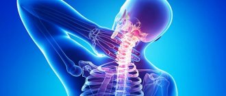
Cervical spine - retrolisthesis
The most anatomically complex and easily injured part of the spinal column, the cervical spine, is distinguished by the following features:
- weak muscle corset;
- the absence of an intervertebral disc at the junction of the skull and the subsequent vertebra, and at the junction of the first and second vertebrae;
- the vertebral arteries pass through a canal formed by the transverse vertebral processes;
- The cervical spine has the greatest mobility.
Vertebral arteries and nerve endings play a large role in feeding the brain and its vital posterior sections. With retrolisthesis, the cervical vertebrae are displaced and compressed, which can lead to hypertension and the development of signs of cerebrovascular accident that are difficult to treat.
With the development of retrolisthesis in the cervical spine, the following symptoms may appear:
- headache,
- dizziness;
- the appearance of tinnitus;
- pain in the cervical area;
- elevated and unstable blood pressure;
- visual impairment;
- manifestations of nausea;
- the appearance of weakness and numbness of the hands;
- Symptoms intensify when moving the head.
The development of retrolisthesis in the cervical region C3 - C5 leads to a long, protracted course, stable functional disorders, which may result in the patient’s disability.
Diagnosis of retrolisthesis
Retrolisthesis has one feature when we compare this disease with other pathologies of the spinal column, which is characterized by a high level of vertebral slippage. The examination must be carried out carefully, establishing the exact size and nature of the vertebral displacements and associated damage. The initial examination is based on the patient's complaints. The doctor, at the same time, checks for any neurological abnormalities that are present. To confirm the disease, the following research methods are used:
- X-ray - shows the presence of osteophytes and characteristic bone growths
- MRI - magnetic resonance imaging - to detect morphological changes in ligaments, discs, nerve roots, and spinal cord. Retrolisthesis is associated with protrusion of the vertebra, invisible on X-ray examination, but magnetic resonance imaging allows it to be detected. This type of study serves to better predict the development of processes and find the most adequate tactics in the treatment of retrolisthesis.
- Electroneuromyography - this diagnostic records and analyzes bioelectrical activity, reflects the state of the fibers when they are subject to muscle tension and at rest. The method is used when it is necessary to determine the area and degree of damage to the nervous system.
- Ultrasound Dopplerography of blood vessels is a study used to identify the condition of blood vessels.
To choose an effective method of treating retrolisthesis, the doctor at the appointment determines the stage of the disease, the intensity of pain, functional disorders in the muscles and internal organs.
What it is?
The diagnosis of retrolisthesis is made in cases where the vertebrae are displaced relative to the structure of the spinal column itself . The fifth lumbar vertebra is more at risk because there is a certain curvature in the area of this vertebra.
Of course, the pathology has an extremely negative effect on the integrity of the spinal sections and increases the possibility of pinching nerve roots and pressing on the spinal cord.
In fact, any of the vertebrae can slip, but in fact, as a rule, it is the lumbar region that is affected, that is, L4 and L5 , as well as the cervical region. That is why the L5 vertebra with restrolisthesis is the most important section for studying the disease.
Retrolisthesis is mostly a “male” disease, since the male part of the population is more susceptible to physical stress associated with the spinal column. The main percentage of patients are adults, due to the fact that the disease is mainly associated with pathologies already formed in adulthood.
However, there are exceptions in the form of traumatic factors that also affect children. In the case of injury at a young age, it is much easier to displace and thus cure retrolisthesis than to carry out this procedure in old age.
Picture of the course of the disease
lumbar pains will clearly indicate that you have retrolisthesis . They can either remain localized in this one area or spread further to the limbs. During the examination, the doctor should immediately prevent the formation of scoliotic deformity of the spine and smoothing of lordosis. At the same time, the lumbar muscles experience unnatural tension, which can be recorded by a doctor as asymmetric muscle tone.
Even during the examination, special symptoms of tension may be revealed , or, if scientifically speaking, Lasegue’s symptoms are when, when stretching your leg in a supine position, you experience a sharp pain that disappears as soon as you bend it at the knee again; Wasserman's symptoms are also revealed - a lumbago occurs when raising the leg vertically, in a supine position.
When the patient ambulates, he or she has a condition defined as neurogenic or caudogenic intermittent claudication. Such a patient has “short steps” when walking. The functioning of joints and ligaments is impaired, skin-muscular sensitivity weakens. The patient begins to experience pain if he sits in the same place for too long.
With retrolisthesis of the L5 vertebra, a disorder occurs in the nervous and muscular systems
A special problem and a significant worsening of symptoms is if, as a result of displacement of the vertebrae, the spinal cord is affected , if it is compressed or pinched. In this case, paresis of the limbs, various digestive disorders, and spontaneous acts of defecation and urination may be added to the overall picture.
Sometimes intervertebral discs are also involved, and ligaments and cartilage are at constant risk. All this is associated with ongoing pain.
Of course, the disease also affects internal organs. Features of the course of the disease are disorders of the kidneys, liver functions, and disruption of the gallbladder. Against this background, shortness of breath and constant cough appear. Sometimes a peptic ulcer develops.
Degrees and classification (if any)
- The first stage of retrolisthesis is characterized by the fact that the vertebra is displaced in relation to the underlying one by 1/4, or 25%.
- The second degree is when half of the vertebra has already disappeared.
- At the third degree, the spine descends by 75%.
- At the last, fourth degree, the vertebra falls out completely.
It should be noted that moving disks takes a long time and occurs over many years. For example, the onset of pathology can be a genetic deviation in the development of the spinal column - its curvature towards the sacrum.
In addition, vertebral retrolisthesis has its own classification:
- the transverse processes of the vertebrae may be fractured, and this will be called spondylolysis spondylolisthesis;
- if retrolisthesis began due to injury, then this is, accordingly, called the traumatic form;
- in the case of osteochondrosis, degenerative retrolisthesis is usually recorded;
- if there is inflammation or swelling, then this is pathological spondylolisthesis;
- and sometimes, a negative result of incorrectly performed surgical operations is post-surgical retrolisthesis.
ICD code
ICD-10 (International Classification of Diseases) diagnosis code: M43.2. Disease "Other fusions of the spinal column."
Prevalence
In most cases, it is the l5 vertebra that is subject to displacement.
l4 is less at risk only if retrolisthesis of the l5 vertebra has already developed.
l2 and l3 are affected only in very rare cases. These are usually traumatic cases.
Conservative treatment method
When the disease is detected in a timely manner, a conservative treatment method for the initial degree of retrolisthesis gives good results. Treatment of retrolisthesis may include the following methods and recommendations:
- it is important to limit physical activity;
- wear an orthopedic corset;
- drug treatment;
- application of soft manual therapy techniques;
- acupuncture;
- laser therapy;
- therapeutic massage course;
- physiotherapy;
- autogravitational therapy - therapeutic traction of the spine;
- physiotherapy;
Treatment tactics are determined by the patient’s condition; it is also taken into account that retrolisthesis is a traumatic curvature of the spinal column. It is necessary to carefully prescribe therapeutic exercises, for the reason that it is possible to provoke a deterioration in the patient’s well-being due to the load on the diseased segment.
In case of illness, special collars, corsets or belts are prescribed that relieve stress from the muscles of the neck and back, which are an orthopedic correction. They need to be worn for a certain, short period of time, so as not to contribute to muscle atrophy.
Autogravitational therapy (therapeutic spinal traction) is one of the effective measures that promotes the release of intervertebral tissues, their nutrition, and prevents destructive dystrophic and degenerative processes in them.
Drug therapy
Drug treatment is prescribed to patients with muscle spasms and severe pain, using painkillers and anti-inflammatory drugs, and drugs for neurological symptoms. If there are concomitant diseases, the doctor may prescribe medications to improve the condition of the joints.
Medications prescribed for retrolisthesis:
- analgesic drugs - to reduce and eliminate pain;
- NSAIDs (non-steroidal anti-inflammatory drugs), to relieve inflammation and reduce pain;
- the appointment of muscle relaxants helps reduce muscle tone and reduce pain;
- neuropathic drugs treat the nervous system with organic lesions.
In difficult cases, a course of epidural steroid injections, consisting of two or three procedures, can be added to the course of treatment for retrolisthesis to eliminate pain, swelling, irritation and numbness. A steroid drug (cortisone) is injected into the epidural space.
Classification
The distance by which the vertebra is displaced downward allows us to distinguish five degrees of retrolisthesis:
- 1 degree with a displacement of no more than 25%;
- 2nd degree with a displacement of no more than 50%;
- Grade 3 varies from 50 to 75%;
- 4 degree with displacement more than 75%;
- Grade 5 with 100% displacement, very rarely diagnosed.
According to etiology, retrolisthesis is divided into:
- degenerative: occurs during the process of wear and aging of the spine and its discs;
- dysplastic: formed as an intrauterine pathology and refers to birth defects;
- traumatic: consequence of injury to intervertebral joints and arcuate plates;
- isthmic: develops as a concomitant disease with spondylolysis or as a congenital defect in the vertebral arch;
- pathological: formed when a bone tissue defect appears due to a tumor.
Retrolisthesis is also classified according to the level of localization and the structure of displacement:
- complete displacement: relative to two adjacent segments, the vertebra is completely displaced backwards;
- stepwise displacement: relative to the upper vertebra, the damaged segment is displaced backward, relative to the lower vertebra - forward;
- partial displacement: a vertebra is displaced backward relative to one segment above or below.
Surgical treatment of retrolisthesis
Surgery is used if the patient:
- severe retrolisthesis of a high degree;
- persistent neurological symptoms;
- Conservative treatment did not bring the desired effect.
Surgery depends on how much the vertebra is displaced. The operation aligns and fixes the vertebrae in the correct position. When vascular or nervous structures are compressed by bone growths or tumors, surgery is performed to remove them. Special implants strengthen the vertebra in case of displacement caused by an unstable ligamentous-muscular system.
The postoperative recovery period, during which it is prohibited to lead an active lifestyle, lasts several months and can last up to 1 year. Currently, modern methods of surgical treatment of spinal retrolisthesis are used - in one of them, displaced vertebrae are connected using special screws, attaching them to a metal structure.
How is the treatment carried out?
The entire treatment takes 7-10 days. The patient is hospitalized 1 day before surgery with tests ready.
The operation is performed under general anesthesia, i.e. For the patient, the entire operation takes place in 1-2 seconds; for the neurosurgeon, it takes 3-4 hours to perform the operation.
The day after the operation you can get up and walk around the room, the day after the operation you can walk along the corridor in a corset.
Before discharge, detailed recommendations are given for the immediate and long-term postoperative period.