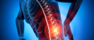
Median hernia (median, medial) is a degenerative disease of the intervertebral disc, in which its shell is destroyed and the inner part falls out into the middle of the spinal canal to the central line of the vertebra. It is dangerous due to compression of the nerve roots and spinal cord. Most often it occurs in the lumbar spine, less often in the cervical and thoracic spine.
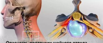
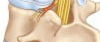
Cervical hernia on MRI.
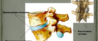
What is it - a median hernia?
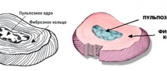
The formation of a median herniation of the intervertebral disc occurs when it is deformed. The internal semi-liquid contents of the disc (nucleus pulposus) breaks out through the dense fibrous membrane and protrudes into the center of the spinal canal.
The pathology causes severe pain due to pinched nerve endings; due to compression of the spinal cord, the patient may lose mobility.
A median hernia can appear in different places of the spine, but most often the lumbar or lumbosacral region suffers, since it bears the maximum load during a person’s physical activity. The vertebrae affected are L5-S1, L4-L5, L1-L2. The L3-L4 vertebrae are less commonly affected, accounting for only 4% of hernial protrusions.
In the cervical region, a median hernia most often forms at the level of the C5-C6, C6-C7 vertebrae. This pathology is rare in the thoracic region. The reason is the maximum fixation of the thoracic vertebrae by the rib bones, as well as the minimum load on this area.
Features of paramedian hernia
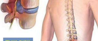
A distinctive feature of this hernial defect is the protrusion of disc contents to the left or right of the dural sac, into the spinal canal. This leads to compression of the spinal cord on one or both sides, compression of the nerve roots and/or rupture of the fibrous ring, which prevents the destruction of the disc.
Because a paramedian hernia puts pressure on the spinal cord and can increase in size over time, there is a risk of damage to spinal structures. Most often, this pathology is a consequence of a natural degenerative process in the spinal column, which results in increased pressure on the intervertebral discs.
The physiological purpose of the intervertebral disc is to act as a shock absorber that prevents friction between the vertebrae. It also takes part in ensuring flexibility and mobility of the spine. Therefore, when it is exposed to strong pressure from a paramedian hernia, it becomes deformed and gradually goes beyond its anatomical boundaries. An inflammatory process may begin at the site of its formation. If left untreated, it spreads throughout the body.
This diagnosis is considered one of the most dangerous, because in the absence of timely medical intervention, the pathological process becomes irreversible, leading to disability of the patient.
Types of median hernias
The disease is classified based on the location of the hernial protrusion, its direction and location relative to the spinal canal. There are the following types:
- Median. Protrusion towards the center line of the spine.
- Paramedian. There are right-handed and left-handed. The protrusion is directed towards the center of the spinal canal and slightly to the side - to the left or right.
- Median-paramedian. The bulging disc is located both toward the center of the midline and to the side of it.
- Median-paramedian bilateral. The hernial protrusion is directed towards the center and to the side, occurring on both sides of the vertebra.
- Paramedian sequestered. The hernia is directed to the center of the spinal canal, the pulpous ring partially or completely falls out.
- Paramedian-foraminal. The protrusion is directed to the place where the bundle of nerve roots emerges.
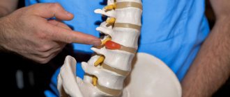
Surgery for lumbar hernia L3–L4
Surgical intervention is a last resort measure necessary in the absence of positive changes in the patient’s condition during conservative treatment carried out for more than 1–3 months. It is also prescribed to patients whose MRI images show an intervertebral hernia of L3–L4 greater than 12 mm or severe pain, even if the size of the protrusion is small.
In certain situations that threaten serious complications, emergency surgery is required. We are talking about sequestered hernias. But such interventions are the most technically complex and require maximum care and skill from the neurosurgeon.
Removal of L3-L4 disc herniation is possible today by:
- nucleoplasty;
- endoscopic surgery;
- microdiscectomy.
These operations to remove L3–L4 intervertebral hernia differ not only in price. The main difference is the indications for their implementation and the existing limitations. Therefore, the technique is selected separately for each patient.
All types of modern surgical interventions for the removal of intervertebral hernias have in common a low degree of risk, a good cosmetic effect and a short rehabilitation period, but only if an experienced neurosurgeon is selected.
After endoscopic surgery and microdiscectomy, a disc defect occurs and sometimes it is necessary to remove it completely. In the first case, the Barricaid mesh implant is used to prevent relapse. The second requires the use of a transpedicular fixation technique or the installation of an M6 prosthesis.
Nucleoplasty
The most modern technique for removing intervertebral hernia of the lumbar region L3–L4 is nucleoplasty. It can be performed through the use of various techniques, on the basis of which they distinguish:
- laser nucleoplasty;
- cold plasma nucleoplasty;
- radio wave nucleoplasty;
- hydroplastics.
Hydroplasty using the SpineGet device is considered one of the most effective and safe techniques. The essence of the method is to introduce a special thin instrument into the nucleus pulposus through a pinpoint puncture of soft tissue. A saline solution is pumped through it under pressure, which leads to mechanical destruction of part of the gelatinous contents of the disc. At the same time, the liquefied material is pumped out, which creates all the conditions for the protrusion to be retracted and pain eliminated.
Nucleoplasty is possible for uncomplicated hernias up to 7 mm in size. The postoperative wound does not require suturing. It is covered with a sterile bandage.
Relief occurs almost immediately or the patient’s condition gradually improves over 2 weeks. A huge advantage of puncture surgery is the absence of the need for hospitalization and complex rehabilitation, so the patient can independently leave the clinic a few hours after surgery.
Endoscopic surgery
Removal of a hernia using endoscopic equipment is carried out through a pinpoint puncture of soft tissue, up to 1 cm in diameter. A hollow endoscope equipped with a video camera is inserted through it, which transmits the image to the monitor. Through the endoscope, the surgeon inserts a special manipulator with which he can resect the protruding part of the disc.
Endoscopic surgery is possible for large hernias, as well as those located in the foraminal openings or other narrow areas of the spinal canal. Because access to the disc is created using a shaver, the risk of nerve damage is dramatically reduced.
Microdiscectomy
Microdiscectomy is a microsurgical operation performed through an incision of up to 3 cm. It is performed in the presence of neurological disorders or in the diagnosis of a sequestered hernia, since it is impossible to completely remove it in a less traumatic way.
Causes
The main reason for the development of a median hernia is spinal osteochondrosis, in which degenerative destruction of the cartilage tissue of the intervertebral discs occurs.
Factors that can provoke pathology:
- weak back muscles due to a sedentary lifestyle;
- poor posture;
- incorrect placement of the foot when walking (for example, flat feet) - disrupts the balance of the distribution of physical load on the spine;
- excess weight, which increases the load on the intervertebral discs;
- an unbalanced diet, as a result of which the body does not receive all the necessary vitamins and minerals;
- metabolic disorders, insufficient consumption of drinking water, which leads to dehydration of cartilage tissue and its gradual destruction;
- smoking and alcohol abuse.
Rehabilitation
How long the rehabilitation will last and how easily it will proceed depends on the type of intervention performed, as well as its volume. Naturally, when several hernias are removed, for example L3–L4 and L4–L5 or L5–S1, the body will take longer to recover.
Patients are almost always prescribed exercise therapy and short-term drug therapy. After nucleoplasty, patients only need to slightly limit physical activity and avoid lifting heavy objects. Performing endoscopic removal of a hernia requires avoiding prolonged sitting; sometimes physical therapy and wearing a corset are additionally prescribed.
After microdiscectomy, recovery is a little more difficult, so patients are discharged from the hospital only after 5–7 days. They also need to adhere to a gentle regime for a longer time.
Thus, modern operations to remove L3–L4 hernia are extremely safe and low-traumatic. They allow you to quickly get rid of debilitating pain and allow you to return to normal life.
Symptoms of pathology
The main symptom of a median intervertebral hernia is a painful syndrome in the part of the spine where the protrusion is localized. The pain is weak at first, intensifies over time, and can be constant or intermittent.
Other symptoms during the formation of a median hernial protrusion in the cervical spine:
- headache;
- dizziness;
- darkening of the eyes;
- noise in ears;
- blood pressure surges;
- lack of coordination;
- numbness, tingling in the arms, shoulders and shoulder blades.
Manifestations when the median protrusion is located in the thoracic spine:
- intercostal neuralgia;
- dyspnea;
- arrhythmia;
- disorders of the gastrointestinal tract.
Symptoms of a hernia in the lumbosacral region:
- acute pain in the lumbar region;
- renal failure;
- loss of sensation in the legs;
- cauda equina syndrome - shooting in the thigh up to the calf muscle, intestinal dysfunction and genitourinary disorders.
What is the danger?
Paramedian disc herniation requires treatment, preferably in the early stages of development. This is due to the fact that in the absence of timely therapy, pathologies of the musculoskeletal system develop, the ability to carry out full movements is lost and, as a result, the inability to care for oneself independently (disability). Doctors at our clinic make every effort to prevent such an outcome, making life easier for patients.
Each type of intervertebral hernia has serious complications, so you should not delay treatment.
See how easy it is to get rid of a hernia in 10 sessions
One of the main consequences of this pathology is severe and debilitating pain in the back, which is difficult to respond to painkillers. This pathological change affects not only locally, so there is general weakness, fatigue, loss of mood and sleep problems. With further progression, paresis, muscle atrophy and paralysis of the limbs are possible. Paramedian disc herniation L5-S1 leads to the appearance of cervicogenic dizziness. They do not cause a major impact on health, but they do affect a person’s quality of life.
A serious and dangerous consequence of this disease is compression and mechanical damage to the spinal canal, since nerve endings responsible for the innervation of internal organs originate from it.
If they become pinched (especially in the lumbar spine), cauda equina syndrome may occur, which is characterized by:
- intense pain in the lower back, spreading to one or two lower limbs;
- impaired sensitivity of the perineum and inner thigh, leading to numbness or tingling of the skin;
- weakness in the legs;
- absence of tendon reflexes in the legs, deterioration of the tone of the anal sphincter.
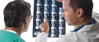
Diagnostic methods
To make a diagnosis, identify the location of the hernial protrusion, its size, in addition to a visual examination, the patient is prescribed the following diagnostic studies:
- X-ray;
- computed tomography (CT);
- magnetic resonance imaging (MRI);
- electroneurography.
The x-ray does not show a hernial protrusion, but in the pictures you can see degenerative processes in the bone tissue and changes in the size of the interdisc space.
The most informative method is MRI. With its help you can get a complete picture of the pathology. If it is impossible to do magnetic resonance imaging, a CT scan is prescribed, but its images are not of high quality. Electroneurography allows you to evaluate the speed of nerve impulses.
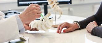
Treatment at the Paramita Clinic
We have developed our own approach to the treatment of paramedian hernias. It includes:
- examination of the patient using modern instrumental and laboratory tests, including MRI;
- development of an individual treatment plan for each patient, including effective Western techniques and traditional Eastern methods.
We use non-surgical hernia treatment techniques
Read more about our unique technique
This helps not only eliminate the hernia, recover faster after surgery, but also restore the health of the body as a whole.
How is a hernia treated?
At the initial stage of the disease, complex conservative treatment is prescribed, which is aimed at relieving pain and relieving the inflammatory process.
For acute pain, bed rest is indicated. This helps relax cramped back muscles, which reduces pressure on the nerve endings of the spinal cord. The patient is prescribed drug therapy. After the acute period has passed, physiotherapy and physical therapy are required.
If conservative treatment carried out for 6 months or more fails and severe complications develop, radical methods in the form of surgery are required.
Medications
When treating a median intervertebral hernia, the patient is prescribed the following groups of medications:
| Group of drugs | How they work | Titles |
| Analgesics | Reduce pain, relieve inflammation | Ketorol, Ketanov |
| Nonsteroidal anti-inflammatory drugs (NSAIDs) | Relieve pain, suppress inflammation, reduce fever | Ibuprofen, Diclofenac |
| Muscle relaxants | Relieves muscle spasm in the affected area | Mydocalm, Baclofen |
| Chondroprotectors | Restore damaged cartilage tissue, improve metabolism in it (not proven) | Chondroxide, Chondrolone, Artra, Alflutop |
| Blockades | Relieves acute pain, prescribed in the absence of effect of analgesics, NSAIDs | Novocaine, Lidocaine |
| B vitamins | Improve metabolic processes, restore damaged tissues | Milgamma |
A study was conducted on the treatment of intervertebral hernias with the drug Ketorol. The total duration of therapy is no more than 5 days. For the first days, patients were prescribed the drug in the form of intramuscular injections of 30 mg 2 times a day, for the next 3 days in the form of tablets of 10 mg 2 times a day. During therapy, other analgesics and non-steroidal anti-inflammatory drugs were not used. Evaluation of the results showed a reduction in pain by 66% on the third day of treatment, and by 82% by the end of treatment.
The intensity of pain after injections decreased by 2.3 times, after tablets - by 4 times compared to the original. The safety of Ketorol was also assessed. Excellent and good tolerability of the drug was observed in 83% of patients, side effects were detected in 16% (source - scientific article “Possibilities of optimizing analgesic and anti-inflammatory therapy in patients with acute back pain syndrome”, authors N.A. Shostak, N.G. Pravdyuk and others).
Massage and exercise therapy
Drug treatment of a median hernia must be supplemented with a complex of physical therapy – exercise therapy. Regular exercise strengthens the muscle corset, restores muscle tone, and relieves stress on the affected vertebrae.
In parallel with physical therapy, after the exacerbation is relieved, massage is prescribed. Manual therapy is possible. These procedures relieve muscle spasms and return the vertebrae to a healthy anatomical position.
Actions must be carried out as carefully as possible so as not to cause even greater displacement of the vertebral structures.
Physiotherapy
Physiotherapeutic procedures are prescribed only after acute pain has been relieved. When treating a median intervertebral disc herniation, the following methods are effective:
- magnetic therapy;
- mud baths;
- laser therapy;
- electrophoresis;
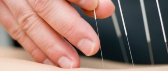
- hirudotherapy;
- acupuncture.
These procedures relax the back muscles, which removes the load from the diseased disc, improve metabolic processes and blood circulation in the tissues, and the conductivity of nerve endings.
Surgical intervention
If conservative therapy for a median hernia does not provide relief, the patient may be prescribed surgery.
Operations are performed using minimally invasive methods:
- cold plasma nucleoplasty;
- laser vaporization;
- microdiscectomy.
After the operation, a long rehabilitation period is required. During this period, it is important to follow all medical recommendations to avoid postoperative complications.
Studies were conducted to analyze the results of surgical treatment in 100 patients with intervertebral hernia L4-L5 and L5-S1 at the Institute of Neurosurgery named after. acad. A.P. Romodanova. A standard microdiscectomy was performed on patients whose hernia was caused by excessive physical activity (heavy lifting), strength sports, or spinal injuries (falls). Evaluation of the results of surgical treatment makes it possible to judge the adequacy and effectiveness of the operation - excellent result in 58% of patients, good result in 41%, satisfactory in 1% (source - scientific article "Analysis of the results of surgical treatment of patients depending on the location of lumbar intervertebral hernias disks”, authors N. E. Polishchuk, I. S. Brinkach, E. I. Slynko).
Folk remedies
Many traditional medicine methods help reduce pain and relieve inflammation. But they can only be used as an addition to drug therapy, massage, physical therapy, and they cannot replace treatment prescribed by a doctor.
Self-medication is prohibited, so before using various herbs, infusions, decoctions, compresses, you should consult a doctor. Some medications can worsen the condition and harm already affected tissues.
Symptoms of intervertebral hernia L4–L5
The main manifestations of the disease are the occurrence of pain in the area of the affected spinal motion segment. They can be constant or occur periodically, be sharp or nagging, or be of a different nature.
Pain usually appears or intensifies with sharp turns of the body, bending, prolonged standing, walking or performing physical work. They usually subside when taking a supine position. Also, many patients note that the lower back reacts to straining during bowel movements.
A kind of test for the presence of a L4–L5 hernia is raising a straight leg in a lying position. If the lesion is observed at this level, such an exercise will lead to the immediate appearance of acute pain and its elimination after bending the raised leg at the knee.
With a fully formed hernia, pain can radiate to the lateral surfaces of the thighs and lower legs, since the nerve fibers located at this level are responsible for their innervation. They often become numb, which can affect gait. You may also experience:
- swelling of the legs;
- limitation of movements in the lower back;
- increased sweating;
- dry skin;
- weakness;
- increased fatigue.
We must not forget about vegetative disorders. Since the portion of the spinal cord at the level of the 4th and 5th lumbar vertebrae is responsible for the proper functioning of the prostate gland and lower extremities, if the condition of the spine deviates from the norm, pain in the knees and feet, as well as urination problems, may occur.
If the hernia strongly compresses the nerve, severe neurological symptoms may occur. In such situations, the patient may:
- encounter paresis or paralysis;
- atrophy of the leg muscles;
- decreased sensitivity;
- complete loss of control over the process of urination;
- persistent erectile dysfunction.
Depending on the type of L4–L5 hernia, both legs or only one of them may be affected.
Recovery prognosis
Early diagnosis of pathology significantly increases the chances of recovery. Therefore, if you experience back pain or other symptoms, you should contact the clinic immediately.
If the median hernia was not diagnosed in time and the patient did not receive treatment, irreversible consequences and paralysis of the limbs occur.
To speed up recovery, it is better to treat a median intervertebral disc herniation in the Czech Republic. Clinics offer complex therapy aimed at relieving pain and inflammation, restoring sensitivity, and reducing the size of the hernial protrusion.
Preventive recommendations
Basic preventive measures for median hernia:
- lack of strong physical activity;
- balanced diet;
- active lifestyle.
Regular exercise is necessary, it strengthens the muscles and stabilizes the spine. This could be swimming, walking, morning jogging.
It is necessary to monitor your posture, choose the right and comfortable shoes without high heels, you can wear an orthopedic back corset if you are constantly forced to sit or stand.
A proper balanced diet is required, which will provide the body and the affected spinal structures with the necessary vitamins and minerals. Also, following a diet will prevent excess weight gain, which increases the load on the spine.
Prevention and prognosis
As practice shows, if you contact our specialists early, the prognosis is favorable for recovery. Correct use of medications and compliance with medical recommendations allows you to restore damaged tissue and prevent the development of severe complications (including disability). To prevent the development of a recurrence of a paramedian hernia, you will need regular exercise therapy, acupressure sessions, and sleeping on an orthopedic mattress and pillow.
After completion of treatment, physical activity should be dosed, including reducing the time of sedentary work, swimming, and paying special attention to proper nutrition, with a sufficient amount of vitamins and microelements