Each segment of the spine is of great importance for the normal functioning of the entire spinal column and spinal cord, since the stability of each segment depends on other vertebrae and discs and, only in this way, the spine can function fully. Over time, the spine is subject to constant stress, injury or other impacts, and is susceptible to various diseases such as disc degeneration, vertebral degeneration, arthritis, etc. These conditions can cause pain and dysfunction.
spinal diseases , but the most common are a number of diseases that are clinically significant.
Ankylosing spondylitis (Bechterew's disease). This disease is a type of arthritis in which chronic inflammation of the joints of the spine and sacroiliac joints occurs. Initially, inflammation occurs in the sacroiliac joints, then moves to the spine, leading to stiffness and limited mobility. With prolonged inflammation of the joints of the spine (spondylitis), calcium deposits are formed in the ligaments around the intervertebral discs, which leads to weakening of the discs and a decrease in their shock-absorbing and support functions. As calcium deposits accumulate in the ligaments, there is a significant decrease in both range of motion and flexibility in the spine. The disease can progress to fusion of the vertebrae, which is called ankylosis. As a result of ankylosis, the spine loses mobility, the vertebrae become fragile, and the risk of vertebral fracture increases. In addition to damage to the spine, ankylosing spondylitis leads to disruptions in the functioning of other organs, since the disease is systemic.
Osteochondrosis
Over time, the spine is subjected to daily stress and minor injuries, which ultimately leads to wear and tear of the intervertebral discs and their degeneration. The fibrous ring of the intervertebral disc is damaged under load, micro-tears of fibrous tissue occur, and then the damaged area is replaced by scar tissue, the elastic properties of which are much worse than those of fibrous tissue. Such changes in the annulus fibrosus lead to decreased shock-absorbing function of the disc and a greater risk of recurrent disc ruptures. As the annulus fibrosus becomes scarred, the gelatinous part of the disc (nucleus) also gradually shrinks, which in turn leads to a decrease in disc height. As the height of the disc and shock-absorbing functions decrease, the vertebrae begin to exert more pressure on each other under load, which leads to the formation of bone growths (osteophytes). Violation of the integrity of the fibrous ring leads to the formation of disc herniations. Disc herniations and osteophytes can cause compression or stenosis, leading to neurological symptoms.
Spinal stenosis
Stenosis is a narrowing of the space in the spine where the spinal cord and spinal roots pass. The space of the spinal canal, as a rule, is initially not very large, especially in the cervical and thoracic spine, but with various pathological changes in the spine it becomes critically small. These can be either degenerative changes in the spine or injuries. Significant narrowing (stenosis) of the spinal canal leads to a compression effect on the spinal cord, which will be manifested by pain, weakness in the limbs, sensory disturbances, and in severe cases, dysfunction of the bladder and intestines. Many older people have spinal canal stenosis to varying degrees. Unlike a disc herniation, in which compression of one or two nerves occurs and a picture of radiculopathy occurs, with stenosis, compression occurs on many nerves simultaneously and this condition is called myelopathy. For stenosis, conservative treatment is possible if the symptoms are moderate. If there are severe neurological symptoms, then surgical treatment is usually recommended, the purpose of which is to decompress the spinal cord.
Diagnostics
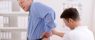
If your spine hurts in the lumbar region, you should make an appointment with a neurologist or vertebrologist. At the appointment, the doctor initially collects anamnesis, asking questions about the nature of the pain, the circumstances of its occurrence, the duration of its persistence, the presence of other symptoms, lifestyle, etc.
Then the specialist conducts an examination. As part of it, he not only palpates the spine, determines the localization of pain, evaluates the gait and posture that the patient takes unconsciously, but also conducts functional tests. With their help, you can detect signs of ankylosing spondylitis, neurological deficits, assess the degree of mobility of the spine and obtain other diagnostic data.
Based on this, the doctor can already suggest possible causes of pain. To clarify them, as well as to accurately determine the extent of damage, instrumental and sometimes laboratory diagnostic methods are additionally prescribed. Most often they resort to help:
- X-rays in frontal and lateral projections, sometimes with functional X-ray tests;
- CT scan – allows better visualization of bone structures, therefore it is more often used to diagnose spondylosis, fractures, bone tumors, etc.;
- MRI – makes it possible to most thoroughly assess the condition of cartilaginous structures and soft tissues, therefore it is more often used to diagnose osteochondrosis, protrusions, intervertebral hernias, spinal cord lesions, etc.;
- electromyography - indicated for neurological disorders of unknown origin, as well as to assess the degree of nerve damage;
- radioisotope osteoscintigraphy – used for the diagnosis of malignant tumors and metastases;
- X-ray densitometry is the optimal method for diagnosing osteoporosis;
- myelography - used to identify signs of compression of the spinal cord and nerves of the cauda equina.
Herniated disc
A herniated disc occurs when the annulus fibrosus that surrounds the intervertebral disc ruptures. This rupture causes the central portion of the disc, which contains a substance called nucleus pulposus, to be released. When pressure is applied to the vertebrae above and below, the nucleus pulposus comes out, puts pressure on nearby nerve structures and causes severe pain and nerve damage. Disc herniations most often occur in the lumbar spine and are sometimes called disc extrusion.
Ankylosing spondylitis (ankylosing spondylitis)
This is a systemic inflammatory disease that affects all spinal structures. Inflammation begins in the sacroiliac joints, then spreads higher, developing in an ascending line. Over time, calcium deposits appear in the ligaments, which leads to a decrease in mobility and flexibility. Without treatment, ankylosing spondylitis progresses, and gradually the vertebrae fuse together: ankylosis occurs. The first symptoms are dull pain and stiffness in the lower back, which intensifies in the morning and decreases after physical activity and a hot shower. During the day, the pain is stronger at rest; when a person moves, it subsides. In the later stages, posture noticeably deteriorates.
Radiculopathy
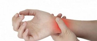
The term radiculitis (radiculopathy) is widely used and means root compression. Radiculitis can occur in both the lumbar and cervical spine, or much less frequently in the thoracic spine. Root compression occurs when there is excess pressure on the nerve root. Excessive pressure can come from both bone tissue and soft tissue (muscles, cartilage, ligaments). This pressure disrupts nerve function, causing pain, tingling, numbness, or weakness.
Osteoporosis is a disease in which bone tissue, including the vertebrae, weakens, which increases the risk of vertebral fracture, even with minor loads.
Vertebral compression fractures are the most common type of fracture caused by osteoporosis, and hip and wrist fractures are also possible with osteoporosis. These vertebral fractures can change the shape and strength of the spine, especially in older women, who often develop spinal deformity as a result of such fractures. The spine becomes excessively tilted in the thoracic region (kyphosis) and the shoulders bulge forward. With severe osteoporosis, even simple movements such as bending forward can lead to vertebral fractures.
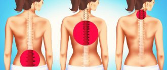
The lower back is a special risk area
A healthy spine has a curved shape: it should not be absolutely straight, as many people believe. Physiological curves in the neck and lower back are especially noticeable - these areas are especially vulnerable due to their mobility. The thoracic region is firmly fixed by the ribs, so it is much less likely to undergo dystrophic changes. Osteochondrosis, hernias and other pathologies most often develop in the lower back, since this section is the most loaded - it accounts for the weight of the entire upper body. The risk of various injuries increases with congenital and acquired deformities, injuries, and excess weight.
Spinal fractures
The vertebrae have great strength and can withstand a lot of pressure, while at the same time the spine does not lose flexibility. But like other bones in the body, they can break under extreme excess pressure, injury or disease. In such cases, damage or fractures to the vertebrae can range from minor to severe.
Compression fractures
As the name suggests, compression fractures occur from excessive axial loads that compromise the integrity of the vertebral body. Osteoporosis is one of the leading causes of compression fractures, as there is a decrease in the ability of the vertebrae to withstand stress. In such cases, even a slight fall or even a cough can lead to a compression fracture. People often perceive back pain as a normal part of aging, and sometimes compression fractures go unnoticed. Repeated compression fractures can lead to a decrease in spinal height. Another common cause of a compression fracture is trauma, such as a fall.
Often, vertebral compression fractures will eventually heal on their own (without treatment). NSAIDs (for example, aspirin) may be prescribed to relieve pain. For severe fractures, surgical methods (vertebroplasty and kyphoplasty) may be used.
Burst fractures
Blowout fractures usually occur after severe trauma (for example, a car accident or a fall from a height). Blast fractures are significantly more dangerous than compression fractures because the anterior and middle portion of the vertebral body is fractured into multiple fragments and is more likely to result in spinal cord injury. Additionally, as the vertebral body loses its integrity, the spine becomes unstable. In some cases, with burst fractures, if there is no impact on the spinal cord, conservative treatment can be performed. If there are loose fragments or damage to the nerve structures, then surgical treatment is necessary.
Flexion-extension fractures
Such fractures are sometimes called Chance fractures and occur during sudden flexion-extension. Most often, this type of injury occurs in car accidents, in people wearing a seat belt, and not only fractures of the vertebrae, but also ligaments, discs, and sometimes internal organs. Such fractures are usually unstable and require surgical treatment. This type of fracture occurs in 5-10% of cases of spinal fractures.
Vertebral fracture with dislocation. Such fractures occur when exposed to great force, and not only the integrity of the vertebral body is violated, but also its displacement (due to rupture of ligaments and discs). Such fractures often require surgical intervention.
Fractures are also divided into stable and unstable. Compression fractures are generally considered stable and do not require surgery. In contrast, unstable fractures (eg, burst or Chance fractures) typically require surgical treatment, often as an emergency.
Prevention measures
There are special measures to prevent diseases of the back and spine that a person can perform himself. As noted above, half of all back diseases occur as a result of poor nutrition and a sedentary lifestyle. Therefore, all diseases can be avoided completely or in the early stages of development. So follow the rules and it will help you avoid a lot of surprises in the future.
A properly selected diet is considered a good means of prevention, and can also alleviate the condition of an already developed disease.
It is important to know that fatty meats, spicy foods and sugar and salt should not be consumed in large quantities.
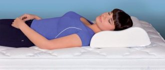
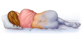
To prevent spinal diseases, you should know about the correct body position. The best option in this case would be a properly selected mattress and a flat bed. Correct body position will bring the spine into good shape. And you need to remember, you should never sleep on your back if you have any diseases of the spine.
And of course, do not forget about the correct position of the back during a sedentary lifestyle. Uneven load on the back when sitting can aggravate the situation. Therefore, it is worth choosing a comfortable chair for work, as well as taking breaks and stretching your spine a little.
Only through prevention can complications and the development of back diseases be prevented, as well as their treatment facilitated. During his lifetime, a person cannot insure himself against injuries or any infectious diseases. Even a minor injury to the spine can lead to serious illness. Therefore, the spine, back, pelvis and neck are considered the most vulnerable places.
Important information
The human body and all joints are designed in such a way that after a while they begin to wear out. After age 40, joints produce less lubricant, which helps reduce friction between joints and vertebrae. But for those who lead a sedentary lifestyle, this problem appears even earlier. Therefore, back diseases can appear at a young age. And with the advent of old age, a person can no longer avoid problems with joints.
But there are ways to help prevent this problem. The most important thing is to seek help from a specialist in time and start treatment, and also eat products containing glucosamine and chondroitin. Products with these substances are excellent for preventive purposes, as well as for those who already suffer from joint pain.
Spinal deformities
Spinal column deformity means any significant deviation from the normal curves of the spine. The most common are
- Scoliosis
- Hyperkyphosis
- Hyperlordosis
There are various causes of pathological curvature of the spine. Some children are born with congenital scoliosis or congenital hyperkyphosis.
Sometimes nerve and muscle diseases, injuries, or other conditions cause spinal deformities (such as cerebral palsy).
Most often (up to 80-85%) scoliosis is “idiopathic” (without an obvious cause). Idiopathic scoliosis develops gradually but can progress rapidly during the growing years of adolescence.
Scoliosis
The term scoliosis was first used to describe this spinal deformity by Hippocrates in 400 BC. It is a progressive disease with an unknown cause (idiopathic) in 80% of cases, although there is evidence that genetics and nutrition play a role. Women are 10 times more likely to develop scoliosis than men. Scoliosis is often accompanied by twisting of the spine, which leads to deformation of the costal arches and chest. Scoliosis usually begins to appear during adolescence. Conservative treatment is quite effective for grade 1-2 scoliosis. In cases of severe deformation (grade 3-4) and in cases of progressive scoliosis in adolescence, surgical treatment is recommended (the earlier surgical treatment is performed, the better the long-term results).
Hyperkyphosis
Mild kyphosis is the natural curvature of the thoracic spine, while hyperkyphosis is an excessive forward tilt of the thoracic spine (slouching). Hyperkyphosis is common in older people and is usually associated with the presence of osteoporosis and previous vertebral compression fractures. Causes of hyperkyphosis can also be injuries, diseases of the endocrine system and other diseases. In adolescence, hyperkyphosis may occur, such as Scheuermann-Mau disease, which is characterized by a wedge-shaped deformation of three or more vertebrae in the thoracic spine. As a rule, conservative treatment for Scheuermann-Mau disease is quite effective, but when the angle of deviation from the axis is more than 60 degrees, surgical treatment is recommended.
Hyperlordosis
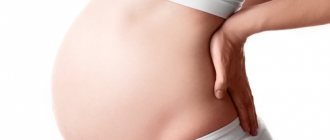
Lordosis is a natural inward curve in the lumbar spine, and hyperlordosis is a pathological increased curve in the lumbar spine. Hyperlordosis is usually accompanied by an abnormal anterior tilt of the pelvis and is often accompanied by excessive protrusion of the buttocks. Symptoms may include pain and numbness if there is compression of nerve structures. As a rule, hyperlordosis is caused by weakness of the back muscles, hyperextension, for example, in pregnant women, in men with excessive visceral fat. Hyperlordosis is also associated with puberty.
Treatment for hyperlordosis is usually not required unless nerve structures are affected.
Non-surgical treatment methods:
- Various types of reflexotherapy - influencing active points of the body in various ways (helps relieve pain and muscle spasms): Acupuncture - influencing biologically active points with microneedles.
- Pharmacopuncture is the introduction of medicinal drugs of natural origin to the source of the problem.
- Tsubotherapy is a gentle effect on the reflex points of the body using metal balls.
Spinal tumors
Spinal tumors are quite rare. Tumors can be benign or malignant. Primary malignant tumors of the spinal cord are very rare. Malignant spinal tumors are usually metastatic in nature and have a primary focus in other organs and tissues.
From a clinical and anatomical point of view, tumors can be classified as epidural, intradural extramedullary and intramedullary tumors.
Metastatic tumors of the spine are the most common for bone metastases.
The most common solid tumors secondary to the spine are breast, prostate, and renal carcinoma, which account for almost 80% of spinal metastases. Tumors of unknown primary origin account for about 5% -10% of cases. Metastases of neoplasms of the hematopoietic system account for about 4% -10%.