Orthotics is a conservative method of treatment and prevention of diseases of the musculoskeletal system, the essence of which is to preserve and restore the functions and shape of the affected parts with the help of special medical devices - orthoses. The use of such devices is indispensable for people suffering from arthritis and arthrosis, as well as in other situations requiring orthotics.
The Yusupov Hospital offers both individual orthosis - custom-made orthosis, which ensures maximum functionality of the device, and the use of ready-made devices that will be selected individually for the patient.
What is orthosis and orthotics?
In order for a person who has lost the motor function of his limbs to be able to return to a normal lifestyle, orthotics are used in medicine.
This is a conservative method of treating diseases of the musculoskeletal system, involving the use of professional orthoses.
What is an orthosis? This is a special device of a certain shape and materials, intended for the purposes of:
- unloading and fixing the spinal column, damaged limbs or their components (joints, tendons, muscles);
- activation and correction of their functions;
- correcting the direction of damaged parts of the limb;
- complete and partial immobilization of a limb for its restoration;
- to straighten a curved area of the hand or foot;
- reducing pain, swelling and inflammatory processes;
- prevention of fractures, dislocations, cracks;
- assistance in movement.
Medical products are used in rehabilitation clinics and hospitals, but in sports schools and fitness centers.
Orthoses are purchased in pharmacies, orthopedic clinics and stores. There are devices of mass production and individual development.
Who is indicated for orthotics?
Wearing orthoses (corsets, bandages, special shoes or orthopedic bodies) is indicated in the following situations:
- problems with ligaments (congenital instability, weakness, damage);
- post-traumatic conditions (after sports injuries);
- arthrosis and arthritis of the joints;
- recovery period after surgical interventions.
The main purpose of using orthotics is to provide the damaged organ with a state of rest. Orthoses help reduce the load on a damaged joint or limb, fix it, correct movements and increase activity. However, the effectiveness of the orthosis depends on its correct selection.
Types of materials
Depending on the purpose of the medical device, different materials are chosen for them. The orthoses are based on elastic fabric or fixing, rigid structures made of plastic, silicone, and metal. The orthopedist selects a device based on the problem and the person’s needs.
The material with which the orthosis is covered is considered important:
- Cotton combined with synthetics. Synthetic fibers act as an elastic element. Cotton makes it possible to wear an orthopedic product for a long time without removing it. They allow the skin to “breathe” and do not cause allergic reactions.
- Wool. In combination with other textile options it gives a micro-massage effect. Used for corsets and bandages.
- Lycra, spandex or elastane. It has breathability and cooling effect. Products made only from these materials provide the limbs with stable fixation.
- Neoprene. Provides light fixation to the limb, warming it.
- Nylon. It is characterized by high wear resistance. A product made from this material will last a long time.
During manufacturing, a specific material or a combination of several is used to achieve the required properties.
Classification of devices
Orthopedists use several types of classification when choosing an orthopedic product. First of all, you need to know why an orthosis is needed. The classification by purpose looks like this:
- Preventive. Applicable for people with chronic degenerative diseases of the musculoskeletal system (arthrosis, arthritis, osteochondrosis, scoliosis), pathologies of bone and cartilage tissue (osteomyelitis, osteoporosis).
- Therapeutic, rehabilitation. Used after surgical treatment, injuries to joints and spine.
- Functional (permanent). Necessary for restoring limb function after a progressive disease (destruction of cartilage tissue, growth of bone calluses).
According to this classification, products of varying degrees of hardness are produced: light (prevention), medium (constant wear for stabilization) and high (for medicinal purposes).
It is important to take into account the design features of the devices:
- Tutor. A soft or semi-rigid orthosis is a sleeve that encircles a limb. Prevents movement of a joint or several joints.
- Orthopedic splint. A rigid type of device for complete or partial fixation of a limb; the injury will heal faster.
- Bandage. Supportive type, made of elastic and soft material.
- Hardware systems. Several splints are connected by hinges, allowing complex patients to perform various movements.
- Corsets. Used to reduce pain, relieve stress and other problems of the spine.
- Longuet. Used for fractures and dislocations of limbs. They are more convenient than plaster casts - they can be removed and, as you recover, the degree of fixation can be reduced.
- Reclinators. The devices correct the spinal column during stooping, the first stage of scoliosis.
- Head holders. They are used after surgery on the cervical spine or in cases of central nervous system disorders, when the patient cannot hold his head up on his own.
There is a classification based on design features. The cuts are made in one piece - elastic bandages or wide tapes - or segmented - with separate inserts made of plastic, leather, metal.
Serial designs of orthopedic devices are assembled according to models. Prefabricated - in modules, depending on the needs of the patient. Individual devices are made based on a cast of the patient’s limb, taking into account his wishes regarding the type of materials.
AREAS OF USE
- Orthotics are used in rehabilitation for neurological diseases. For example, spinal orthosis for scoliosis and more serious lesions of the musculoskeletal system allows, during complex treatment, to fix any part of the spine, thereby supporting the muscular corset and stopping the progression of the pathological process.
- Orthotics for cerebral palsy help keep muscle groups with increased tone in the desired position, relieving them of excessive stress. Orthosis for cerebral palsy helps maintain range of motion and prevent joint contractures that develop if muscles with increased tone are in a shortened position for a long time.
- Orthotics for stroke are used in the early stages of rehabilitation, when the patient is just beginning to master a vertical position and the legs bend under the weight of the body.
Services for prosthetics and orthotics for disabled people are paid for by the state!
Citizens who contact Doctor Ost LLC for orthotic services have the right to receive full reimbursement of the funds spent on treatment at the expense of the Social Insurance Fund if they have an IPRA. Find out more from our administrators!
Rules for using orthoses
Strict adherence to the rules for using orthoses will guarantee effective elimination of the problem: restoration of motor function, elimination of pain, and others. Proper use of orthopedic devices will prolong their service. You need to pay attention to the recommendations of the doctor and the manufacturer.
First of all, the wearing period is important. The period of use of the orthopedic device is determined by the doctor depending on the established diagnosis and identified concomitant pathologies.
Reducing the rehabilitation time will lead to complications of the disease, and prolonging the established period will lead to the development of complications (thrombosis, circulatory disorders, muscle weakness).
Preventive devices must be removed every 8 hours, 2 hours are given to rest, then the device is put on again. Elastic types of orthoses must be removed at night, articulated models are worn on an ongoing basis.
Any orthosis must be put on correctly. At an appointment with an orthopedist, ask to show the optimal wearing option if the device implies a preventive effect. The specialist will indicate the following parameters:
- what should be the degree of fixation;
- in which area to install the product;
- how to fix it correctly: fasten with straps, Velcro or clips.
Do not put the prosthesis on an area where the skin is injured or inflamed. Varicose veins and swelling will become absolute or relative contraindications to the use of orthopedic devices on the extremities.
The rules for caring for models are indicated on the packaging or label. Some types of orthoses can be washed in a washing machine on a delicate cycle. Other types will have to be washed by hand.
Treatment of fractures and false joints of bones still occupies a large place in scientific research in traumatology. But despite the known achievements, the results often leave much to be desired.
In recent years, various types of operations have been well developed: extraosseous, intraosseous and compression-distraction osteosynthesis, if necessary with autoplasty. But, despite all the methods of surgical treatment used, to achieve a positive effect in the future, a long period of external immobilization is often required.
Reliable and functionally beneficial immobilization of fractures and false joints of extremity bones is still relevant. It is known that long-term immobilization of a limb leads to severe muscle wasting, stiffness in adjacent joints and long-term circulatory disorders in the injured limb, which ultimately adversely affects the full and rapid recovery of the victim and sharply increases the period of disability [2], [4] ,[5], [9],[11].
The main material for making fixing bandages is still gypsum. The disadvantages of these bandages include: heavy weight; fragility during use, especially under load (lower limb); contamination of the dressing both outside and inside with skin secretions; When water gets on the bandage, it gets wet and loses its fixing properties. All this requires repeated replacements over long periods of treatment.
The term “therapeutic prosthetics” is discussed in the monograph by N.A. Shenk [10], in which she proves the feasibility of using orthopedic devices for therapeutic purposes and draws the attention of doctors to the fact that the achievements of the prosthetic industry are not fully used in the treatment of orthopedic and trauma patients. According to many modern authors, this problem remains relevant to this day [7], [5], [6], [8], [1], [12], [9], [3].
At the same time, foreign specialists pay great attention to orthosis therapy in the treatment of these categories of patients. D.Clement [13] published the results of treatment of injuries of varying complexity and location in athletes. The authors came to the conclusion that only the use of orthopedic devices made it possible to quickly restore their normal level of motor activity in 75% of cases.
Recently, domestic specialists have begun to pay attention to the possibilities of orthoses in the treatment of fractures. Thus, M. Barakat [2] for segmental fractures recommends the use of orthoses for the next year after the cessation of therapeutic immobilization, considering that with this nature of the pathology, the process of mineralization and bone remodeling increases significantly.
Considering the above, we decided to study the therapeutic and design characteristics of existing models of orthoses and evaluate the effectiveness of orthosis therapy in the immediate and long term in patients with limb fractures and their consequences.
Classification of orthoses
– according to manufacturing technology
are divided into individual and mass-produced products. Orthoses for individual purposes are made according to measurements or plaster casts, and serial orthoses are made according to standard models, patterns, templates or assembled from standard modules.
– by appointment
orthoses can be: preventive – used in “at-risk” groups and in the initial stages of development of skeletal deformities; therapeutic - temporarily used at the stages of treatment after a limb injury and permanent - prescribed for persistent loss of function and shape of the limb;
– by function
Orthoses are divided into: fixation, unloading, corrective and functional (training the development of joints). In practice, they are often manufactured in combination, performing two or three functions at once: for example, fixation-unloading, fixation-corrective or fixation-unloading-corrective;
– according to design features
limb orthoses are divided into splints, splints and devices;
– according to materials used
orthoses are divided into splint-leather, plastic, textile and combined.
– by degree of hardness
they can be hard, semi-hard, soft and elastic.
Material and methods
A clinical and radiological analysis of the results of orthosis therapy was carried out in 1950 patients aged 12 to 78 years with fractures, pseudarthrosis and bone defects. Of these, 52 were diagnosed with a fracture of the clavicle in the s/w, the acromial end of the clavicle and a partial rupture of the clavicular-acromial joint, in 216 patients - a fracture of the distal metaepiphysis of the radius; 52 had a fracture of the internal condyle of the tibia; 35 had a fracture of the lateral condyle of the tibia, 18 had transcondylar fractures of the tibia. All fractures of these locations were without displacement or it did not violate congruence in the joint. 43 had damage to the knee ligaments; 4 had a fracture of the fibula in the upper third, and 134 people had a fracture of the lateral malleolus. In 1076 cases, a pseudarthrosis of the upper and lower extremities was detected, and in 320 cases, a bone defect extending from 2.0 to 4.5 cm was detected. Moreover, in 171 patients, the pathological focus occurred simultaneously in 2 segments. Also, in 443 patients, a pseudarthrosis or bone defect was complicated by chronic osteomyelitis.
Both serial and custom orthoses were used. The inclusion of a particular hinge device in the design was determined by the nature of the destruction of bone tissue, the proximity of the pathological focus to the joint, as well as the absence or presence and maturity of the regenerate between the fragments.
When determining the indications for choosing a product, data from an earlier clinical, physiological and biomechanical examination of 42 patients with consequences of injuries to the musculoskeletal system, equipped with various designs of orthoses, were also taken into account. It turned out to be important and has been proven that muscle training while walking in lockless devices helps restore their motor function, both upper and lower extremities.
SERIAL ORTHOSES
For clavicle fractures, a clavicular bandage has been developed (RF patent No. 2185130) (Fig. 1), consisting of a flexible dorsal pad and a system of straps with Velcro strips sewn onto them.
The basic geometric relationships of the bandage are as follows: length of the pad L = 1, radius of concavity of the pad R = 0.75 L, radius of convexity of the pad r = 0.5 L, width of the straps h = 0.2 L, width of the stripes H = 0.3 L. Fig. .1. Clavicular bandage
Indications for use were fractures of the clavicle in the s/w, fractures of the acromial end of the clavicle, and partial rupture of the acromial clavicular joint.
The bandage was applied as follows. A pad was placed on the patient’s back in the area between the shoulder blades slightly below the neck. The straps were thrown over the shoulder girdle and axillary areas, and their ends were passed through the buckles, pulling them until the tension created the required repositioning force, sufficient to correct the clavicle fragments and hold them in the achieved position. After this, the ends of the straps were fixed using Velcro tapes sewn onto the straps.
To immobilize the forearm and fixate the hand (Fig. 2), we created and used a posting orthosis (RF patent No. 2231335).
Fig.2. Forearm and hand fixator
Posting is the precise angular curvature that the orthopedic device must impart to the limb being fixed.
Indications for use were fractures of the distal radius and ulna without displacement, styloiditis and tendovaginitis. The device was also used for immobilization in the postoperative period for unstable fractures of the forearm bones in the lower third.
The orthosis consists of a base, to one corner of which a belt with Velcro tape is sewn. On one of its sides there are fastening buckles sewn opposite to which, on the other side of the base, the same belts with Velcro tapes are sewn. On the outside of the base there is a pocket for a stiffening rib, which is made of a metal strip. The rib is bent in such a way that it provides the required position in the fracture area and the wrist joint.
The orthosis was used as follows. The cuff for the corresponding hand (right or left) was placed on a smooth surface. The patient's wrist joint was placed on it with the dorsal surface. They tightly covered the forearm and joint with the base and fixed this coverage with belts passed through the buckles. Next, the upper strap was thrown between the first and second fingers, and its end was also secured with Velcro tapes. When fragments were displaced in the fracture area, manual reduction was performed. The stiffener was bent so that it would fix the achieved position, and, inserting it into the pocket, if necessary, it was bent to the required configuration.
To reduce swelling in the acute period, the forearm in the orthosis was additionally placed in a fabric bandage (RF patent No. 2187982), made in the form of a trench with one blind edge and a system of length-adjustable straps that were fixed on the patient.
After radiographically confirmed consolidation of the fractures, immobilization was continued by replacing the posting orthosis with a cuff for the wrist joint (RF patents No. 2177290 and No. 2207088). The orthosis (Fig. 3) is made in the form of an elastic band with a tapering end on one side of which there is a soft, non-stretchable strip with a Velcro fastener. Its other end is equipped with a locking loop, which is necessary to simplify the independent use of the cuff. The opposite part of the fastener is sewn along the tape, making the orthosis universal. Patients used the cuff continuously for one month. In the next 2 to 6 months, its use was individual and depended on the degree of increasing loads.
Fig.3. Wrist cuff
In order to immobilize injuries to the knee joint area, we 8
Fig.4. Knee splint
In order to simultaneously immobilize the lower limb and maintain or restore motor function in the knee joint, we used various designs of devices (brace) for the knee joint (Fig. 5).
Fig.5. Knee joint device
Indications for their use were fractures of one of the tibial condyles without displacement, partial damage to one of the lateral or cruciate ligaments, after the manifestations of an acute injury subsided.
The designs of knee joint devices have elastic sleeves for the lower leg and thigh, made of dense thickened fabric, such as cuffs. They are equipped with tires with polycentric knee joints, which have degree graduations and special locking devices that allow you to regulate the range of motion in the knee joint. Fasteners made of Velcro tapes attach the splints to the cuffs, and on the thighs the connector and fasteners are made, as a rule, along the back surface, and on the lower leg segment - along the front surface. Such orthoses are more comfortable and cosmetic, since at the moment of greatest effort during a rear push, the load is absorbed by the smooth front surface of the thigh socket.
To fix the ankle joint, a semi-rigid textile ankle joint orthosis has been developed (RF patent No. 2223725) (Fig. 6).
Fig.6. Semi-rigid (textile) splint for the ankle joint
Indications for use were fractures of the lateral malleolus without displacement, isolated fractures of the fibula in the lower part, partial injuries of the ankle ligaments, consolidating fractures of the ankles after elimination of displacements, as a stage after previous treatment.
When constructing and using the orthosis, great importance was given to the possibility of restoring and maintaining the shape of the subtalar joint in it, since one of the conditions for ideal functioning is to achieve a neutral position in which the central axes of the tibia, talus and calcaneus would be in strict correspondence, and the subtalar angle would approach to 2-3 degrees of varus curvature. Also, the design of the orthosis included the possibility of conservative posting to facilitate adaptation and prevent excessive correction.
The cut of the product consists of a “boot” cut at the front, which continues at the bottom with a cut toe. In the back of the “boot” there is an oval heel hole. On the outer side surfaces of the “boot” there are special fabric pockets sewn for stiffening ribs. The rear stiffening ribs are located parallel to the axis of the tibia, and the front ones run along the outer surface from the lower third of the tibia to the projection of the base of the head of the fifth metatarsal bone, and along the inner surface to the first sphenoid bone. There are lacing holes along the edges of the boot. At the top back, the heel hole is half closed with a curtain made of wide knitted rubber, sewn from the inside. The front valve - the “tongue” of the orthosis is a plate made of the same material that tapers downwards. On the lower narrow part of the plate there are two lacing holes, similar to the holes on the girth. Along the edges of the “tongue” there are pockets for curved flat stiffening ribs. The stiffening ribs are made of various shapes and stiffness, the appropriate selection of which was used for optimal fixing effect on the ankle joint. The orthosis is laced with a regular long flat cord.
It was used as follows. The appropriate standard size of the fixator was selected according to the size of the patient’s leg, and also selected according to the stiffness and shape of the stiffener. They were inserted into the appropriate channels. The lace was passed through the opening of the cover and from the inside of the tongue. Next, lacing was carried out “crosswise” from the bottom to the top of the fastener with a lace. After successively tightening the lace from bottom to top, it was tied. During the treatment process, the required replacement of the stiffeners in the girth and tongue was performed (without removing the orthosis), as well as tightening and loosening of the lacing to increase the therapeutic effect.
CUSTOM ORTHOSICS
These structures must meet the following medical and technical requirements:
— the ability to perform fixation and dynamic correction of damaged segments in a functionally correct position;
— the possibility of partial unloading of the damaged segment with transfer of the load to healthy areas of the body to restore limb function;
— the required safety margin;
- optimal hygiene - the product must be removable and subject to wet processing;
- aesthetics - if possible, the orthosis should protrude little from under clothing;
— maximum technical optimization of the design;
- light weight;
- the ability to fit the patient, adjust, and apply effort to control the amplitude and direction of movements;
— the ability to change linear and volumetric dimensions.
Individual orthoses often use elastic structures and materials: springs, rubber straps, a bracelet splint made of vinyl plastic, elastic steel. To control movements in articulated joints, various mechanical rods and electric drives are used.
All this, combined with modern high-tech polymer materials, allows us to provide complete treatment and rehabilitation of the patient in extraordinary, complex, but specific situations by creating a special orthosis.
For simultaneous immobilization, providing support for the lower limb and unloading it during walking, we used a collapsible splint for the entire leg (Fig. 7), consisting of a base and an overlay made of plastic according to the shape of the patient’s limb. The base at the top has an oval seating ring along the contour of the thigh with a platform for resting on the ischial tuberosity, and at the bottom there is a support pad. On both sides, along the edges of the base, the ends of Velcro tapes are secured, and the overlay has corresponding loops for them. This design ensures a tight fit of the product over the entire surface of the limb.
Fig.7. Full leg splint
Indications for the use of a collapsible splint were consolidating fractures of the lower third of the femur or upper third of the leg, fractures of the same location after osteosynthesis, non-union of fractures, false joints and defects, osteomyelitis and inflammatory diseases of the bones forming the knee joint.
Using similar materials and technologies, a splint for the shin and ankle joint is made.
Indications for its use were consolidating fractures of the ankles, lower and middle third of the leg, fractures of the same location after osteosynthesis, non-union of fractures, false joints and bone defects of the same location, osteomyelitis and inflammatory diseases of the bones forming the ankle joint.
To soften the orthosis sockets, thin sheets of foamed polythene were used. The sleeves themselves were made of high-density polyethylene by vacuum molding and lamination based on an acrylic composite, including reinforcing layers of Perlon textile sleeve and carbon fabric.
First, a polyvinyl alcohol protective film (PVA) is placed on the treated gypsum positive. One or two perlon covers are put on top of it, then alternating glass sleeves with one or two layers of carbon reinforcement. After this, again one or two layers of perlon sleeve and a PVA bag on top of them. Next, under a vacuum of 0.9 atm. acrylic resin binders are poured.
The splint design (Fig. 8) was a sleeve covering the foot and lower leg and equipped with Velcro fasteners along the front surface. The sleeve, depending on the location and nature of the pathological focus, ended from the lower part to the lower part of the leg. The lower part of the sleeve was made in the form of an insole, exactly matching the contour of the patient’s foot. This construction ensured the correct support of all the soles on a horizontal plane and at the same time maintained the correct ratio of the axis of the lower leg to the foot.
Fig 8. Rigid splint for the ankle joint
To solve the problem of maintaining movements in the ankle joint during immobilization, the orthosis is equipped with a mechanical device. We believe it is important that the connection axes be placed as close as possible to the inclined anatomical axis of movement. Considering the fact that no mechanical joints can completely reproduce movement in all planes and there is no rotation of the foot support relative to the shin support when walking, to install it on the inner surface, we used the lower border of the medial malleolus as a starting point, a horizontal line passing through it on the same height relative to the ground was used to install the hinge device along the lateral surface.
We would like to note that the mechanical axes of artificial joints must be parallel to each other and correspond to the vertical axis of the lower leg. If there is an incompatibility between the anatomical and mechanical axes, the movement of the limb increases within the orthosis and the mobility in the hinges of the device deteriorates. The amplitude of movements in the ankle joint to the dorsal or plantar side was determined in each specific case.
Splint for rigid fixation of the hip joint with a semi-corset (Fig. 9).
Fig.9. Rigid splint for the hip joint with a semi-corset
The indications were: immobilization of the hip joint and thigh in a given position; with instability after endoprosthetics; during the period of consolidation of fractures of the hip joint and upper third of the thigh; for ununited fractures and false joints of the upper and middle third of the thigh, complicated by chronic osteomyelitis; for osteoplastic replacement of femoral diaphysis defects; in the presence of ischemic distraction regenerate after femoral lengthening.
The splint provides rigid immobilization of the damaged area under vertical loads and thus allows you to begin early measured walking with support on the affected limb. If necessary, in a lying position, the patient can easily remove the splint on his own and develop movements in the hip and knee joints.
The device for the arm (Fig. 10) consists of plastic sleeves on the forearm and shoulder, connected to each other by a special hinge to ensure measured mobility in the elbow joint; there are soft-elastic valves on the front surface of the sleeves. Fixation to the segment is carried out using Velcro tapes. The orthosis is made from a plaster cast.
Fig. 10. Device on hand
Indications for use were delayed consolidation of fractures of the forearm bones in the upper and middle third, non-union of fractures and false joints, when surgical intervention is impossible, inflammatory diseases in the elbow joint.
When issuing the finished product, they ensured a tight fit of the sleeves and checked the absence of compression of soft tissues and circulatory disturbances, and the possibility of the required range of movements in the elbow joint.
RESULTS
The results of orthosis therapy were studied over a period of 3 months to 5 years.
Patients with fractures of the clavicle and distal metaepiphysis of the radius with slight displacement simultaneously underwent manual reposition of the fragments and immobilization with orthoses.
Patients with various fractures without displacement of fragments, partial rupture of the acromioclavicular joint, and damage to the ligaments of the knee joint received only fixation for the entire required period.
During the treatment process, patients in both groups, after completing half the standard period of rigid immobilization (in the presence of radiological signs of developing bone callus), without removing the orthosis, loosened the fixation (part of the stiffening ribs were removed) and began dosed functional rehabilitation. In this case, no secondary displacement of bone fragments was noted. Consolidation was achieved in all cases, except for 4 patients with fractures of the distal metaepiphysis of the radius. The lack of fusion in them was associated with early spontaneous cessation of immobilization.
In these categories of patients, mass-produced orthoses were mainly used. I would like to note that after removing the orthoses, function was restored in 90%, which made it possible to reduce the period of general disability from 1 to 3 weeks. In patients treated using this technique, Volkmann's ischemic contrarhy and Sudeck's syndrome were not observed.
Orthosis therapy for patients with pseudarthrosis and bone defects was used in cases where more active surgical tactics were not possible for various reasons. Therefore, the first main goal of using orthoses was to restore or improve the functional suitability of the segment and the entire limb as a whole, and only the second was to achieve consolidation with the maximum possible correct orientation of the fragments and eliminate the initial deformity.
Improvement and normalization of the function of the injured limb occurred in 1345 patients (96%). This was achieved with the least effort on the upper limb. Moreover, we did not observe any dependence of the effect on the location and degree of bone tissue destruction. The treatment mainly used individual orthoses, which made it possible to “open” the unaffected joints.
Consolidation in patients with injuries to the upper limb occurred in only 12% of cases (168 people) over a period of 7 months to 2 years, and in 10% (140 patients) “tight” pseudarthrosis formed, which subsequently allowed patients to actively use painlessly limb. All other patients required surgical treatment for complete recovery. Despite this, 15 people refused further treatment and used the injured limb in an orthosis (follow-up up to 5 years).
Normalization of lower limb function occurred much more slowly and to a lesser extent. While the process of consolidation in this category of patients was more active, complete and in a shorter time. Both of these circumstances are associated with patients performing the maximum possible motor and support functions in the affected limb.
The reasons for the unsatisfactory outcomes of orthosis therapy in this category of patients (51 observations) were the incorrect choice of orthosis design, violations of treatment principles, the initial severe functional state of the victim and the degree of bone destruction.
Discussion
In our opinion, the positive features of universal orthotics are:
— accessibility for both the doctor and the patient;
- relatively low cost;
— simplicity in the technique of application;
- the ability to change the shape, configuration, and, to some extent, the size of the product without stopping treatment;
- design.
All this can contribute to the widespread use of orthotics and will make it possible to provide the necessary assistance to patients not only in large cities, but also in small towns where there are no prosthetic and orthopedic enterprises, and doctors in clinics and hospitals have little orthotic therapy skills.
In cases where, due to the anatomical and physiological characteristics of the damaged segment, it is impossible to use serial products, individual express orthotics are performed. New technologies in the manufacture of orthopedic devices make it possible to receive an individual device on the same day or, in extreme cases, one or two days from the date of appointment. Express orthosis is carried out in the presence of specially developed semi-finished products and modern materials that allow modeling of an individual orthosis and bringing its configuration as close as possible to the anatomical shape of the body segment. A device manufactured in this way ensures adequate immobilization of the injury site, accelerates the patient’s transition to a vertical position, creates the opportunity to carry out early functional loading of the injured limb, accelerates the time to begin self-care, and reduces the overall period of disability. For express orthotics, polymer bandages can be used, both to create splints with complete blocking of movements in the joint, and with the installation of mechanical hinges.
However, the variety of options for fractures and pseudarthrosis according to their nature and location does not always allow for optimal orthosis with the currently available and individual designs.
The current circumstances prompted us to create a whole series of products that differ in their properties, capabilities and functions, allowing them to be used with the greatest efficiency in each specific case, and the results of the study gave reason to continue to develop an orthotic therapy system for trauma patients and determine its place in the treatment complex for this category patients.
The experience gained allowed us to develop the basic principles of the therapeutic effects of orthoses:
— fixation of the affected segments or the entire limb in a functionally correct position;
— complete or partial unloading of the damaged limb segment, to create optimal conditions for the functioning of the limb as a whole;
— ensuring rest in the acute period of injury or inflammatory disease, which is the prevention of the development of deformities at the stages of treatment;
— development of contractures and stiffness of joints with simultaneous rigid fixation of the limb is achieved by installing various hinges in the orthosis using limiting springs, elastic rods or graduated devices with a measured opening of the range of necessary movements.
The results of the study gave grounds to develop a system of orthotic therapy for trauma patients and determine its place in the complex of treatment for this category of patients.
In conclusion, we would like to share the statistical results of a study conducted at one of the clinical bases of the Department of Traumatology, Orthopedics and Military Surgery of the Moscow State Medical University - City Clinical Hospital No. 59 (for the period of 1997 - 300 traumatology and orthopedic beds). Until 1997, orthoses in the hospital were used only in exceptional cases, when treatment became futile and the function of the limb was significantly impaired (primarily this concerned the supporting function of the lower limbs). Since this period, doctors of the departments and staff of the department began to be constantly informed about the possibilities and achievements of orthosis therapy. As a result, after some time there appeared a positive trend in the use of orthoses in various categories of trauma patients, presented in the diagram. As can be seen from it, if in 1997 only 5 people per thousand treated patients used orthosis therapy, in 1998 already 24, in 1999 - 47, in 2000 - 73, in 2001 - 102, 2002 - 134, in 2003 – 169.
Fig. 11. Dynamics of growth of orthotics in City Clinical Hospital No. 59
There has also been a significant increase in the number of orthotics for trauma patients in specialized institutions over the past five years, both in absolute units and in relation to orthotics for orthopedic pathology. Thus, according to INPO "PARISO", the use of mass-produced orthoses by patients with various fractures and their consequences increases annually by 40% - 50%. Individual orthosis for trauma patients, according to the Federal State Unitary Enterprise "CITO", increases annually by 10% - 15% and reached 3,400 people per year by 2003.
Fig. 12 Orthotics in a specialized institution
Conclusions:
1. The use of modern orthoses in trauma patients allows, in many cases, in comfortable conditions, to improve treatment results, reduce rehabilitation time and normalize the functional state of the victim.
2. In case of stable fractures without displacement or slight displacement of bone fragments, and partial ruptures of the ligamentous apparatus, mass-produced orthoses allow the doctor to carry out therapeutic immobilization and begin early functional rehabilitation.
3. Modern materials and vacuum molding technology make it possible, through individual modeling of products, to bring their configuration as close as possible to the anatomical shape of a body segment and to ensure adequate immobilization of the place of the false joint due to precise anatomical and physiological adaptation to the patient’s body, which makes it possible to carry out early loading of the injured limb.
4. Posting orthoses allow you to accurately measure the position of the injured limb and correct it during treatment
5. The use of thermoplastic allows us to fundamentally change the method of conservative treatment and rehabilitation of patients in the postoperative period. The manufactured orthosis allows, at stages before consolidation of the fracture, to return the lost function to the limbs. For patients with deformities of the musculoskeletal system, dynamic correction should be carried out during treatment in the same orthosis, taking into account the restoration of lost function.
6. It is necessary to continue familiarizing medical practitioners with the possibilities of orthosis therapy. When studying traumatology and orthopedics at medical universities and academies, as well as during postgraduate training, it is necessary to pay more attention to the problems and achievements of orthotics.
LITERATURE
1. Abdulkhabirov M. A. Orthotics and prosthetics: realities and reserves. Abstracts of the Russian scientific and practical conference “Orthotics. The path to perfection", Moscow, 2002. pp.13-15.
2. Barakat M. Possible outcomes of treatment of segmental fractures. IX Russian National Congress “Man and His Health” November 22-24, 2004, St. Petersburg, Russia. Congress materials, pp. – 10.
3. Voronyanskaya L.K., Trofimenko T.A., Fedotova V.S. The use of technical means in the rehabilitation of disabled people with pathologies of the musculoskeletal system. IX Russian National Congress “Man and His Health” November 22-24, 2004, St. Petersburg, Russia. Congress materials, pp. – 209.
4. Garkavenko Yu.E., Pozdeev A.P. Errors and complications when lengthening the lower limb in children. and. “Bulletin of the Guild of Prosthetists and Orthopedists” St. Petersburg, 2002, No. 3, pp. 61-63.
5. Keyer A.N., Rozhkov A.V. Guide to prosthetics and orthotics. St. Petersburg, 1999
6. Orthotics (translation from German, edited by Z. Heim and V. Kafingst) BUFA-GTZ, 1992, pp. 27-82.
7. Spivak B.G., Skoblin A.A.. Medical and technical indications for orthotics in pathologies of the musculoskeletal system. and. “Bulletin of the Guild of Prosthetists and Orthopedists” St. Petersburg, 2003, No. 4, pp. 14-16.
8. Shenk N.A. Therapeutic prosthetics in orthopedics. M., 1975
9. Shikhmagomedov A.A., Sokolin A.F. Upper limb orthosis. and. “Bulletin of the Guild of Prosthetists and Orthopedists” St. Petersburg, 2000, No. 2, pp. 17-21.
10. Shikhmagomedov A.A., Roskov R.V. Orthotics for injuries and diseases of the musculoskeletal system. Abstracts of the Russian scientific and practical conference “Orthotics. The path to perfection", Moscow, 2002. pp.62-64.
11. Clement DB, Taunton JE, Smart GW et McNicol KL A survey of overuse running injuries Phys. Sports Med. – 1981 – 9 – 47 – 58.
Doctors' recommendations
The type of orthosis should be chosen by the attending physician: vertebrologist, neurologist, traumatologist. He will prescribe the use of the device depending on the purposes:
- Relief of pain in the spine and limbs. Suitable options are made of elastic material without plastic or metal inserts. Such models do not restrict movement, but support the problem area.
- Treatment of degenerative changes in the joints of the spine. Models of medium rigidity of fixation are required; if an arm or leg is injured, the doctor will determine the degree of bending, the number of ribs and splints of the product.
It is important to choose the correct size of the orthosis. Due to the manufacturer's sometimes use of a non-standard sizing grid, you may purchase an unsuitable orthopedic device. It is important to take measurements based on the purpose of the product:
- Corsets and bandages: measure the circumference of the waist, lower back, and chest.
- Head supports and neck splints: neck circumference.
- Hip: circumference of the waist, hips and the problem area of the limb.
- Knees: leg circumference at the kneecap, above and below it by 15 cm.
- Ulna: the circumference of the upper limb at the joint.
It is important to consider the material of the devices; an allergic reaction test should be performed before installation.
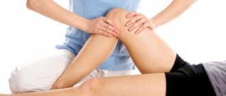
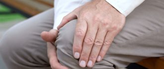
What causes knee pain?
If you clearly understand the nature of the pain syndrome, choosing a knee brace will not cause difficulties. Causes of knee pain:
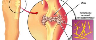
- Inflammatory processes: tendinitis, synovitis, chronic inflammation of the bursa, periarthritis, arthritis.
- Injuries: rupture or sprain of ligaments, bruise, dislocation of a joint, crack or fracture of the cup.
- Malignant and benign tumors: Baker's cyst.
- Vascular pathologies: venous diseases of the legs.
- Degenerative-dystrophic changes: bone tuberculosis, Osgood-Schlatter and Koenig diseases, gonarthrosis, arthrosis.
Before buying a knee brace, you need to undergo an examination. Only a doctor can make the correct diagnosis and tell you whether knee pads are needed or not, and will also help you choose the type.